

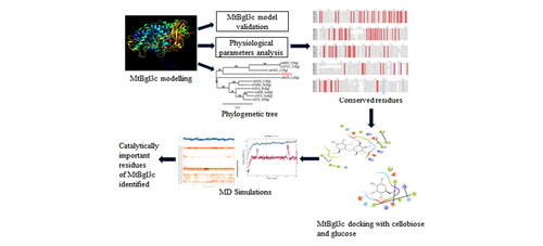
Abstract
Cellulases are the enzymes with diverse range of industrial applications. Cellulases degrade cellulose into monomeric glucose units by hydrolysing β-1,4-glycosidic bonds. There are three components of cellulases: a) endoglucanase, b) exoglucanase and c) β-glucosidase which act synergistically in cellulose bioconversion. The cellulases are the third largest industrial enzymes with a great potential in bioethanol production. In this investigation, a β-glucosidase of a thermophilic fungus Myceliophthora thermophila (MtBgl3c) was analysed for its structural characterization using in silico approaches. The protein structure of MtBgl3c is unknown, therefore an attempt has been made to model 3D structure using Modeller 9.23 software. The MtBgl3c protein model generated was validated from Verify 3D and ERRAT scores of 89.37% and 71.25%, respectively derived from SAVES. Using RAMPAGE the Ramachandran plot was generated, which predicted the accuracy of the 3D model with 91.5% amino acid residues in the favored region. The ion binding and N-glycosylation sites were also predicted. The generated model was docked with cellobiose to predict the most favorable binding sites of MtBgl3c. The key amino acid residues involved in cellobiose bonding are Val88, Asp106, Asp287, Tyr255, Arg170, Glu514. The catalytic conserved amino residues of MtBgl3c were identified. The dock score of cellobiose with MtBgl3c is much lower (–6.46 kcal/mol) than that of glucose (–5.61 kcal/mol), suggesting its high affinity for cellobiose. The docking data of MtBgl3c with glucose illustrate its tolerance to glucose. The present study provides insight into structural characteristics of the MtBgl3c which can be further validated by experimental data.
3D structure of β-glucosidase (MtBgl3c) of Myceliophthora thermophila is being proposed based on computational analyses
The amino acid residues Asp106, Asp287, Tyr255, Arg170 and Glu514 have been identified to play catalytically important role in substrate binding
Docking and interaction of MtBgl3c with cellobiose and glucose has been confirmed
Docking analysis of MtBgl3c with glucose suggested its glucose tolerance
The data would be useful in engineering enzymes for attaining higher catalytic efficiency
Highlights
Communicated by Ramaswamy H. Sarma
Graphical abstract
Acknowledgements
TS is grateful to DBT-Indo-US Science & Technology Forum (File No. IUSSTF/JCERDC-SGB/IUABC-UDSC/2016) and University Grants Commission, New Delhi [File No. 18-1/2011 (BSR) dated Feb, 15, 2016] for providing financial assistance. AD wishes to express her gratitude to DST-Indo-US Science & Technology Forum, New Delhi for the award of fellowship under WISTEMM Overseas Student Internship Program (2020_02085). We duly acknowledge Dr. Amaresh Kumar Sahoo (Assistant Professor), Department of Applied Sciences, Indian Institute of Information Technology Allahabad (India) for his guidance in revision of the manuscript and providing computational facilities for docking and molecular dynamics simulation studies.
Disclosure statement
The authors do not have any conflicting, competing and financial interests.