

Abstract
The main objective of the present study is to investigate the molecular structure and DNA binding interaction of the tyrosyl-lysyl-threonine (YKT) tripeptide, which has anticancer, antioxidant and analgesic properties, using various in silico (MD, QM, molecular docking), spectroscopic (UV, FT-IR, FTIR-ATR, Raman, gel electrophoresis) and in vitro (MCF-7 and HeLa cancer cell lines and BEAS-2B cell line) methods. The optimized geometry, vibrational wavenumbers, molecular electrostatic potential (MEP), natural bond orbital (NBO) and HOMO-LUMO (highest occupied molecular orbital- lowest unoccupied molecular orbital) calculations were carried out with Density Functional Theory (DFT) using B3LYP/6-311++G(d,p) basis set to indicate conformational, vibrational and intramolecular charge transfer characteristics. The assignment of all fundamental theoretical vibration wavenumbers was performed using potential energy distribution analysis (PED). DNA is a significant pharmacological target of drugs in several diseases such as cancer. For this reason, molecular docking calculation was used to elucidate the binding and interaction between YKT tripeptide and DNA at the atomic level. Also, the dynamic behaviors of YKT and DNA was examined using MD simulations. Besides, the interaction of YKT with DNA was experimentally examined by UV titration method and agarose gel electrophoresis method. Experimental results showed that YKT was intercalatively and electrostatically bound to CT-DNA (Calf thymus DNA) and cleavage pBR322 DNA in the presence of H2O2. The pharmacokinetic profile of YKT was also obtained. Cytotoxic effect of YKT was evaluated on MCF-7, HeLa and BEAS-2B cell lines. Hence, these studies about YKT tripeptide may pave the way for the development of various cancer drugs.
Communicated by Ramaswamy H. Sarma
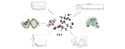
Graphical Abstract
Acknowledgments
This study consists of the results of the doctorate thesis study and was supported by the Scientific Research Project Coordination Unit of Istanbul University [FDK-2018-32253, ONAP-2423] and it was supported by International Research Scholarships for Research Assistants scholarship of The Council of Higher Education. In this study, the infrastructure of Applied Nanotechnology and Antibody Production Laboratory established with TUBITAK support (project numbers: 115S132 and 117S097) was used. Authors would thank to TUBITAK for their support. MCF-7 and HeLa cell lines were kindly gifted by Dr. M. Topçul and Dr. I. Cetin (Stem Cell and Biomolecular Technology Research Laboratory, Istanbul University) and BEAS-2B cell lines were kindly gifted by Dr. Nazlıhan Aztopal (Istinye University).
Disclosure statement
The authors have no conflicts of interest to declare.
Ethical approval
No animals were used in this study. Ethics approval was not required.