

Abstract
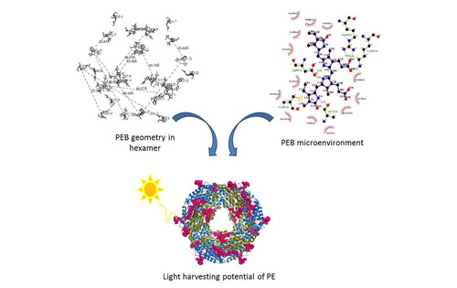
Phycoerythrin (PE) is green light-absorbing pigment-protein that assists in efficient light harvesting in cyanobacteria and red-algae. PE in cyanobacteria stays less studied so far as compared to that in red algae. In this study, PE from marine cyanobacteria Halomicronema sp. R31DM is purified and subjected for its structural characterisation by X-ray crystallography in order to understand its light-harvesting characteristics. The crystal structure is solved to a resolution-limit of 2.21 Å with reasonable R-factors values, 0.16/0.21 (Rwork/ Rfree). PE forms hexamer of hetero-dimers made up of two peptide chains, α- and β-subunits containing 2 and 3 phycoerythrobilin (PEB) chromophores covalently attached to them, respectively. Geometry of five chromophores is analysed along with their relative position within the PE hexamer. Also, their interactions with the surrounding microenvironment are analysed. The plausible energy transfer pathways in hexamer structure have been predicted based on relative position and geometry of chromophores. This structure enriches the structural information of cyanobacterial PE in order to understand its light-harvesting capacity.
Communicated by Ramaswamy H. Sarma
Acknowledgements
Authors thank Dr Ravindra D Makde, BARC for his help in diffraction data collection. DM and SNP are thankful to PDPIAS, CHARUSAT and Bhartiben and P. R. Patel Biological Research Laboratory for providing instrumentation facility to conduct this research. RRS acknowledges National Postdoctoral fellowship grant (N-PDF) (funded by Science and Engineering Research Board, New Delhi) and NAWA-Ulam fellowship grant (grant PPN/ULM/2019/1/00175 funded by Polish National Agency for Academic Exchange, Poland).
Disclosure statement
The authors declare that they have no competing interests.
Accession numbers
The coordinates and structure factors have been deposited to the Protein Data Bank (PDB) with PDB ID, 7F86.