Abstract
Introduction: Face transplantation (CT) is a complex and innovative treatment that restores function, aesthetics and allows the social reintegration of patients with severe facial deformities (1). TC involves not only the development of anatomy-surgical techniques, but also the manufacture of a mask congruent with the donor site. Our experience in the manufacture of the mask with a third dimension (3D) technology for the dignification of the donor is presented. Presented at the 3rd World Congress of the UNESCO Chair for Teaching and Research in Digital Anatomy Paris Descartes at Egas Moniz University Institute - Almada, Portugal.
Materials and methods: Stereo photometry technology was used, combining the obtention of geometric and photogrammetry calculations. With this, the donor site was scanned in a fraction of a second. Subsequently, various softwares were used, the surface mesh was calculated and a 3D model was generated. It was printed on PLA and ABS materials. Once the physical model was obtained, it was defined artistically. 24 hours later, the “mask” was placed on the donor site.
Results: With the use of stereo photometry, the completion times were reduced from 2.5 hours to 1 minute to obtain the model, and 10 minutes to obtain the 3D printing file. The total time was 24 hours, also allowing a process of minimizing invasion for the 3D scanning of the donor site. A mask of punctual proportion was obtained to the donor site while retaining facial features.
Figure 1. A,D) 3D facial reconstruction with stereophotometry technique. B,E) Representation of the 3D file optimized for 3D printing. The orange line shows the thickness of the printing layer. C) 3D printed donor site prosthesis, PLA material, unpainted. F) Final prosthesis. G) Donator of facial tissues. H) Dignification of cadaver with 3D printed mask.
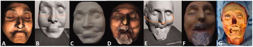
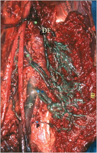
Figure 2. Venous arcades of the semi-membranosus muscle, bypassing the Hunter canal by their lower arcades (blue dots) connected into the popliteal vein (P) and their higher arcades (green dots) connected into the deep femoral vein.