

Abstract
Introduction: Teaching of the morphological sciences suffers today from the lack of human corpses for dissection (due to ethic or religious issues), worsened by an increasing demand for educational anatomy.
Fortunately, the technological revolution now put at our disposal educational tools to teach and learn anatomy: virtual dissection table, virtual reality techniques, and 3D printing.
The network of partners of the Unesco Chair of Digital anatomy [Citation1] in Paris Descartes University collaborates to setup educational models of the human body in order to build 3D databases for virtual dissection.
This can be done by 2 different techniques producing 3d vectorial models:
• Manual segmentation of the anatomical slices of “visible human” projects: US VHP [Citation2] Korean visible human [Citation3] and Chinese projects [Citation4,Citation5] as well as modeling of the uro-genital system [Citation6]
• Modeling of angio-CT by semi-automatic segmentation. Can be achieved on patients exams by the use of Horos software. [Citation7]
Material and methods: 250 students preparing the master of anatomy of Descartes University (DUACN) have worked since 2015 on this project.
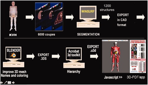
They used the winsurf software version 3.5 [Citation8,Citation9] to make the segmentation of the main anatomical structures on the 5650 slices of the Korean visible woman (thickness 0.2 mm) Then, the whole 3d vectorial model has been exported into Blender software version 4.79 [Citation10] to improve the meshes and correct mistakes. Finally the model was converted into .u3d format, in order to take advantage of the powerful interface of 3dpdf Acrobat file [Citation11] The main steps are shown on .
Figure 1. Methodology of building the 3dpdf file from the anatomical slices.
Results: The display device is a 65 inches touch screen: all the 3d functions could be easily controlled with only 3 fingers. The final 3D model includes 1200 anatomical structures. The 3dpdf Acrobat interface makes possible to move, zoom, rotate each of them. Their transparency could be modified.
The selection is possible by system (bones, ligaments, muscles, vessels, nerves, organs) or by region. The original slices can be displayed within the 3d model.
The name of a selected structure is displayed in 4 languages.
A window on the left is available to drop and display anatomical drawings pictures and texts, so that anyone could easily build a course or a lesson of anatomy.
Discussion and conclusion: The Korean team has already provided 3d vectorial models of the man and woman [Citation12]. The Unesco Chair of Digital Anatomy aims to develop and share these new tools in the laboratories of anatomy around the world.