Abstract
Introduction: Students learn and process information in different ways. In macroscopic anatomy, certainly the visual part plays an important role [Citation1]. The increasing prevalence of technology means that medical educational software applications have an increasingly important role in medical education. We come up with our experience of 5 years in the creation of models and three-dimensional (3D) materials applied to the teaching and understanding of macroscopic anatomy. Presented at the 3rd World Congress of the UNESCO Chair for Teaching and Research in Digital Anatomy Paris Descartes at Egas Moniz University Institute - Almada, Portugal.
Materials and methods: Surface scanning technologies such as 3D scanners and photometry software were used to obtain 3D models made from anatomical dissection models in cadaveric material. The models were loaded in the free Access online platform (www.proyectohdm.com) applied to online 3D visualization systems, anaglyph, augmented reality, virtual reality, as well as advanced prototypes of mixed reality and 3D printing in solid and flexible materials to obtain surgical educational simulators.
Results: An online platform, easily accessible and multiplatform, was created with 50 three-dimensional topographic models of different anatomical structures, which allow users to view all angles of the anatomical piece. 3D anatomical models can be visualized in technologies such as anaglyph, augmented reality and virtual reality. The 3D printing allowed obtaining replicas of the cadaveric model of dissection, in various types of materials at low cost for its anatomical study. Printing on flexible materials enabled the possibility of creating educational neurosurgical simulators. The platform currently has more than 100 thousand monthly visitors from around the world.
Discussion and conclusions: The use of 3D technologies applied to the field of anatomical study has been previously demonstrated [Citation2]. The anatomical models presented in this work to be generated from the cadaverous structure allow a high level of detail and precision. Through ts incorporation into visualization systems in anaglyph technologies, virtual reality and increased reintegration of the virtual model to a tangible plane, using mixed reality and 3D printing, students and academics have a better understanding of the three-dimensional disposition of the anatomical structure manifested by the academic acceptance of the online platform, based on the growing number of visitors.
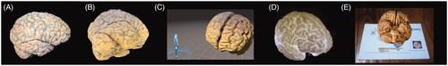
Figure 1. A) Brain photography B) Reconstruction 3D brain C) Virtual reality D) 3D printing flexible material E) Augmented reality.