

Abstract
Introduction: Periodontitis presents complex bacterial biofilms and multifactorial mechanisms, being its management a major challenge [Citation1–3]. Throughout periodontitis treatment period, patients’ oral hygiene habits play a key role on periodontal therapy success. The aim of this study was to assess pocket depth (PD) variation levels of upper and lower teeth at 3, 6 and 12 months’ follow-up in patients that underwent non-surgical periodontal treatment.
Materials and methods: 24 patients, from the Periodontology Department of the Egas Moniz Dental Clinic, were retrospectively analyzed over the period of 2013–2017. The patients had moderate to severe periodontitis according to Page and Eke’s case definitions [Citation4], and were treated by dentists at various levels in their specialist training. All patients received non-surgical periodontal treatments and follow-up visits at 3, 6 and 12 months, with plaque and dental calculus control. Six sites per tooth were measured (mesiobuccal, buccal, distobuccal, mesiolingual, lingual and distolingual) and recorded, excluding third molars. PD was measured with a CP-12 SE (Hu-Friedy, Chicago, IL, USA). This study was approved by the Egas Moniz Ethics Committee (IRB approval number: 595).
Results: The upper and lower teeth initial mean (± standard deviation) PD were 4.85 (±1.13) and 4,93 (±1.23), respectively, and they were not statistically different (p = 0.2146, independent t-test). displays radar plots for PD recovery at 3, 6 and 12 months’ follow-up of non-surgical periodontal treatment.
Figure 1. Radar plot for 3, 6 and 12 months’ follow-up of non-surgical periodontal treatment.
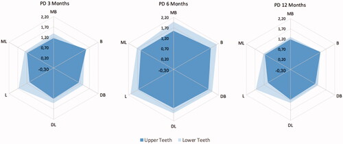
Discussion and conclusions: Overall, lower teeth presented PD healing more increased than upper teeth. Furthermore, 6 months’ therapy follow-up represented the recovery peak. At 12 months, the recovery levels were similar to the 3 months’ values. The lingual site from lower teeth presented the greatest recovery. Future research is necessary to unveil this specific interval raised variation.