Abstract
Introduction: The avulsion of a tooth is defined as a complete displacement from its alveolus.[Citation1] Dental avulsion of permanent teeth is considered one of the most severe and aggressive dental injuries. According to dental trauma studies in different populations, it comprises from about 0.5-7.75% of the total of dental injuries, being more frequent between 9 and 10 years of age [Citation2–6]. The purpose of this study was to evaluate the success of existing protocols when dealing with dental avulsion.
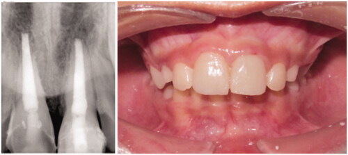
Materials and methods: The informed consent in use was signed at the pediatric service of Clinica Universitária Egas Moniz. A 7-year-old female patient suffered craniofacial trauma with avulsion of the upper right central incisor (URCI) and enamel-dentin coronary fracture of the upper left central incisor (ULCI). The avulsed tooth, with open apex, was transported in milk and reimplanted after 16 hours. At Clinica Universitária Egas Moniz pediatric emergency service, the URCI was carefully cleaned from non-viable tissue. It underwent extra-oral endodontic treatment with an apical MTA plug, filled with thermoplastic gutta-percha and later closed with composite resin. Subsequently, under local anesthesia, the clot was removed from the alveolus and irrigated with saline solution. The tooth was reimplanted into the socket and stabilized with semi-rigid splint to adjacent teeth for 4 weeks. The ULCI was sealed with glass ionomer and later restored with composite resin. The patient was medicated with amoxicillin and clavulanic acid for 8 days, being instructed to a soft diet and to avoid sports practice. Follow-up appointments at 7 days, 4 weeks with splint removal, 3, 6 and 9 months. At 3 months, the ULCI presented with necrosis, so endodontic treatment was performed. Last follow-up was at 18 months.
Results: Clinical evaluation after more than one year showed absence of clinical symptoms and radiographic images compatible with periapical health.
Discussion and conclusions: The URCI suffered ankylosis with radiographic loss of periodontal ligament space, and slight root resorption. These findings are in agreement with the expected complications in the literature. Similar studies agree that the prognosis of the treatment of this trauma is deeply influenced by on-site actions and care taken shortly after the accident [Citation1–8]. However, the patient was allowed to maintain aesthetic, functional and physiological function, as well as preservation of the alveolar contour.
Figure 1. vulsion of the URCI; non-complicated fracture the ULCI.

Figure 2. Removal of the cloth and cleaning of the alveolus; pulpal aspect of the URCI before endodontic treatment.
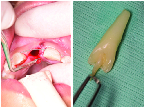
Figure 3. Reimplantation of the URCI and splinting.
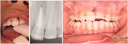
Figure 4. Endodontic treatment of the ULCI.
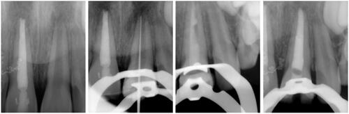
Figure 5. Follow-up at 18 months.