Abstract
A 44-year-old man with acute renal failure and antineutrophil cytoplasmic antibodies (ANCA) positivity was described. The first renal biopsy specimen showed tubulointerstitial nephritis (TIN) with normal glomeruli. However, delayed recovery of renal function with low-dose steroid treatment for TIN prompted a second renal biopsy 1 month later; and the specimen demonstrated a dramatically different morphology, with necrotizing and crescentic glomerulonephritis. Improvement in renal function occurred, together with reduction of ANCA titers, following intensive immunosuppressive therapy. This case illustrates an unusual presentation of TIN in ANCA-associated renal vasculitis. The possible pathogenetic mechanism are discussed.
INTRODUCTION
The characteristic renal lesion of antineutrophil cytoplasmic antibodies (ANCA)-associated vasculitic disorders, such as microscopic polyangiitis, Wegener's granulomatosis (WG), Churg-Strauss syndrome, and renal-limited vasculitis, is pauci-immune necrotizing and crescentic glomerulonephritis. Renal biopsy must be performed early to make the diagnosis if successful treatment is to be implemented. However, some renal biopsy specimens from patients with ANCA-associated diseases only have tubulointerstitial inflammation without glomerular lesions. Such expression of renal disease is supported by the observation of a few patients with a clinical course that is consistent with tubulointerstitial nephritis (TIN) rather than glomerulonephritis.[Citation[1]] Therefore, it has been speculated that TIN is a more benign presentation form within the spectrum of ANCA-associated renal vasculitis. However, we report an unusual case of acute renal failure (ARF) and ANCA positivity presenting with transformation from TIN to crescentic glomerulonephritis. To our knowledge, only one case of WG with this unique presentation has been described in the literature.[Citation[2]]
CASE REPORT
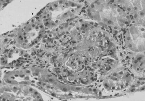
A 44-year-old man was admitted with a 4-week history of profound malaise. Two weeks after the onset of symptoms, he had noticed lower-extremity edema for which he started taking herbal preparation, but otherwise he did not receive any medications from a medical doctor or over the counter. There were no complaints of arthralgia, epistaxis, hemoptysis, or upper respiratory tract symptoms. His past medical history was unremarkable, and he had had no any antecedent illness. He had no known allergies. On admission, physical examinations showed a body temperature of 36.8°C, respiratory rate of 20 breaths/min, pulse rate of 100 bpm, and blood pressure of 160/100 mmHg. Examinations of the lungs, heart, abdomen, and nervous systems were normal. He had 2+ lower-extremity pitting edema. No skin lesion was seen. Laboratory studies revealed hemoglobin 13.4 g/dL, hematocrit 38.9%, platelet count 244,000/uL, and white blood cell count 9000/uL, with 77.9% neutrophils, 13.3% lymphocytes, 3.8% monocytes, and 4.8% eosinophils. A chemistry panel revealed blood urea nitrogen 100.2 mg/dL, creatinine 9.6 mg/dL, sodium 122 mmol/L, potassium 6.3 mmol/L, calcium 6.3 mg/dL, phosphorus 11.9 mg/dL, glucose 112 mg/dL, uric acid 10.2 mg/dL, albumin 2.5 g/dL, and globulin 2.8 g/dL. Erythrocyte sedimentation rate was 103 mm/h and C-reactive protein was 2.69 mg/dL. Urinalysis showed 3+ blood and 4+ protein, with 20 to 25 red blood cells and 1 to 3 white blood cells/high-power field in the urinary sediment. Urine culture was negative. Both serum and urine protein electrophoresis were negative for paraproteins. A chest radiograph was normal. Renal ultrasound demonstrated normal-size kidneys and no hydronephrosis. Serologic studies showed antinuclear antibody, anti-double-stranded DNA antibody, hepatitis B surface antigen, and hepatitis C antibody to be negative. An indirect immunofluorescence test for ANCA was positive, with a perinuclear pattern at a titer of 1:320. Serum C3 and C4 complement levels were normal. After control of uremia by hemodialysis therapy, a renal biopsy was performed on hospital day 5. The tissue contained 25 glomeruli. All had a normal appearance. However, diffuse infiltrate of mononuclear cells was present in the interstitium and tubular epithelium (). The renal vessels were essentially normal. A diagnosis of TIN with unknown etiology was made and low-dose oral prednisolone (0.5 mg/kg/day) was administered beginning on hospital day 6. However, the renal function failed to improve, and the patient remained dialysis dependent over the ensuing 1 month. A second renal biopsy was performed on hospital day 30 and demonstrated dramatically different morphology. Six of 10 examined glomeruli showed necrotizing lesion with cellular crescents (). Interstitial mononuclear infiltrate was still present but less predominant. No vasculitis or granuloma was found. Immunofluorescence showed no deposition of immunoglobulin or complement in the glomeruli and tubular basement membranes. No electron-dense immune complex deposition was seen by electron microscopy. The patient was treated with 1 g/day pulse intravenous methylprednisolone for 3 consecutive days beginning on hospital day 31, followed by 1 mg/kg/day oral prednisolone and 2 mg/kg/day oral cyclophosphamide. On hospital day 43, increased urine output was noted, and dialysis support was stopped. Over the following days, his renal function steadily improved and serum creatinine levels declined to 2.5 mg/dL when he was discharged home on hospital day 51. Over the ensuing 1 month, his renal function continued to improve, with serum creatinine levels returning toward normal, and serologic follow-up ANCA titers had fallen to 1:40.
Figure 1. First renal biopsy specimen showing diffuse interstitial infiltrate of mononuclear cells with normal glomeruli (hematoxylin and eosin stain ×100).
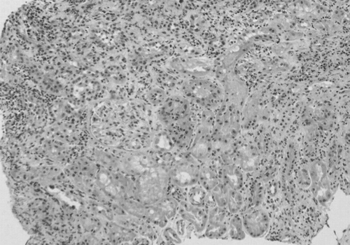
Figure 2. Second renal biopsy specimen showing glomerular necrosis and circumferential cellular crescent (hematoxylin and eosin stain ×400).
DISCUSSION
Although the pathogenesis of ANCA-associated vasculitis remains unclear, the presence of ANCA suggests an autoimmune basis. Diseases due to an immune mechanism generally affect primarily the glomeruli, with secondary involvement of the tubules and interstitium. It has been demonstrated that renal lesion of ANCA-associated systemic vasculitis is characterized by pauci-immune necrotizing glomerulonephritis. However, predominant TIN occurring in patients with ANCA-associated diseases has been anecdotally reported in the literature. Geffriaud-Ricouard et al. described one WG case with antimyeloperoxidase ANCA presenting with granulomatous interstitial nephritis without glomerular lesions.[Citation[3]] Fannin et al. and Ernam et al. each reported one case of WG with cytoplasmic ANCA positivity in which the renal biopsy specimen showed isolated TIN.[Citation[4],Citation[5]] Furthermore, Lockwood described four cases of ANCA-associated acute interstitial nephritis, all without evidence of extrarenal vasculitis.[Citation[1]] More interestingly, Banerjee et al. described another case of WG and cytoplasmic ANCA positivity presenting with acute interstitial nephritis. However, a second renal biopsy 9 weeks later demonstrated dramatically different morphology, with necrotizing and crescentic glomerulonephritis. The patient remained dialysis dependent despite treatment with corticosteroids, and cyclophosphamide was commenced thereafter.[Citation[2]] In our patient, the first renal biopsy, with adequate tissue analyzed to exclude the possibility of focal glomerulonephritis that was not sampled in the biopsy specimen, demonstrated isolated TIN without glomerular lesions. However, there were no detectable common causes of TIN. The presence of ANCA was a surprise, and we did not consider it relevant to the development of TIN at the time of the diagnosis of TIN. Because the renal function did not improve with low-dose steroid treatment for TIN, a second renal biopsy showed characteristic renal lesions of ANCA-associated glomerulonephritis, with pauci-immune necrotizing and crescentic glomerulonephritis. Improvement in renal function occurred, along with reduction of ANCA titers, after intensive immunosuppressive therapy was given. Therefore, we conclude that ANCA-associated renal vasculitis was the underlying disease and was responsible for the TIN in our patient.
The mechanism of ANCA-associated TIN is enigmatic. Although the possibility cannot be ignored that the tubulointerstitial changes may be caused or modified by necrotizing angiitis, this direct relationship remains to be established because the identification of vasculitis in vessels other than glomerular capillaries is found in less than 10% of renal biopsies. It is now recognized that immune-mediated mechanisms are important in initiating or amplifying interstitial damage. The presence of mononuclear cell infiltrates in the tubules and interstitium without antibody deposition suggest a cell-mediated injury. The observations of elevated ANCA titers in drug-induced TIN and in TIN and uveitis syndrome have implicated that ANCA may have some relevance to the development of TIN.[Citation[6],Citation[7]] More recently, it was demonstrated that ANCA-mediated neutrophil activation may play an important role in the pathogenesis of pauci-immune vasculitis or nephritis. Activated neutrophils, releasing lysosomal enzymes and oxygen radicals, were shown to be present not only in affected glomeruli, but also in the renal interstitium, which could contribute to a direct cell-mediated injury to the interstitium and the glomeruli.[Citation[8],Citation[9]] However, further studies are necessary to investigate the pathogenic role of cell-mediated immunity in ANCA-associated diseases. In contrast, it is not clear whether the transformation process from predominant tubulointerstitial lesions to glomerular lesions is a natural history or a variant of the disease.
In conclusion, ANCA could be present in tubulointerstitial and glomerular nephritis in patients with ARF. The prognosis of ANCA-associated TIN is not always favorable. The possibility of transformation to crescentic glomerulonephritis should be kept in mind in patients with TIN and ANCA positivity. Follow-up renal biopsy is recommended when the clinical history is inconsistent with the relatively benign course of TIN.
REFERENCES
- Lockwood CM. Antineutrophil cytoplasmic autoantibodies: the nephrologist's perspective. Am J Kidney Dis 1991; 18: 171–174, [PUBMED], [INFOTRIEVE]
- Banerjee A, McKane W, Thiru S, Farrington K. Wegener's granulomatosis presenting as acute suppurative interstitial nephritis. J Clin Pathol 2001; 54: 787–789, [PUBMED], [INFOTRIEVE]
- Geffriaud-Ricouard C, Noel LH, Chauveau D, Houhou S, Grunfeld JP, Lesavre P. Clinical spectrum associated with ANCA of defined antigen specificities in 98 selested patients. Clin Nephrol 1993; 39: 125–136, [PUBMED], [INFOTRIEVE]
- Fannin SW, Hagley MT, Seibert JD, Koenig TJ. Bronchocentric granulomatosis, acute renal failure, and high titer antineutrophil cytoplasmic antibodies: possible variants of Wegener's granulomatosis. J Rheumatol 1993; 20: 507–509, [PUBMED], [INFOTRIEVE]
- Ernam D, Atikcan S, Yilmaz A, Atalay F, Demirag F, Memis L. An unusual renal presentation of Wegener's granulomatosis. Tuberk Toraks 2003; 51: 193–196, [PUBMED], [INFOTRIEVE]
- Okada K, Okamoto Y, Kagami S, et al. Acute interstitial nephritis and uveitis with bone marrow granulomas and anti‐neutrophil cytoplasmic antibodies. Am J Nephrol 1995; 15: 337–342, [PUBMED], [INFOTRIEVE]
- Montoliu J, Amoedo ML, Panades MJ, Ramos J. Lessons to be learned from patients with vasculitis. Nephrol Dial Transplant 1997; 12: 2781–2786, [PUBMED], [INFOTRIEVE], [CROSSREF]
- Brouwer E, Huitema MG, Mulder AHL, et al. Neutrophil activation in vitro and in vivo in Wegener's granulomatosis. Kidney Int 1994; 45: 1120–1131, [PUBMED], [INFOTRIEVE]
- Weidner S, Carl M, Reiss R, Rupprecht HD. Histologic analysis of renal leukocyte infiltration in antineutrophil cytoplasmic antibody-associated vasculitis: importance of monocyte and neutrophil infiltration in tissue damage. Arthritis Rheum 2004; 50: 3651–3657, [PUBMED], [INFOTRIEVE], [CROSSREF]