Abstract
Tubulointerstitial nephritis and uveitis (TINU) syndrome is a rare entity first described in 1975, affecting mainly young women and adolescents. We present a case of a 52-year-old female patient (one of the oldest in the literature) who complained of fever, anorexia, nausea, and vomiting. After she was admitted to our hospital, laboratory tests revealed tubular proteinuria, elevated erythrocyte sedimentation rate (ESR), anemia, and renal insufficiency (serum creatinine 4.2 mg/dL) with metabolic acidosis. Ophthalmologic examination revealed anterior uveitis (iritis) and renal biopsy showed acute tubulointerstitial nephritis. The diagnosis of TINU syndrome was established and the patient was treated with oral corticosteroids. All symptoms and ophthalmologic abnormalities disappeared after 6 weeks of treatment. Renal function also recovered completely and remained stable at follow-up. TINU syndrome should be considered in the differential diagnosis of unexplained tubulointerstitial nephritis, especially in the presence of ocular findings. Corticosteroid therapy is still controversial, but it helps in the quick resolution of renal and mainly eye abnormalities.
INTRODUCTION
Tubulointerstitial nephritis and uveitis syndrome, also known as TINU syndrome,Citation[[1]] was first reported in 1975 by Dobrin et al.Citation[[2]] and since then more than 100 cases have appeared in the literature, mainly in ophthalmology and pediatric nephrology journals.Footnote[[3]] The pathogenesis of the syndrome is not well understood, although delayed-type hypersensitivity and suppressed cell-mediated immunity have been implicated.Footnote[3–7]
Both conditions, interstitial nephritis and uveitis, are common; their concurrence, however, is quite rare. A high degree of suspicion is, therefore, essential for an early identification of patients and a prompt treatment of the disease. The condition should be familiar to many specialists, including pediatricians, nephrologists, ophthalmologists, and rheumatologists.
So far, the majority of cases presented included young adolescents, while few reports exist regarding older patients.Citation[[8]] We present here the case of a 52-year-old female patient with TINU syndrome, one of the oldest reported in the literature.
CASE REPORT
A 52-year-old previously healthy woman was admitted to our hospital with a 7-month history of fever (max. temperature 38°C). Before admission she had taken no medications apart from paracetamol for enhanced temperature. In the last 4 weeks before admission she had suffered from anorexia, fatigue, loss of weight, and for the last 10 days, nausea and vomiting. Furthermore, she complained of bilateral ocular pain and somewhat blurred vision.
Except from pale skin and mucosae, physical examination was normal with no evidence of lymphadenopathy. Blood pressure was 135/80 mmHg.
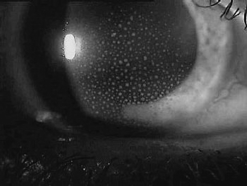
Ophthalmologic examination revealed a visual acuity of 10/10, a tone of 18/11 mmHg, and a reduced break-up time (mild xerophthalmia). Slit lamp examination revealed anterior uveitis (iritis) of the left eye with corneal precipitates ().
Figure 1 Anterior uveitis (iritis) with multiple keratic precipitates seen on the corneal endothelium.
White blood cell (WBC) count was 9,500/mL, with normal differential. Hematocrit was 30.1%, hemoglobin 10.4 g/dL, platelet count 330,000/mL, and the erythrocyte sedimentation rate (ESR) 85 mm/h. On admission, serum urea measured 90 mg/dL and serum creatinine was 3.5 mg/dL and 4.2 mg/dL 2 days later. Creatinine clearance was estimated 12 mL/min. Arterial blood gases analysis revealed a metabolic acidosis with a pH value of 7.28 (pCO2 24 mmHg, HCO3− 11 meq/L). Urine examination showed pyuria (8–10 white blood cells per HPF) and low molecular weight (tubular) proteinuria of 0.9 g/24 h. Both urine and blood cultures came out negative. Ziehl-Nielsen stain and bone marrow culture were negative as well. Ferritin, vitamin B12, and folate levels were normal. ANA, AMA, and ANCA were negative. Enhanced levels of IgG (1600 mg/dL, normal range 500–1300) and CRP (5.4 mg/dL, normal range <0.5) were determined, while IgA, IgM, C3, C4, RF, and ASTO were within the normal limits. The concentrations of tumor markers (CA 15–3, CA 125, CA 19–9, CEA, and aFP) were normal. The patient was euthyroid (TSH 0.85 μIU/mL, T3 0.2 ng/mL, FT4 1.42 ng/dL). A micronodular goiter was assessed and cervical lymphadenopathy was excluded on cervical ultrasound.
ECG and chest x-ray were normal. Endoscopy of the upper gastrointestinal tract revealed a sliding diaphragmatocele and gastritis. On abdominal ultrasound, the kidneys appeared normal (11 cm in longitudinal diameter), while the findings of computed tomography of the chest and abdomen were unremarkable.
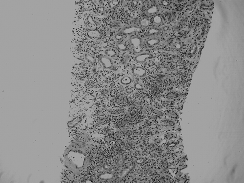
A renal biopsy was conducted and the diagnosis of acute tubulointerstitial nephritis was established. Light microscopy revealed expansion of the interstitium due to interstitial edema and infiltration of inflammatory cells composed of lymphocytes, plasma cells and few polymorphonuclear leucocytes (). Renal tubules were involved with tubulitis, disruption of the tubular basement membrane, and exudation of Tamm-Horsfall protein. Glomerular and vascular structures were preserved apart from a mild intimal fibrosis and hyalinosis of an interlobar artery. Immunofluorescence microscopy showed granular mesangial deposition of C3.
Figure 2 Diffuse, severe interstitial inflammation mainly with lymphocytes and plasma cells accompanied by tubulitis (H/E stain × 100).
During hospitalization, the patient manifested a night fever of 37.4°C and she received isotonic saline for hydration. Bicarbonate was administered as symptomatic treatment for metabolic acidosis. She was further treated with a course of oral corticosteroids (48 mg of methylprednisolone daily for 7 days followed by a quick dose tapering to 16 mg), omeprazole and rHuEPO (erythropoietin beta). For uveitis corticosteroids (g. Maxidex, dexamethasone 0.1% eye drops) and cycloplegics (g. Cyclogyl, cyclopentolate 1% eye drops) were applied topically both in tapering dose over 4 weeks and iritis resolved without sequelae.
Under the above treatment all clinical manifestations, including fever, nausea, and vomiting, disappeared promptly. The patient was discharged 20 days after admission free of symptoms, on oral methylprednisolone (Medrol 16 mg o.d.). WBC count remained well below 10,000/mL, while renal function was improved (serum urea 40 mg/dL, serum creatinine 2.1 mg/dL). At discharge, urinary protein excretion was 500 mg/24 h and blood gases (i.e., pH value) were normalized without further need for bicarbonate treatment.
Corticosteroids (Medrol 4 mg o.d.) were continued for 6 months and the patient remained in follow up for 12 months. During that time, ocular manifestations and all laboratory findings of renal dysfunction subsided completely, with normal serum creatinine (0.95 mg/dL) and no proteinuria (less than 50 mg/24 h).
DISCUSSION
Tubulointerstitial nephritis and uveitis remains a rare syndrome and is mainly a diagnosis of exclusion. It presents with symptoms and signs of uveitis and interstitial nephritis, as well as with systemic findings, such as fever (53%), rash, arthralgia and malaise (44%), anorexia (28%), and weight loss (47%). Uveitis is predominantly anterior (80%) and bilateral (77%), however, it may sometimes be posterior.Citation[[9]] In the majority of cases, nephritis appears first (65%), but there are cases in which uveitis precedes renal disease (21%), or develops concurrently (15%).Citation[[8]]
Renal symptoms are typical for acute interstitial nephritis and include flank pain (28%), pyuria (mainly sterile), hematuria, and proteinuria (below nephrotic range). A degree of renal impairment is also frequent, which can progress to acute renal failure or end-stage renal disease (ESRD).Citation[[10]], Citation[[11]]
The syndrome occurs more often in adolescents and young women (median age 15 years),Citation[[12]] although reports exist regarding adults.Citation[[7]], Citation[13–15] It is three times more frequent in females and there appears to be no racial or familial predisposition.Citation[[16]] True incidence of the syndrome is difficult to calculate, but can range from 2–8% of patients with uveitis in a tertiary hospital setting.Citation[[17]] In a review of three series that totaled 128 patients of acute interstitial nephritis, a 4.7% incidence of TINU syndrome was reported.Citation[[18]] There is a tendency for TINU syndrome to occur earlier in males than in females (mean age 16.8 years vs. 28 years).Citation[[9]] It's the rarity of concurrence of interstitial nephritis with uveitis that makes the true incidence hard to define.Citation[[9]]
Laboratory findings are not typical and may include eosinophilia, anemia (mainly normocytic, normochromic), elevated erythrocyte sedimentation rate (ESR), and C‐reactive protein (CRP) and abnormal liver enzymes. Occasionally, patients may present with antineutrophil cytoplasmic antibodies (ANCA), antinuclear antibodies (ANA), rheumatoid factor (RF), or reduced complement levels.Citation[19–21] The determination of β2-microglobulin in the urine is helpful for the early discovery of acute interstitial nephritis, particularly in those patients without proteinuria and pyuria.Citation[[7]], Citation[[13]] Apart from decreased glomerular filtration rate (GFR), patients usually present with various distal or proximal tubular defects (i.e., aminoaciduria, glucosuria, urine-concentrating defects, acidosis, Fanconi's syndrome).Citation[[6]], Citation[[22]], Citation[[23]]
Histologic findings on light microscopy include tubulointerstitial edema with infiltrating lymphocytes, plasma cells, and histiocytes. The glomeruli are normal or exhibit mild proliferation of the mesangial matrix, with no signs of vasculitis. Sometimes noncaseating granulomas and eosinophilic infiltrates may be observed in the renal interstitium. Electron microscopy findings are nonspecific.Citation[[22]]
Differential diagnosis of TINU syndrome includes numerous disorders, like sarcoidosis,Citation[[4]], Citation[[13]], Citation[[24]] Sjögren syndrome,Citation[[25]], Citation[[26]] Behçet disease, Wegener granulomatosis, systemic lupus erythematosus (SLE), hyperthyroidism,Citation[[27]] primary hypoparathyroidism,Citation[[28]] and certain infectious diseases (tuberculosis, brucellosis, toxoplasmosis, herpes simplex).Citation[[23]], Citation[[29]] The various characteristic clinical and laboratory findings help to distinguish those entities from TINU syndrome. Nevertheless, it seems that the syndrome is underdiagnosed, as the condition is often overlookedCitation[[30]] and the patients are not always admitted to the hospital. The diagnosis of TINU syndrome still remains a diagnosis of exclusion. Sometimes a gallium scintigraphy is needed to confirm the diagnosis, but this is not always needed.Citation[[31]]
Although efforts have been made to ascribe TINU to drugs (which are known to cause interstitial nephritis), no causality could be documented, partly because these drugs are not known to cause uveitis and partly because patients usually do not exhibit rash, the most common feature of drug-induced reactions.Citation[[2]], Citation[[32]]
Uveitis is an intraocular inflammation, whose symptoms depend upon the extent of the uveal tract involved. Anterior uveitis usually presents with redness, pain, and various degrees of visual loss, while posterior uveitis is usually painless, but presents with floaters. Anterior uveitis can usually be distinguished from other causes of the red eye syndrome by the fact that redness is confined to the limbus (or the junction between the sclera and the cornea), as well as the presence of pain and constricted pupil.
The differential diagnosis of the “red eye syndrome” includes keratitis, conjunctivitis, episcleritis, scleritis, and acute closed angle glaucoma. The best way to distinguish between the different causes of red eye syndrome from anterior uveitis is by the slit lamp eye examination, where, in the case of anterior uveitis, leukocytes are found in the aqueous humor, that fills the space between the cornea and the lens. In comparison, signs of active chorioretinal inflammation and the presence of leukocytes in the vitreous humor are suggestive of posterior uveitis.
Uveitis can have a multiple etiology: inflammatory, systemic, masquerade syndromes, and syndromes confined primarily to the eye. Infectious causes of uveitis include CMV infection (especially in the immunocompromised host),Citation[[33]] toxoplasmosis (usually a reactivation of a congenitally acquired infection or latent disease),Citation[[34]] cat scratch disease, chlamydia,Citation[[32]] and, rarely, syphilis and tuberculosis (especially when uveitis worsens despite adequate steroid therapy). Under the term “masquerade syndromes” researchers post uveitis secondary to lymphoma of central nervous system (CNS), typically the B cell type. Systemic diseases that are associated with uveitis include spondyloarthropathies (ankylosing spondylitis and reactive arthritis) that are twice as common in males, unilateral, and with a great prognosis and inflammatory bowel disease (Crohn's disease and ulcerative colitis) that is more common in females, bilateral, insidious in onset, and chronic in duration. Apart from systemic diseases, several uveitis syndromes are restricted to the eye, such as sympathetic ophthalmia, pars planitis (which can be a manifestation of multiple sclerosis or sarcoidosis), and birdshot choroidopathy.Footnote[[35]]
Reports are somewhat confusing regarding the need for therapy for renal disease. Although many have reported great results using steroids,Citation[[1]], Citation[[2]], Citation[[4]], Citation[[36]] there are reports of spontaneous recovery.Citation[[1]], Citation[[5]], Citation[[6]], Citation[[10]], Citation[37–39] Apart from steroids, other immunosuppressive agents were used with success. While steroids do not seem necessary for all patients, evidence exists that renal function improvement is more rapid in patients receiving steroids.Citation[40–43] There are no guidelines to follow, but it seems reasonable to follow the patient for 4 to 6 weeks, with full clinical and laboratory assessment. If the first biopsy is not diagnostic, then perhaps a second one should be scheduled, to exclude other causes.Citation[[44]] Prognosis, while great for adolescents, remains unfavorable for adults who haven't received treatment.
Contrary to renal disease, patients with uveitis should receive therapy, which consists of cycloplegics (to alleviate pain and discourage synechiae formation), topical steroids (to reduce local inflammation), and systemic steroids (in refractory cases). Relapses of uveitis were not unusual (41%), but they responded well to a new trial of steroids.Citation[[30]], Citation[[45]] Intraocular complications of TINU syndrome were reported in 21% of patients, with posterior synechiae being the most frequent.Footnote[32–44] The risk of vision loss due to TINU remains low, although data are sparse.Citation[[45]]
TINU syndrome should be considered as a possible diagnosis in patients presenting with unexplained idiopathic interstitial nephritis. It still remains a diagnosis of exclusion, requiring a kidney biopsy and a thorough ophthalmologic examination. Diagnosis should be followed by prompt initiation of corticosteroid therapy and close monitoring of renal and eye function.
Notes
3. Lee G, Ashfaq A. Tubulointerstitial nephritis and uveitis (TINU syndrome). Up to Date 2004.
35. Rosenbaum J. Uveitis: etiology, diagnosis and treatment. Up to Date; 2004.
REFERENCES
- Vanhaesebrouck P, Carton D, De Bel C, Praet M, Proesmans W. Acute tubulointerstitial and uveitis syndrome (TINU syndrome). Nephron. 1985; 40: 418–422, [INFOTRIEVE], [CSA]
- Dobrin RS, Vernier RL, Fish AL. Acute eosinophilic interstitial nephritis and renal failure with bone marrow lymph node granulomas and anterior uveitis. A new syndrome. Am. J. Med. 1975; 59: 325, [INFOTRIEVE], [CSA], [CROSSREF]
- Gafter U, Kalechman Y, Zevin D, Korzets A, Livni E, Klein T, Sredni B, Levi J. Tubulointerstitial nephritis and uveitis: association with suppressed cellular immunity. Nephrol. Dial. Transplant. 1993; 8: 821–826, [INFOTRIEVE], [CSA]
- Yoshioka K, Takemura T, Kanasaki M, Akano N, Maki S. Acute interstitial nephritis and uveitis syndrome: activated immune cell infiltration in the kidney. Pediatr. Nephrol. 1991; 5: 232–234, [INFOTRIEVE], [CSA], [CROSSREF]
- Birnbaher R, Balzar E, Ausfricht C, Schmaldienst S, Woloszczuk W, Foster E. Tubulointerstitial nephritis and uveitis: an immunological disorder?. Pediatr. Nephrol. 195; 9: 193–195, [CSA], [CROSSREF]
- Dincer A, Dincer E. A case of tubulointerstitial nephritis and uveitis in an adult male. Int. Urol. Nephrol. 2004; 1: 123–127, [CSA]
- Mandeville J, Levinson R, Holland G. The tubulointerstitial nephritis and uveitis syndrome: major review. Surv. Ophthalmol. 2001; 46(3)195–208, [PUBMED], [INFOTRIEVE], [CSA], [CROSSREF]
- Levinson RD, Mandeville JT, Holland GN, Rosenbaum JT. Tubulointerstitial nephritis and uveitis syndrome: recognizing the importance of an uncommon disease. Am. J. Ophthalmol. 2000; 129: 798–799, [PUBMED], [INFOTRIEVE], [CSA], [CROSSREF]
- Igarashi T, Kawato H, Kamoshita S. Acute tubulointerstitial nephritis and uveitis syndrome presenting as multiple tubular dysfunction including Fanconi's syndrome. Pediatr. Nephrol. 1992; 6: 547–549, [INFOTRIEVE], [CSA], [CROSSREF]
- Kindler J, Kemper R, Helmchen U. Acute tubulointerstitial nephritis and uveitis syndrome (TINU syndrome). Occurrence of uveitis after stopping steroids. Nephrol. Dial. Transplant. 1998; 13: 1892–1893, [INFOTRIEVE], [CSA], [CROSSREF]
- Sessa A, Meroni M, Battini G. Acute renal failure due to idiopathic tubulointerstitial nephritis and uveitis: “TINU syndrome.” Case report and review of the literature. J. Nephrol. 2000; 13: 377–380, [INFOTRIEVE], [CSA]
- Takemura T, Okada M, Hino S, Fukushima K, Yamamoto S, Miyazato H, Maruyama K, Yoshioka. Course and outcome of tubulointerstitial nephritis and uveitis syndrome. Am. J. Kidney Dis. 1999; 34: 1016–1021, [INFOTRIEVE], [CSA]
- Alkhalil C, Tanvir F, Ahmed A, Lowenthal D. A case report of tubulointerstitial nephritis and uveitis (TINU syndrome) and follow-up for one year. Int. Urol. Nephrol. 2002; 34: 577–579, [INFOTRIEVE], [CSA], [CROSSREF]
- Welzl-Hinterkorner E, Tholen A. Bilateral cystoid macular edema in an older female patient with tubulointerstitial nephritis and uveitis (TINU syndrome). Klin. Monatsblt. Augenheilkd. 2000; 216(2)116–117, [CSA], [CROSSREF]
- Burnier M, Jaeger P, Campiche M, Waulters JP. Idiopathic acute interstitial nephritis and uveitis in the adult. Report of one case and review of the literature. Am. J. Nephrol. 1986; 6: 312–315, [INFOTRIEVE], [CSA]
- Rosenbaum JT. Bilateral anterior uveitis and interstitial nephritis. Am. J. Ophthalmol. 1988; 105: 534–537, [INFOTRIEVE], [CSA]
- Baker RJ, Pusey CD. The changing profile of acute interstitial nephritis. Nephrol. Dial. Transplant. 2004; 19: 8–11, [INFOTRIEVE], [CSA], [CROSSREF]
- Conz PA, Milan M, Bragantini L. TINU syndrome associated with reduced complement levels. Nephron. 2001; 89: 340–341, [INFOTRIEVE], [CSA], [CROSSREF]
- Simon AH, Alves-Filho G, Ribeiro-Alves MA. Acute tubulointerstitial nephritis and uveitis with antineutrophil cytoplasmic antibody. Am. J. Kidney Dis. 1996; 28: 124–127, [INFOTRIEVE], [CSA]
- Chen HC, Sheu MM, Tsai JH, Lai YH. Acute tubulointerstitial nephritis and uveitis with anti-neutrophil cytoplasmic antibodies in an adult: an autoimmune disease?. Nephron. 1998; 78: 372, [INFOTRIEVE], [CSA], [CROSSREF]
- Azar R, Verove C, Boldron A. Delayed onset of uveitis in TINU syndrome. J. Nephrol. 2000; 13: 381–383, [INFOTRIEVE], [CSA]
- Vohra S, Eddy A, Levin A, Taylor G, Laxer R. Tubulointerstitial nephritis and uveitis in children and adolescents. Pediatr. Nephrol. 1999; 13: 426–432, [INFOTRIEVE], [CSA], [CROSSREF]
- Cacoub P, Deray G, Le Hoang P. Idiopathic acute interstitial nephritis associated with anterior uveitis in adults. Clin. Nephrol. 1989; 31: 307–310, [PUBMED], [INFOTRIEVE], [CSA]
- Vidal E, Rogues AM, Aldigier JC. The TINU syndrome or the Sjogren syndrome?. Ann. Intern. Med. 1992; 116: 93, [INFOTRIEVE], [CSA]
- Retornaz F, Niamkey E, Kadjo K, Durand JM, Soubeyrand J. Acute tubulointerstitial nephritis and uveitis with angiotensin-converting enzyme increase. Nephron. 1999; 83: 284, [INFOTRIEVE], [CSA], [CROSSREF]
- Paul E, Van Why S, Carpenter TO. Hyperthyroidism: a novel feature of tubulointerstitial nephritis and uveitis syndrome. Pediatrics 1999; 104: 314–317, [INFOTRIEVE], [CSA], [CROSSREF]
- Catalano C, Harris PE, Enia G, Postorino M, Martorano C, Maggiore Q. Acute interstitial nephritis associated with uveitis and primary hypoparathyroidism. Am. J. Kidney Dis. 1989; 14: 317–318, [INFOTRIEVE], [CSA]
- Ljutiac D, Glavina M. Tubulointerstitial nephritis with uveitis syndrome following varicella zoster reactivation. Nephron. 1995; 71: 485–486, [CSA]
- Gallego N, Estepa R, Mampaso F, Garcia F, Reche A, Ortupo J. Tubulointerstitial nephritis and asymptomatic uveitis. J. Nephrol. 2000; 13: 373–376, [INFOTRIEVE], [CSA]
- Helms E, Servilla K, Hartshorne M, Harris A, Nichols M, Tzamaloukas A. Tubulointerstitial nephritis and uveitis syndrome: use of gallium scintigraphy in its diagnosis and treatment. Int. Urol. Nephrol. 2000; 37(1)119–122, [CSA], [CROSSREF]
- Stupp R, Mihatsch MJ, Matter L, Streuli RA. Acute tubulointerstitial nephritis with uveitis (TINU syndrome) in a patient with serologic evidence for chlamydia infection. Klin. Wochenschr. 1990; 68: 971–975, [INFOTRIEVE], [CSA], [CROSSREF]
- Gion N, Stavrou P, Foster CS. Immunomodulatory therapy for chronic tubulointerstitial nephritis uveitis. Am. J. Ophthalmol. 2000; 129: 764–768, [PUBMED], [INFOTRIEVE], [CSA], [CROSSREF]
- Guignard JP, Torrado A. Interstitial nephritis and toxoplasmosis in a 10-year-old child. J. Pediatr. 1974; 85: 381–382, [INFOTRIEVE], [CSA]
- Bunchman TE, Bloom JN. A syndrome of acute interstitial nephritis and anterior uveitis. Pediatr. Nephrol. 1993; 7: 520–522, [PUBMED], [INFOTRIEVE], [CSA], [CROSSREF]
- Van Acker KJ, Buyssens N, Neetens A, Lequesne M, Desmet N. Acute tubulointerstitial nephritis with uveitis. Acta Pediatr. Belg. 1980; 33: 171–177, [CSA]
- Burghard R, Brandis M, Hoyer PF, Ehlrich JHH, Galaske RG, Brodehl J. Acute interstitial nephritis in childhood. Eur. J. Pediatr. 1984; 142: 103–110, [INFOTRIEVE], [CSA], [CROSSREF]
- Gianviti A, Greco M, Barsotti P, Rizzoni G. Acute tubulointerstitial nephritis occurring with 1-year lapse in identical twins. Pediatr. Nephrol. 1994; 8: 427–430, [INFOTRIEVE], [CSA], [CROSSREF]
- Fried T. Acute interstitial nephritis. Postgrad. Med. J. 1993; 93: 105–120, [CSA]
- Van Leusen R, Assmann KJM. Acute tubulointerstitial nephritis with uveitis and favorable outcome after five months of continuous ambulatory peritoneal dialysis (CAPD). Neth. J. Med. 1988; 33: 133–139, [INFOTRIEVE], [CSA]
- Suzuki K, Nakahata T, Tanaka H, Waga S. Repeat renal biopsy in tubulointerstitial nephritis and uveitis syndrome: report a case. Nippon Jinzo Gakkai Shi 2003; 45(5)445–448, [INFOTRIEVE], [CSA]
- Sanchez-Burson J, Garcia-Parrua C, Montero-Granados R, opnzalez-Escribano F, Gonzalez-Gay A. Tubulointerstitial nephritis and uveitis syndrome in southern Spain. Semin. Arthr. Rheumat. 2002; 32: 125–129, [CSA], [CROSSREF]
- Waeben M, Boven KD, Heer B, Tassington MJ. Tubulointerstitial nephritis- uveitis (TINU)-syndrome with posterior uveitis. Bull. Soc. Belge. Ophthalmol. 1996; 261: 73–76, [CSA]
- Manjon MT, Sanchez-Burson J, Montero R, et al. Two cases of acute tubulointerstitial nephritis associated with panuveitis (TINU syndrome). J. Rheumatol. 1999; 26: 234–236, [PUBMED], [INFOTRIEVE], [CSA]