Abstract
Heart diseases are responsible for death in hemodialysis patients. The aim of this study was to determine whether we can assess the degree of calcification of the heart and great vessels in hemodialysis patients by non-gated conventional computed tomography (CT) without contrast media. Thirty patients were included in the present study. The hemodialysis group comprised 15 patients and the age-matched control group comprised 15 patients without hemodialysis or cardiac diseases who underwent CT scanning. Axial cross-sectional images were taken from the aortic arch to the diaphragm to detect calcification of the aorta and coronary arteries. Eleven patients in the hemodialysis group showed calcification in 1.9 ± 1.4 coronary vessels, a frequency significantly greater than that of the 0.3 ± 0.2 coronary vessels in the control group (p < 0.01). Fourteen patients in the hemodialysis group showed calcification of the aorta with a mean score 9.7 ± 7.2, significantly greater than mean score in the control group (3.5 ± 2.2; p < 0.01). These results suggest that we can assess an increase in the incidence of calcification of the coronary arteries and the aorta by conventional CT scanning without contrast media in patients undergoing hemodialysis.
Introduction
Advance in hemodialysis therapy has made possible long-term survival in patients with end-stage kidney disease. However, the prevalence of ischemic heart disease has increased and shown to be responsible for a third of the deaths in hemodialysis patients.Citation[1] Several studies on electron beam computed tomography (CT) for the evaluation of the heart and great vessel calcification have been reported in patients undergoing hemodialysis.Citation[2–4] Recently, Raggi et al.Citation[4] reported using electron beam CT so that the coronary artery calcium scores are directly related to the prevalence of myocardial infarction and angina in hemodialysis patients. However, many hospitals are not always equipped with electron beam CT. The present study used conventional CT without contrast media to examine the calcification of the coronary arteries and the aorta in hemodialysis patients and compared the findings against those in non-hemodialysis patients without coronary artery disease.
Patients and Methods
The hemodialysis group comprised 15 patients (8 men and 7 women; mean age 55 ± 12 years) with a 4.5 ± 6.2-year duration of dialysis. Eight patients had diabetes. The control group comprised 15 non-hemodialysis patients (9 men and 6 women; mean age 55 ± 8 years) who underwent CT examinations for conditions other than heart disease. Although coronary angiography was not performed in the control group, none of these patients had a history of angina pectoris or myocardial infarction. Eight patients in the control group had diabetes. There was no significant difference in age between the two groups. This study was approved by the human research ethics committee. Informed consent was obtained from each participant.
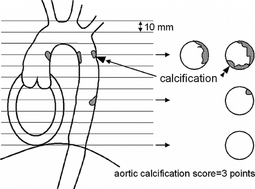
To detect the calcification of the aorta or the coronary arteries, non-gated conventional CT scanning was used without contrast media at 1 cm slice thickness from the upper margin of the aortic arch to the diaphragm. The extent of calcification in the thoracic aorta was expressed as the sum of the aortic cross-sectional slices with calcification (aortic calcification score; see ). For example, if aortic calcification was present in three aortic cross-sections, as shown in , the calcification score was 3. In addition, the number of calcified coronary vessels was totaled (0–3 vessels). The hematocrit and serum calcium, phosphorous, and parathyroid hormone levels were measured concurrently with the CT scanning before dialysis.
Results were expressed as mean ± SD. Significant differences were determined by an unpaired Students t-test. The association between variables was determined by linear regression analysis. Results were considered statistically significant at p < 0.05.
Results
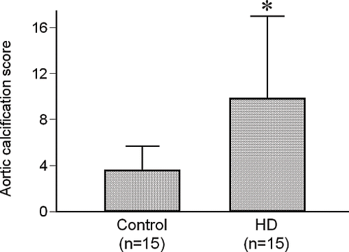
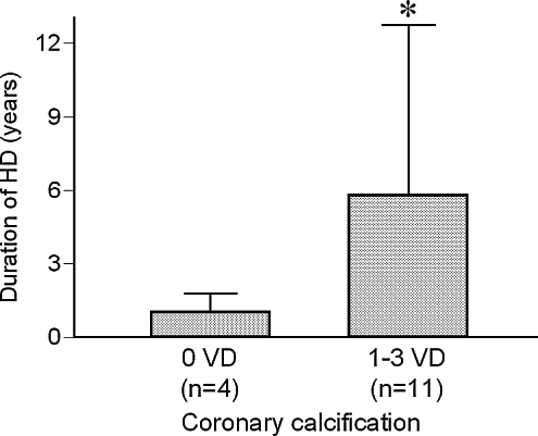
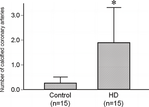
Coronary artery calcification was observed in 11 of the 15 hemodialysis patients (1.9 ± 1.4 vessels per subject) and in 2 of the 15 control patients (0.3 ± 0.2 vessels per subject). A typical case with hemodialysis is shown in . The hemodialysis patients had a significantly greater number of calcified coronary arteries in comparison to the control patients (p < 0.01; see ). Aortic calcification was found in 14 of the 15 hemodialysis patients (mean score 9.7 ± 7.2, range 0–26) and in 6 of the 15 non-hemodialysis patients (mean score 3.5 ± 2.2, range 0–8). The hemodialysis patients had significantly higher aortic calcification scores than the control group (p < 0.01; see ). There was no significant difference in the calcification scores of the coronary arteries and aorta between the diabetic hemodialysis patients (coronary calcification, 2.4 ± 1.2; aortic calcification, 11.5 ± 7.7) and the non-diabetic hemodialysis patients (coronary calcification, 1.4 ± 1.5; aortic calcification, 7.6 ± 6.4). No patients had only coronary calcification whereas 4 patients had only aortic calcification. Moreover, there were significantly more patients with calcification of both aorta and coronary arteries in the hemodialysis group than in the control group (p < 0.01). The patients with coronary calcification (n = 11) had spent significantly longer time on dialysis (mean, 5.7 ± 6.9 years; range, 0.2–22.0 years) than the patients without coronary calcification (n = 4, mean, 1.0 ± 0.6 years; range, 0.3–1.8 years, p < 0.01; see ). However, there was no significant correlation between the dialysis duration and the aortic calcification score. There was no significant association between coronary artery calcification and serum calcium, phosphorous, or parathyroid hormone levels in the present study.
Figure 2. X-ray computed tomography image without contrast media at the level of (a) aortic arch, (b) aortic root, and (c) left ventricle. Arrow heads show calcification of the aorta and the coronary arteries. Abbreviations: Ao = aorta, LA = left atrium, LV = left ventricle, RV = right ventricle, LAD = left anterior descending artery, LCX = left circumflex, RCA = right coronary artery.
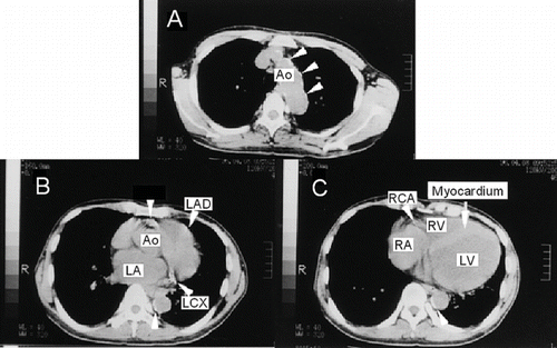
Figure 3. The numbers of calcified coronary arteries in the control group and the hemodialysis group. Abbreviation: HD = hemodialysis. *p < 0.01.
Discussion
Dialysis therapy has provided long-term survival for patients with end-stage renal disease. However, long-term dialysis patients face the complication of arteriosclerosis, which negatively affects their prognosis and quality of life. Cardiovascular deaths are reported to account for 35–50% of all deaths in dialysis patients.Footnote[5] It is therefore important to diagnose atherosclerosis of the coronary arteries and the great vessels by noninvasive methods. One characteristic finding in the atherosclerotic coronary artery lesion is the calcification of plaque-containing cholesterol. Coronary calcification is thought to be a highly sensitive marker of underlying atherosclerotic diseaseCitation[6],Citation[7] and associated with cardiovascular events in asymptomatic individuals.Citation[8],Citation[9] Coronary calcium provides independent incremented information in addition to traditional risk factors in the prediction of all-cause mortality.Citation[10]
In the present study, non-gated conventional CT scanning was used for the evaluation of the coronary and aortic calcificationCitation[11–14] because it was simple to obtain clear images. No attempt was made to quantify the extent or severity of vascular calcification in each vessel. CT was performed with slices at 1 cm intervals. Smaller calcification levels can be detected using 5 mm slices; however, as the level of radiation exposure would be greater, 1 cm slices seemed appropriate. The number of calcified coronary arteries (0–3 vessels) were counted for coronary calcification score, which were somewhat inadequate in expressing the calcification levels. In conventional CT, however, the calcification density is modified by the artifacts resulting from heartbeats. Thus, scores using the density were considered inaccurate. In contrast, the level of aortic calcification was expressed as the number of calcified aortic cross-sections. In conventional CT, the density of calcification is modified by the artifacts resulting from aortic pulsation. Therefore, density was not used in the scoring of aortic calcification.
Calcification had barely occurred in the coronary vessels of the control group, whereas coronary calcification was observed in an average of 1.9 vessels in the hemodialysis group. Furthermore, dialysis duration was significantly longer in patients with coronary calcification than in those without coronary calcification. These results are thought to provide insight into the possibility that hemodialysis can be a risk factor of coronary artery disease. Coronary calcification in hemodialysis patients is considered to occur by a combined mechanism of calcification diffused from the media and calcinosis in the intima.Citation[15] Calcification of the coronary arteries probably progresses more rapidly in dialysis patients than in non-dialysis patients. A high blood level of phosphorous, high intake of calcium,Citation[2],Citation[3] secondary hyperparathyroidism,Citation[16] and administration of active vitamin DCitation[17] have been reported as causal factors in coronary calcification. Goodman et al.Citation[3] reported a significant correlation between coronary calcification and serum levels of calcium and phosphorous in 205 hemodialysis patients. However, no statistically significant correlation was found between serum levels of calcium, phosphorous or parathyroid hormone, and vascular calcification.
The relatively short hemodialysis period of 4.5 years is thought to contribute to these results. Furthermore, the presence of diabetes did not affect the incidence of vascular calcification in hemodialysis patients in this study. However, several investigators have reported that diabetes was significantly associated with extent of coronary calcification.Citation[4],Citation[18] The difference may be due to the smaller number of hemodialysis patients in this study or the differences in the scoring method for calcification.
Aortic calcification was also found more frequently in the hemodialysis group than in the control group; however, the calcification score did not increase significantly with the duration of hemodialysis. Although the clinical significance of aortic calcification is not entirely clear, Raggi et al.Citation[4] showed that the aortic calcification scores in hemodialysis patients are directly associated with the prevalence of claudication and aortic aneurysm, while coronary calcification scores are significantly associated with the prevalence of acute myocardial infarction and angina.
The control group and the hemodialysis group were matched for age but not gender. The proportion of female patients was higher in the hemodialysis group. Because men were reported to have a higher incidence of calcification in the general population as well as in hemodialysis population,Citation[4] the higher incidence of calcification in the hemodialysis group in the present study was not likely due to the difference in gender. A larger number of female patients in the hemodialysis group may reduce the calcification scores and consequently reduce the probability of a difference in calcification scores between the two groups.
Notes
5. Agodoa LYC, Held PJ, Wolfe RA, Port FK. Causes of death. In: Renal Data System, USRDS 1998 Annual Data Report. Bethesda, Md.: National Institute of Diabetes and Digestive and Kidney Disease; 1999:79–90.
References
- Lundin AP III, Alder AJ, Feinroth MV, Berlyne GM, Friedman EA. Maintenance hemodialysis. Survival beyond the first decade. JAMA 1980; 244: 38–40
- Braun J, Oldendorf M, Moshage W, Heidler R, Zeitler E, Luft FC. Electron beam computed tomography in the evaluation of cardiac calcification in chronic dialysis patients. Am. J. Kidney Dis 1996; 27: 394–401
- Goodman WG, Goldin J, Kuizon BD, Yoon C, Gales B, Sider D, Wang Y, Chung J, Emerick A, Greaser L, Elashoff RM, Salusky IB. Coronary-artery calcification in young adults with end-stage renal disease who are undergoing dialysis. N. Engl. J. Med 2000; 342: 1478–1483
- Raggi P, Boulay A, Chasan-Taber S, Amin N, Dillon M, Burke SK, Chertow GM. Cardiac calcification in adult hemodialysis patients. A link between end-stage renal disease and cardiovascular disease?. J. Am. Coll. Cardiol 2002; 39: 695–701
- Simons DB, Schwartz RS, Edwards WD, Sheedy PF, Breen JF, Rumberger JA. Noninvasive definition of anatomic coronary artery disease by ultrafast computed tomographic scanning: a quantitative pathologic comparison study. J. Am. Coll. Cardiol 1992; 20: 1118–1126
- Sangiorgi G, Rumberger JA, Severson A, Edwards WD, Gregoire J, Fitzpatrick LA, Schwartz RS. Arterial calcification and not lumen stenosis is highly correlated with atherosclerotic plaque burden in humans: a histologic study of 723 coronary artery segments using nondecalcifying methodology. J. Am. Coll. Cardiol 1998; 31: 126–133
- Arad Y, Spadaro LA, Goodman K, Newstein D, Guerci AD. Prediction of coronary events with electron beam computed tomography. J. Am. Coll. Cardiol 2000; 36: 1253–1260
- Wong ND, Hsu JC, Detrano RC, Diamond G, Eisenberg H, Gardin JM. Coronary artery calcium evaluation by electron beam computed tomography and its relation to new cardiovascular events. Am. J. Cardiol 2000; 86: 495–498
- Shaw LJ, Raggi P, Schisterman E, Berman DS, Callister TQ. Prognostic value of cardiac risk factors and coronary artery calcium screening for all-cause mortality. Radiology 2003; 228: 826–833
- Tamiya E, Matsui H, Nakajima T, Hada Y, Asano K. Detection of coronary artery calcification by x-ray computed tomography and its significance: a new technique. Angiology 1992; 43: 22–31
- Tamiya E, Kishi R, Sugishita K, Ito N, Ikenouchi H, Hada Y. Involvement of lipoprotein (a) in calcification of coronary arteries and thoracic aorta as detected by x-ray computed tomography. Int. J. Angiol 1995; 4: 218–223
- Tamiya E, Hada Y, Ando T, Murota Y, Ito N, Sugishita K, Shimizu T, Asano K. Thoracic aortic calcification measured by x-ray computed tomography vs age and risk factors of arteriosclerosis. Int. J. Angiol 1997; 6: 1–4
- Tamiya E, Imai Y, Ito N, Ikenouchi H, Hada Y, Tanaka K, Murota Y, Ando T, Furuse A, Asano K. Are metallic markers necessary for coronary artery bypass grafts? A study using x-ray computed tomography and selective graft angiography. Int. J. Angiol 1999; 8: 29–32
- Lewin K, Trautman L. Ischaemic myocardial damage in chronic renal failure. Br. Med. J 1971; 16: 151–152
- De Francisco AM, Ellis HA, Owen JP, et al. Parathyroidectomy in chronic renal failure. Q. J. Med 1985; 55: 289–315
- Mallick NP, Berlyne GM. Arterial calcification after vitamin-D therapy in hyperphosphatemic renal failure. Lancet 1968; 2: 1316–1320
- Detrano RC, Wong ND, French WJ, Tang W, Georgiou D, Young E, Brezden OS, Doherty T, Brundage BH. Prevalence of fluoroscopic coronary calcific deposits in high-risk asymptomatic persons. Am. Heart J 1994; 127: 1526–1532
