Abstract
Background. Therapeutic failure in preventing renal disease progression in type 2 diabetic nephropathy (DN) is due to a failure in the early detection of DN by microalbuminuria and the inappropriate correction of renal hemodynamic maladjustment secondary to glomerular endothelial dysfunction. Methods. Thirty patients associated with normoalbuminuric type 2 DN were subject to the following studies: tubular function by means of fractional excretion of magnesium (FE Mg), vascular function by means of determining the circulating endothelial cell, VEGF, VEGF/TGF B ratio, and intrarenal hemodynamic studies. Results. FE Mg, circulating endothelial cells, and TGF B were abnormally elevated, and VEGF/TGF B ratio was decreased in these normoalbuminuric patients. The intrarenal hemodynamic study revealed a hemodynamic maladjustment characterized by a preferential constriction at the efferent arteriole and a reduction in peritubular capillary flow. Following treatment with vasodilators, a decrease in efferent arteriolar resistance and increase in peritubular capillary flow as well as glomerular clearance were observed. Conclusion. FE Mg appears to be a more sensitive marker than microalbuminuria for the early detection of DN. Increased endothelial cell injury is reflected by enhanced circulating endothelial cell loss in conjunction with the increased TGF B and the decreased ratio between VEGF and TGF B. This is further supported by the dysfunctioning glomerular endothelium, which is characterized by hemodynamic maladjustment and a reduction in the peritubular capillary flow. A correction of such hemodynamic maladjustment by multidrug vasodilators effectively improves renal perfusion and restores renal function in type 2 DN.
INTRODUCTION
Diabetic nephropathy (DN) has become the primary cause of end-stage renal disease worldwide.Citation[1] The best of the present therapeutic strategy of DN simply slows the renal disease progression but is unable to restore the renal function. Two specific issues that are relevant to solving such therapeutic failure are indications of a diagnostic marker for early detection of DN and appropriate therapy that is able to restore renal function in DN. With respect to the diagnostic issue of DN, microalbuminuria has been generally accepted to serve such a purpose.Citation[2] However, it has recently been noted that structural as well as functional aberrations are illustrated even in the normoalbuminuric stage of a diabetic patient.Citation[3],Citation[4] Pagtalunan et al. demonstrated a structural change such as increased glomerular basement membrane width and mesangial fractional volume in normoalbuminuric type 2 diabetes mellitus. In this regard, tubulointerstitial disease or fibrosis appears to be the most acceptable marker for the determinant of kidney involvement as well as its disease severity.Citation[5] However, the uniform practice is not to perform kidney biopsy for the histopathologic information in diabetes mellitus. In addition to this specific issue, tubular function study by mean of fractional excretion of magnesium (FE Mg) may be the alternative approach because FE Mg correlates with the magnitude of tubulointerstitial fibrosis observed in the other clinical setting of glomerulonephropathy.Citation[6] FE Mg is used for the ability of the tubular cell to reabsorb the glomerular filtrate of magnesium as well as retain the intracellular portion of tubular magnesium, which is the second most abundant cation next to the potassium. Therefore, FE Mg value remains within normal limit (<2.2%), when the tubular function is intact or there is no tubulointerstitial fibrosis. In contrast, FE Mg is abnormally elevated in association with tubulointerstitial fibrosis or disease. Therefore, it would be interesting to know whether FE Mg is likely to be a suitable and more sensitive marker for early detection of DN than microalbuminuria.
In addition to the diagnostic issue that would delay the initiation of proper treatment, thereby causing therapeutic failure in type 2 DN, the other crucial issue that is also responsible for such a failure is the lack of proper understanding in the pathogenesis of nephronal damage or tubulointerstitial fibrosis in diabetes mellitus. In fact, vascular disease has been substantiated to be the determinant of chronic ischemic or hypoxic injury in other forms of chronic kidney diseasesCitation[7–9] but not as yet proven to be the case in diabetes mellitus. It is therefore the intention of this article to explore the biomarker that would assist in early screening for vascular disease as well as determine the dysfunctional glomerular endothelium by mean of intrarenal hemodynamic study.
MATERIAL AND METHODS
Thirty patients associated with type 2 diabetes (mean ages 46 ± 7 years) were enrolled in the study. Ten age-matched subjects without diabetes served as healthy controls. Diabetic subjects who showed no clinical evidence of heart disease, no obvious renal disease, and complied to follow-up and investigative procedures were also included.
Blood Collection and Preparation of Plasma Samples
One volume of 0.109 M trisodium citrate solution was mixed with 9 volumes of blood, and the mixture was centrifuged at 2500 g for 10 min to collect plasma. The citrated plasma was stored at −20°C for the determination of plasma factors. Clotted blood was also collected for the determinations of serum creatinine and magnesium. Three milliliters of EDTA blood were collected for isolating the circulating endothelial cell (CEC). The enumeration of CEC, ELISA for VEGF, TGF B, intrarenal hemodynamic study, and tubular function (FE Mg) were performed by the previously described methods as indicated.Citation[6],Citation[10]
Multidrug Vasodilators
The therapeutic intervention includes three or more of the following drugs: angiotensin converting enzyme (ACE) inhibitor (Enalapril 20–40 mg/day), AII receptor antagonist (Valsartan 50–100 mg/day or Micardis 80–160 mg/day), calcium channel blocker (Dynacirc 5–10 mg/day), and an antiplatelet agent (aspirin gr I/day). Normoalbuminuric patients with creatinine clearance greater than 80 mL/min/1.73m2 and FE Mg less than 4% were initially started with a combination of low dosage of two drugs, such as ACEI + calcium channel blocker, as monotherapy is usually unable to restore the renal function. However, such a combination usually does not improve the creatinine clearance even with an increase of the dosage of each drug. Therefore, a combination of three or more drugs is put forward by adding AII receptor antagonist and antiplatelet agent. Patients with creatinine clearance lower than 80 mL/min/1.73m2 or FE Mg greater than 4% are started with a higher dose. The increased dosage of vasodilators is hoped to improve renal function, which is reflected by the increase in creatinine clearance. It is noted that by adjusting the doses of such combinations, the creatinine clearance was improved and the FE Mg was reduced.
Statistical Analysis
Comparing the sample mean of two quantitative variables was determined by the non-parametric method using the Mann-Whitney test. The difference between groups was performed by Student's unpaired t test. The difference between pretreatment and post-treatment within groups was performed by Student's paired t test; p values below 0.05 were considered to be significant.
RESULTS
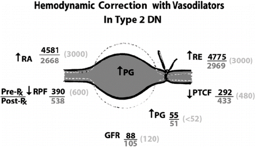
showed demographic profiles of the initial evaluation of the patients. Significant differences were observed in mean arterial pressure (MAP), fasting blood sugar, Hb A1C, FE Mg, creatinine clearance, and peritubular capillary flow. demonstrated abnormal values of circulating endothelial cells, TGF B, and the ratio between VEGF/TGF B. demonstrated the hemodynamic correction with vasodilators. Significant improvements in renal plasma flow, peritubular capillary flow, and efferent arteriolar resistance were observed following the multidrug vasodilators. illustrated the significant improvements in creatinine clearance and FE Mg following the treatment with multidrug vasodilators.
Table 1 Demographic characteristics of type 2 DN
Table 2 Results of biomarker studies
Figure 1. Hemodynamic correction with vasodilator in type 2 diabetic nephropathy. Normal values represent in parenthesis. Abbreviations: GFR = glomerular filtration rate in mL/min/1.73m2, PG = intraglomerular hydrostatic pressure in mmHg, PTCF = peritubular capillary flow in mL/min/1.73m2, RA = afferent arteriolar resistance in dyne.s.cm−5, RE = efferent arteriolar resistance in dyne.s.cm−5, RPF = renal plasma flow in mL/min/1.73m2.

Table 3 Restoration of renal function in type 2 DN
DISCUSSION
Accumulating evidence renders support that microalbuminuria may not be able to detect DN early, and the diagnosis is often delayed when the kidney disease has already been established for quite sometime. The abnormal FE Mg observed in this study reflects the tubulointerstitial disease, and therefore FE Mg is assisting in early screening of DN at a much earlier stage, when most of the renal reserve has been well maintained. An early diagnosis of DN would lead to the early initiation of therapy for preventing renal disease progression.
With respect to the pathogenetic mechanism of renal disease progression, it is interesting to observe crucial evidence that supports the role of vascular disease in DN. The increased number of circulating endothelial cells loss in conjunction with the enhancement of TGF B and the paradoxical decrease in VEGF (endothelial proliferator) and TGF B (endothelial antiproliferator) ratio would reflect injury to the endothelial cell, particularly the glomerular endothelial cell. A dysfunction of the glomerular endothelium is confirmed in this study by the altered intrarenal hemodynamics observed, the so-called hemodynamic maladjustment. This is characterized by a preferential constriction of the efferent arteriole and a significant reduction in peritubular capillary flow that supplies the tubulointerstitial compartment (see ). Such a finding confirms an ischemic stage that has already been established even in normoalbuminuric stage of diabetes mellitus. The hemodynamic maladjustment observed in this normoaluminuric type 2 DN share a common path with that observed in a variety of clinical settings of chronic kidney diseases.Citation[10–13] As peritubular capillary flow reduction attested in other settings of chronic kidney diseases has previous been demonstrated to be the determinant of tubulointerstitial fibrosis, it is likely that the peritubular capillary flow reduction in this study may be responsible for the kidney disease progression associated with type 2 diabetes mellitus.Citation[8]
In accordance with the preceding information, the correction of hemodynamic maladjustment would likely enhance peritubular capillary flow and thus promote the reparative process of tubulointerstitial injury. From previous therapeutic trials in other clinical settings of chronic kidney diseases, it is known that it would usually require more than monotherapy of vasodilator to overcome the sustained constriction at the efferent arteriole. This finding concurs with other expertises who failed to restore renal function with monodrug therapy with a vasodilator.Citation[14–15] The result of this correction of hemodynamic maladjustment with multidrug vasodilators indicates a successful reduction in efferent arteriolar resistance in conjunction with increases in peritubular capillary flow and renal plasma flow. Increase in creatinine clearance and glomerular filtration rate following such therapy correlates with the hemodynamic improvement. It is likely that the failure to restore renal function under conventional therapy results from the late initiation of therapy due to the diagnostic reliance on the insensitiveness of microalbuminuria, inappropriate treatment with monotherapy of the vasodilator, and that those patients with normal blood pressure usually received no vasodilator. The authors also agree with other experience based on experimental study in animals that early treatment in diabetes mellitus would ameliorate or induce regression of the kidney disease process.Citation[16],Citation[17]
In conclusion, a restoration of renal function can be successfully accomplished in type 2 DN under the assistance of early screening of the status of DN with FE Mg and the early initiation of multidrug vasodilators.
ACKNOWLEDGMENT
This study is supported by Thailand Research Fund.
REFERENCES
- Amos AF, McCarthy DJ, Zimmet P. The rising global burden of diabetes and its complications: Estimates and projections to the year 2010. Diabet. Med. 1997; 14(Suppl. 5)S1–S85
- Parving HH, Hommel E, Mathiesen E, et al. Prevalence of microalbuminuria, arterial hypertension, retinopathy and neuropathy in patients with insulin dependent diabetes. Br. Med. J. 1988; 296: 156–160
- Pagtalunan ME, Miller PL, Jumping-Eagle S, et al. Podocyte loss and progressive glomerular injury in type II diabetes. J. Clin. Invest. 1997; 99: 342–348
- Moriya T, Moriya R, Yajima Y, et al. Urinary albumin as an indicator of diabetic nephropathy lesions in Japanese type 2 diabetic patients. Nephron. 2002; 91: 292–299
- Risdon FC, Sloper SA, de Wardener HE. Relationship between renal function and histologic changes found in renal biopsy specimens from patients with persistent glomerular nephritis. Lancet. 1968; 2: 363–366
- Futrakul P, Yenrudi S, Futrakul N, Sensirivatana R, Kingwatanakul P, Jungthirapanich J, et al. Tubular function and tubulointerstitial disease. Am. J. Kidney Dis. 1999; 33: 886–891
- Kang DH, Kanellis J, Hugo C, Truong L, Anderson S, Kerjaschki D, et al. Role of microvascular endothelium in progressive renal disease. J. Am. Soc. Nephrol. 2002; 13: 806–816
- Futrakul N, Yenrudi S, Sensirivatana R, Watana D, Laohapaibul A, Watanapenphaibul K, et al. Peritubular capillary flow determines tubulointerstitial disease in idiopathic nephrotic syndrome. Ren. Fail. 2000; 22: 329–335
- Fine LG, Ong ACM, Norman JT. Mechanism of tubulointerstitial injury in progressive renal disease. Eur. J. Clin. Invest. 1993; 25: 259–265
- Butthep P, Nuchprayoon I, Futrakul N. Endothelial injury and altered hemodynamics in Thalassemia. Clin. Hemorheol. Microcirc. 2004; 31: 287–293
- Futrakul N, Sitprija V, Siriviriyakul P, Futrakul P. Glomerular endothelial dysfunction, altered hemorheology and hemodynamic maladjustment in nephrosis with focal segmental glomerulosclerosis. Hong Kong J. Nephrol. 2004; 6: 69–73
- Futrakul N, Laohaphaibul A, Futrakul P. Glomerular endothelial dysfunction and hemodynamic maladjustment in vesicoureteric reflux. Ren. Fail. 2003; 25: 479–483
- Futrakul P, Pochanugool C, Poshyachinda M, et al. Intrarenal hemodynamic abnormality in severe form of glomerulonephritis: therapeutic benefit with vasodilators. J. Med. Assoc. Thai. 1992; 75: 375–385
- Pisoni R, Faraone R, Ruggenenti P, Remuzzi G. Inhibitors of the rennin angiotensin system reduce the rate of GFR decline and end-stage renal disease in patients with severe renal insufficiency. J. Neprhol. 2002; 15: 428–430
- Ruggenenti P, Perna A, Benini R, Bertani T, Zoccali C, Maggiore Q, et al. In chronic nephropathies prolonged ACE inhibition can induce remission: Dynamics of time-dependent changes in GFR. J. Am. Soc. Nephrol. 1999; 10: 997–1006
- Nagai Y, Yao L, Kobori H, Miyata K, Ozawa Y, Miyatake A, et al. Temporary angiotensin II blockade at the prediabetic stage attenuates the development of renal injury in type 2 diabetes rats. J. Am. Soc. Nephrol. 2005; 16: 703–711
- Hilgers K. Never too early to treat type 2 DN. J. Am. Soc. Nephrol. 2005; 16: 574–575