Abstract
In this study, serum and urinary VEGF levels and VEGF expression in PBMNC were correlated with daily proteinuria, renal function tests, and renal histopathologic findings in untreated patients with different glomerulonephritis and with the course of renal function and proteinuria for one year. Forty-five untreated patients with different glomerulonephritis and 11 healthy persons comprised the study and control groups, respectively. VEGF mRNA expression was detected by RT- PCR in peripheral blood mononuclear cells (PBMNC), and VEGF levels were measured by ELISA in serum and urine samples simultaneously. Male/female ratio was 24/21 and mean ages were 34.49 ± 14.98. Serum and urinary VEGF levels, VEGF expressions in PBMNC, and the ratios of urine VEGF/urine creatinine were found to be similar in patients and controls. There were important correlations between urinary VEGF levels and baseline serum Cr (p = 0.035) and ESR (p = 0.022). There was also a marginal correlation between urinary VEGF levels and baseline CCr (p = 0.072). There was no correlation between serum and urinary VEGF levels and PBMNC mRNA expression and pathological findings such as with or without glomerular sclerosis, tubulointerstitial fibrosis (TIF), periglomerular fibrosis, and mesangial cell proliferation in renal biopsy. Serum and urinary VEGF levels or VEGF expression in PBMNC in patients with renal amyloidosis or proliferative or nonproliferative glomerulonephritis were similar with that of healthy controls and each other. Serum and urinary VEGF levels and PBMNC VEGF mRNA expression in untreated patients with different glomerulonephritis and controls were similar. We found only one important correlation, that between urinary VEGF levels and baseline serum creatinine levels in patients with different glomerulonephritis. Urinary VEGF can be an important pathogenesis of glomerular disease or a simple proteinuria. Serum and urinary VEGF levels and PBMNC VEGFmRNA did not change by periglomerular sclerosis, periglomerular fibrosis, or tubulointerstitial fibrosis on renal biopsy. PBMNC VEGF mRNA expression decreased in patients undergoing remission. In addition to the important correlation between urinary VEGF and serum creatinine, we also found an important correlation between erythrocyte sedimentation rate and urinary VEGF. This finding was interesting because we could not find a similar conclusion in other studies.
INTRODUCTION
Vascular endothelial growth factor (VEGF) is a selective endothelial mitogen and belongs to a family of multipotent cytokines. VEGF stimulates endothelial cell proliferation and differentiation and increases vascular permeability. Epidermal growth factor, transforming growth factor beta, platelet derived growth factor, insulin like growth factor I, angiotensin II, interleukin (IL)-1, and IL-6 have the potential to up-regulate VEGF expression.Citation[1] VEGF can contribute to the relaxing capacity of the renal vasculature. This relaxation capacity is partly mediated by the NO/endothelium-derived relaxing factor pathway.Citation[2]
In human kidneys, VEGF mRNA and /or protein were detected predominantly in glomerular podocytes, distal tubules, and collecting ducts, to a lesser extent in some proximal tubules. In different glomerulonephritis, renal VEGF expression was found to be increased, decreased or unchanged.Citation[1] In this study, serum and urinary VEGF levels and VEGF expression in PBMNC were correlated with daily proteinuria, renal function tests, and renal histopathologic findings in untreated patients with different glomerulonephritis and with the course of renal function and proteinuria for one year.
PATIENTS AND METHODS
The study group consisted of 45 patients undergoing renal biopsy for hematuria and/or proteinuria. Patients did not receive medication while they were biopsied. VEGF mRNA expression was detected by RT-PCR in peripheral blood mononuclear cells (PBMNC), and VEGF levels were measured by ELISA in serum and urine samples simultaneously. Urine and blood samples taken from 11 healthy persons in similar age were used as control group. Blood pressure (BP), hematocrit (hct), white blood cell (WBC), erythrocyte sedimentation rate (ESR), blood glucose, blood urea nitrogen (BUN), creatinine (Cr), total protein, serum albumin, total cholesterol (t chol.), LDL chol, triglyceride, creatinine clearance (CCr), and daily proteinuria were recorded at baseline and at 3, 6, and 12 months. In addition to conservative therapy such as diet and angiotensin-converting enzyme inhibitors, all patients except those with amyloidosis were treated with glucorticoids or a combination of glucorticoid and cyclophosphamide. Colchicine was used in patients with renal amyloidosis. PBMNC VEGF mRNA and serum and urine VEGF levels were repeated in six cases after the disappearance of proteinuria.
Determination of Expression of VEGF mRNA with the Method of Real Time PCR mRNA Isolation
White blood cell (WBC) counts of blood samples were determined in Beckman Coulter Gen S System II. Blood samples which similar volume with solution containing 3 × 10³ WBCs were taken into Eppendorf tubes. Erythrocyte lysing solution (1 mL Roche Applied Science) was added to the whole blood and sat at room temperature for ten minutes. Samples centrifuged in 13000 rpm and WBCs were precipitated. Fragmented erythrocytes were eliminated. 300 μl of MagNa pure LC mRNA isolation kit I (Roche Applied Science) was added to the WBC pellet, and mixed in vortex. Samples were kept −20°C until isolation. Isolation was made automatically at MagNA (Roche machine). Isolation mRNA samples were put in four different Eppendorf tubes and kept at −86°C.
cDNA Synthesis
Isolated mRNAs were changed to cDNA kit using first strand cDNA synthesis kit (Roche Applied Science).
VEGF Primer Design
Primers were designed using Primer Premier Software at METIS Biotechnology (Ankara, Turkey). Primer synthesis was made at TIB-MOLBIOL (Germany). VEGF primer strains were as follows:
VEGF Forward 5′-AAC TTT CTG CTG TCT TGG GTG- 3′,
VEGF Reverse 5′- ACA AAT GCT TC TCC GCT CT-3′.
PCR product was 457 bp and mRNA Genbank Accession number was M32977.
Standard Curve
Light cycler control kit RNA (Roche Applied Science) was used for this aim. Standard curve was between 106 and 103 copy number.
Real Time PCR
VEGF cDNA was amplified in 20 μl reaction volume capillary tubes using VEGF RNA-specific primers.
Light Cycler Fast Start DNA Master
SYBR Gren I (Roche Applied Science) was used for amplification. Contact of PCR product occurred during SYBR Gren I annealing, with double stranded DNA, caused fleurosans so that the amplification can be detected as online on Software of Light cycler. 1 μl cDNA was added to this mixture, which underwent the following:
Denaturation program: 10 min at 95°C
Amplification program: 0 sec at 95°C, 10 sec at 65°C, 19 sec at 72°C, 45 cycles
Melting curve analysis: 0 sec at 95°C, 15 sec at 65°C, and temperature is increased gradually (transition rate 0.1°C / sec).
Fluorescence canal alignments: F1/1 was used. Results were analyzed by Light Cycler Software.
ELISA method for VEGF measurements: ELISA method for VEGF measurements at urine and serum by using R&D System was used. Results were shown as pg/mL.
Renal Biopsy
Specimens were evaluated through light microscopy and immunohistochemistry by an experienced renal pathologist. Electron microscopy was not used. In addition to histopathologic diagnosis, glomerular sclerosis, periglomerular fibrosis, tubulointerstitial fibrosis, and mesangial cell proliferation were also recorded.
Statistical Analysis
Nonparametric, Mann Whitney, and chi square methods were used for statistical analysis. Statistically importance accepted as lower than p = 0.05.
RESULTS
Patients Characteristics
Male/female ratio in study group was 24/21, and mean ages were 34.49±14.98 (range 16–68). In a control group, 7 were male, the mean age was 30.50±14.10, and there was no difference for age between patients and controls. and show the renal histopathology and demographic and biochemical findings on baseline and 3, 6, and 12 months.
Table 1 The diagnosis of renal biopsy
Table 2 Demographic and biochemical findings at baseline and 3, 6, and 12 months
Serum and Urine VEGF Levels and VEGF mRNA Expression in PBMNC
and and show the VEGF mRNA expression in PBMNC and serum and urinary VEGF levels and the ratio of urine VEGF /urine creatinine levels in patients and controls. PBMNC VEGF mRNA and serum urine VEGF levels and urine VEGF/urine Cr were not different between patients and controls (p > 0.05). There was no correlation between serum VEGF and daily proteinuria, BUN, and serum Cr and CCr levels. There was an important correlation between urine VEGF and baseline serum Cr (p = 0.035) and ESR (p = 0.022). There was also a marginal correlation between urine VEGF and baseline CCr (p = 0.072). Urine VEGF/urine Cr ratios were 6.13 ± 5.55 and 3.15 ± 2.89 in patients and the control, respectively. There was no difference for urine VEGF/urine Cr between patients and controls. We did not find an important correlation between PBMNC VEGF mRNA and biochemical parameters. shows the baseline and serial serum-urinary VEGF and PBMNC mRNA levels in six of the patients undergoing remission in the follow-up period. There was an important decrease in VEGF mRNA levels in PBMNC after treatment according to baseline levels.
Figure 1. PBMNC VEGF mRNA expression and serum/urine VEGF levels in patients and controls.
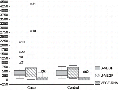
Figure 2. The ratio of urine VEGF/urine creatinine levels in patients and controls.
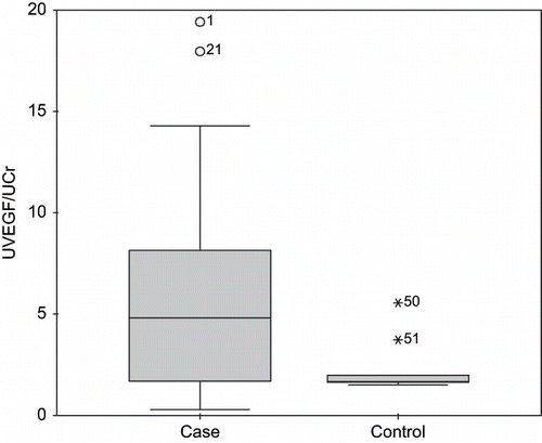
Table 3 PBMNC VEGF mRNA expression and serum/urine VEGF levels in patients and controls
Table 4 Baseline and serial serum and urine VEGF and PBMNC mRNA levels in six of the patients whose proteinuria disappeared in follow-up period
Renal Histopathologic Findings and VEGF Levels
shows the serum and urinary VEGF levels and PBMNC mRNA expression with and without glomerular sclerosis, tubulointerstitial fibrosis (TIF), periglomerular fibrosis, and mesangial cell proliferation in renal biopsy. Serum and urinary VEGF levels or VEGF expression in PBMNC in patients with renal amyloidosis and proliferative and nonproliferative glomerulonephritis were similar with those of healthy controls and each other.
Table 5 The serum and urine VEGF levels and PBMNC mRNA expression according to the glomerular sclerosis, tubulointerstitial nephritis (TIF), periglomerular fibrosis, and mesangial cell proliferation
Follow-Up and VEGF Levels
According to whether or not serum Cr did or did not become normal or improve by more than 50%, baseline serum and urine VEGF and PBMNC mRNA expression were not different. Also, according to the improvement or lack of improvement of proteinuria, baseline serum and urine VEGF and PBMNC mRNA expression were similar. Second measurements of serum and urine VEGF levels were unchanged in six patients undergoing remission, but mRNA expression in PBMNC significantly decreased.
DISCUSSION
VEGF is a multipotent cytokine. In different diseases, including renal diseases, VEGF was evaluated, and renal VEGF expression was found to be increased, decreased or unchanged.Citation[1] In other research, plasma or urine VEGF levels were not different between patients with minimal change in the disease in nephrotic state and those in remission or healthy controls.Citation[3],Citation[4], In contrast, urinary VEGF levels were increased in patients with minimal change in disease with nephrotic syndrome when compared with patients without nephrotic syndrome and healthy controls, and correlated with the degree of proteinuria.Citation[5] In our study group, only one patient had a minimal change in disease, and her serum and urine VEGF levels and PBMNC VEGF mRNA expression were lower than mean levels. In our six patients with membranoproliferative glomerulonephritis, serum and urine VEGF levels were found to be similar to levels in other patients. When we also grouped our patients according to proliferative or nonproliferative glomerulonephritis, serum and urine VEGF levels and PBMNC VEGF mRNA expression were similar. It was reported that in patients with membranoproliferative glomerulonephritis-like lesion, as in POEMS syndrome, serum VEGF levels were increased compared to primary membranoproliferative glomerulonephritis or normal controls.Citation[6]
It was found that in PBMNC VEGF, mRNA expression was higher in FSGS.Citation[7] In type 2 diabetes, mellitus plasma VEGF levels were higher than controls. Plasma VEGF levels was found to be increased when urine albumin excretion increased.Citation[8] Urine VEGF levels correlated with serum creatinine and proteinuria.Citation[9] In our patients with both diabetic and sclerosing glomerulonephritis, serum and urinary VEGF levels and PBMNC VEGF mRNA expression were within the median limits. It was reported that in many glomerular diseases, VEGF expressing cells were decreased or absent in areas of focal or globally sclerosis.Citation[10] Decreased numbers of VEGF expressing cells in glomeruli were also noted in amyloidosis, diabetes, and crescentic glomerulonephritis, as well as diffuse proliferative glomerulonephritis secondary to lupus erythematosus.Citation[10] Serum VEGF levels were similar in patients with FSGS and controls, but urinary VEGF levels was higher in patients with FSGS than controls. Urinary VEGF levels did not correlate with serum VEGF, degree of proteinuria, or renal function.Citation[11] Among our patients, there was only one patient with acute rejection. It was found that in renal biopsies during acute rejection, VEGF expression was almost the same as with normal kidneys.Citation[12] In our six patients with renal amyloidosis, serum and urinary VEGF levels and PBMNC VEGF mRNA expression were lower than that of other patients, but there were no important differences. We also found similar serum VEGF levels in patients with different glomerulonephritis compared to healthy controls. In renal biopsies with or without sclerosis, tubulointerstitial fibrosis, mesangial cell proliferation, and periglomerular fibrosis, serum VEGF levels, urinary VEGF levels, the ratio of urinary VEGF/urinary creatinine and PBMNC VEGF mRNA expression were found similar. However, we did not evaluate for VEGF protein and expression in renal biopsies. In our patients, there was no correlation between serum VEGF and daily proteinuria, BUN, serum creatinine and creatinine clearance levels. We also did not find correlations between urinary VEGF levels and PBMNC VEGF mRNA expression and baseline BUN, daily proteinuria, and creatinine clearance. There were an important correlation between urinary VEGF and baseline serum creatinine and ESR. In the literature, no correlation was found between ESR and urinary VEGF level; perhaps it is related to the relationship between acute inflammation and proteinuria, including urinary VEGF. There was also a marginal correlation between urine VEGF and baseline creatinine clearance. According to our results, urinary VEGF correlated with renal function and daily proteinuria. Urine VEGF/urine creatinine of the patients was higher than that of controls, but there was no statistical difference between patients and controls. We did not find any important correlations between PBMNC VEGF mRNA and biochemical parameters. In the follow-up period, improvement of renal function or daily proteinuria serum suggested that urine VEGF levels and PBMNC VEGF mRNA were similar. During the remission, second measurements of serum and urine VEGF did not change, but PBMNC VEGF mRNA decreased in patients undergoing remission.
SUMMARY
Serum and urinary VEGF levels and PBMNC VEGF mRNA expression in untreated patients with different glomerulonephritis and controls were similar. In the literature, VEGF levels in different glomerulonephritis also have many controversies.
We found only one important correlation between urinary VEGF levels and baseline serum creatinine levels in patients with different glomerulonephritis. Urinary VEGF can be an important pathogenesis of glomerular disease or a simple proteinuria. Thus, it is not easy to evaluate the importance of urine VEGF.
In renal amyloidosis, proliferative and nonproliferative glomerulonephritis serum and urinary VEGF levels or VEGF expression in PBMNC were also similar to that of healthy controls and each other. Also, serum and urinary VEGF levels and PBMNC VEGFmRNA did not change by periglomerular sclerosis, periglomerular fibrosis, or tubulointerstitial fibrosis on renal biopsy.
PBMNC VEGF mRNA expression decreased in patients undergoing remission. In addition to the important correlation between urinary VEGF and serum creatinine, we also found an important correlation between erythrocyte sedimentation rate and urinary VEGF, so that urinary VEGF may be a sign of renal inflammation. We could not find a similar conclusion in the literature.
Related Research Data
REFERENCES
- Schrijvers BF, Flyvbjerg A, De Vriese AS. The role of vascular endothelial growth factor (VEGF) in renal pathophysiology. Kidney Int. 2004; 65: 2003–2017
- Klanke B, Simon M, Röckl W, et al. Effects of vascular endothelial growth factor (VEGF)/vascular permeability factor (VPF) on haemodynamics and permselectivity of the isolated perfused rat kidney. Nephrol Dial Transplant. 1998; 13: 875–885
- Nitta K, Uchida K, Honda K, et al. Serum vascular endothelial growth factor concentration in rapidly progressive glomerulonephritis. Nephron. 1998; 80: 357–358
- Nitta K, Uchida K, Kimata N, et al. Increased serum levels of vascular endothelial growth factor in human crescentic glomerulonephritis. Clin Nephrol. 1999; 52: 76–82
- Matsumoto K, Kanmatsuse K. Elevated vascular endothelial growth factor levels in the urine of patients with minimal-change nephrotic syndrome. Clin Nephrol. 2001; 55: 269–274
- Soubrier M, Sauron C, Souweine B, et al. Growth factors and proinflammatory cytokines in the renal involvement of POEMS syndrome. Am J Kidney Dis. 1999; 34: 633–638
- Cheong HI, Lee JH, Hahn H, et al. Circulating VEGF and TGF-beta1 in children with idiopathic nephrotic syndrome. J Nephrol. 2001; 14: 263–269
- Wasada T, Kawahara R, Katsumori K, et al. Plasma concentration of immunoreactive vascular endothelial growth factor and its relation to smoking. Metabolism. 1998; 47: 27–30
- Cha DR, Kim NH, Yoon JW, et al. Role of vascular endothelial growth factor in diabetic nephropathy. Kidney Int. 2000; 58(Suppl.77)S104–S112
- Shulman K, Rosen S, Tognazzi K, Manseau EJ, Brown LF. Expression of vascular permeability factor (VPF/VEGF) is altered in many glomerular diseases. J Am Soc Nephrol. 1996; 7: 661–666
- Honkanen EO, Teppo AM, Gronhagen-Riska C. Decreased urinary excretion of vascular endothelial growth factor in idiopathic membranous glomerulonephritis. Kidney Int. 2000; 57: 2343–2349
- Gröne HJ, Simon M, Gröne EF. Expression of vascular endothelial growth factor in renal vascular disease and renal allografts. J Pathol. 1995; 177: 259–267