
Abstract
It has been revealed in recent studies that Hypericum Perforatum (HP) is influential on cancer, inflammatory diseases, bacterial and viral diseases, and has neuroprotective and antioxidant properties. In this study, we investigated the effect of HP, which is known to have antioxidant and anti-inflammatory effects, on kidney I/R damage. Male Sprague–Dawley rats were divided into three groups, and each of the groups had eight rats: The Control Group; the Ischemia/Reperfusion (I/R) Group; and the IR + HP Group which was treated with 50 mg/kg of HP. The right kidneys of the rats were removed, and the left kidney developed ischemia during the 45th min, and reperfusion occurred in the following 3rd h. The histopathological findings and also the level of Malondialdehyde (MDA), Glutathione (GSH) and superoxide dismutase (SOD), catalase (CAT), and glutathione peroxidase (GSH-PX) enzyme activations in the renal tissues were measured. Blood Urea Nitrogen (BUN), Creatinin (Cre) from serum samples were determined. The levels of BUN, Cre, and kidney tissue MDA increased at a significant level, and the SOD, CAT, and GSH-PX enzyme activity decreased at a significant level in the I/R group, compared with the Control Group (p < 0.05). In the I/R + HP group, the levels of MDA decreased at a significant level compared to the I/R group, while the SOD, CAT, and GSH-PX activity increased (p < 0.05). In histopathological examinations, it was observed that the tubular dilatation and epithelial desquamation regressed in the IR + HP Group when compared with the I/R Group. It has been shown with the histological and biochemical results in this study that HP is protective against acute renal I/R.
Introduction
Kidney Ischemia/Reperfusion (I/R) Damage is a common clinical problem that has high mortality and morbidity rates, and results in Acute Renal Failure (ARF).Citation1,Citation2 During the interventions applied to the kidneys like kidney transplantation and coronary bypass, the blood-flow to the kidneys may be cut off. The cutoff of the blood leads to damages in the tissues. During the reperfusion in which the blood-flow is established again, the increase in the production of free radicals and inflammatory response may result in more damage.Citation3 These reactions, which occur after I/R, may cause a process that may proceed as bad as necrosis and apoptosis in kidney tubules.Citation4 There are antioxidant defense systems that eliminate the Reactive Oxygen Species (ROS) that are produced in the tissues. Superoxide dismutase (SOD), glutathione peroxidase (GSH-PX), and catalase (CAT), which exists in tissues, remove the ROS in a rapid manner under normal circumstances.Citation5 During the I/R, the antioxidant defense system becomes inadequate, and as a result, oxidative damage occurs in the tissues.Citation6 These events result in cell damage, which is caused by lipid peroxidation. Oxygen-centered free radicals include Hydroxyl Radical (OH), Hydrogen Peroxide (H2O2) and the Superoxide Radical (O2•) and are among well-known radical species that are relevant to reperfusion injury at the highest rate.Citation7
Hypericum Perforatum (HP), which is also known as St. John’s Wort (SJW), is a plant that has been used as a medication for 2000 years. It has spread widely in the world in mild-climatic areas of Asia, Europe, America, and Australia.Citation8 In studies conducted in recent years, it has been demonstrated that HP is influential on cancer, inflammatory diseases, bacterial, and viral diseases; and has neuroprotective and antioxidant properties.Citation9 HP includes bioactive compounds like hypericin and hyperphorin, flavonoids, xanthone derivatives, and biapigenin.Citation10 It has been shown in previous studies that HP prevents the apoptosis and DNA damage occurring due to ROS,Citation11 and has anti-inflammatory influences.Citation12,Citation13 It has also been demonstrated that it has protective effects like antioxidant and anti-inflammatory effects against intestinal and liver I/R damage.Citation14,Citation15
In this empirical study, we investigated the protective effects of HP, which is used commonly as herbal medication in the world, and which has antioxidant and anti-inflammatory properties, against kidney I/R damage.
Material and method
Plant material
Flowering herbs of HP were collected from Malatya (Turkey) in May 2016 and were dried in the shade. The plant was identified by Assistant Prof. Dr. Mustafa Sinan Kaynak and a voucher specimen was stored in the herbarium of İnonu University, Faculty of Pharmacy.
Preparation of the plant extract
The dried and coarsely powdered flowering herbs of HP (100 g) were macerated with 80% ethanol (2 × 300 ml) for two days by continuous stirring at room temperature. The extracts were separated from the pellets by filtering twice through the piled filler paper to increase the yield. Extracts adhering to the wall of the glass bowl were separated by an ultrasonic device and were settled to the bottom. Then it was evaporated to dryness under reduced pressure (EtOH extract, yield 38.7 g) by rotary evaporator vacuum (Rotavapor Buchi r-210, Canada). The extract was stored at −80 °C until use in the experiments. The dried extract was dissolved in 10% Tween 80-saline solution before intraperitoneal (IP) administration to animals.
Animals
The empirical protocol in this study was approved by the Ethical Committee on Animal Research of Inonu University (permit number 2016 A-05). Twenty-four male Sprague–Dawley rats (n = 24), weighing 300–350 g, were obtained from Animals Research Center of Inonu University Laboratory, and were kept in a controlled room. The rats were provided with standard pellet diet and water ad libitum at a temperature of 21 ± 2 °C. The light cycle was established as 12:12-h light/dark.
Experimental protocols
The rats were divided into three groups in random order. In order to anaesthetize/analgesia the animals before surgical operation, Ketamine (Ketalar, Eczacibasi, Istanbul, Turkey) 70 mg/kg and xylazine (Rompun, Bayer, Istanbul, Turkey) 8 mg/kg IP were used. The lumbar areas of the rats were shaved and sterilized with povidine iodine solution. Then midline laparotomies were carried out and right nephrectomy was performed on all the rats in the groups.
Group 1 rats (The Control Group) (n = 8), underwent right nephrectomy. After 3 h, the animals were sacrificed by removing the blood from their hearts.
Group 2 rats (The I/R Group) (n = 8), underwent right nephrectomy. Then, the left renal artery was occluded with a clamp for 45 min followed by reperfusion for 3 h.
Group 3 rats (The I/R + HP Group) (n = 8), underwent the same surgical procedure as in the I/R Group. About 50 mg/kg HP extract was given IP at the beginning of ischemia.
Then, the rats were sacrificed by removing the blood from their hearts, and the left kidneys were removed in a fast manner, decapsulated, and divided into two equal longitudinal parts. One of these parts was placed in formaldehyde solution for histopathological examinations with light microscopy. The other part of the kidney was kept in deep freezer at −80 °C until assayed for Malondialdehyde (MDA), Glutathione (GSH), SOD, CAT and GSH-PX enzyme activity. The blood obtained was separated to evaluate serum levels of Blood Urea Nitrogen (BUN) and Creatinin (Cre) by using an Olympus Autoanalyzer (Olympus Instruments, Tokyo, Japan).
Glutathione measurement
The Glutathione measurement was made in accordance with the Ellman Method in the homogenates.Citation16 The homogenates were mixed with 10% trichloroacetic acid solution in an equal amount, and were centrifuged at 4000 rpm for 10 min. By doing so, an extract that was separated from proteins was obtained. Disodium phosphate solution was added to the non-protein extract. Then, the solution, which was prepared with 5.5-dithiobis-2-nitrobenzoic acid, which itself was dissolved in 1% sodium citrate solution, was added. It was read in the spectrophotometer at 410 nm wavelength and the values were recorded. The calculations were made according to the standard graphics that formed the GSH level.
Malondialdehyde (MDA) assay
MDA was measured according to the directions set in the method by Uchiyama and Mihara.Citation17 The MDA contents of the homogenates were determined as an indicator of lipid peroxidation by monitoring Thiobarbituric Acid (TBA) reactive substance formation. The absorbance was measured by spectrophotometer (UV-1601; Shimadzu, Kyoto, Japan) at 532 and 520 nm. The results were given in nmol/g tissue according to a standard graphic.
Determination of SOD activity
Total SOD activity was determined according to the directions set in the method by Sun et al.Citation18 The principle of the method is the inhibition of Nitro Blue Tetrazolium (NBT) reduction by the xanthine–xanthine oxidase system as a superoxide generator. SOD enzyme activity in the medium creates blue color that is inversely proportional to NBT. The absorbance was measured spectrophotometrically at 560 nm. SOD activity was expressed in U/g protein.
Determination of CAT activity
CAT enzyme reveals H2O2, by converting it into water and oxygen. CAT activity was determined according to the directions set in the method by Aebi.Citation19 The method is based on the decomposition of H2O2 (which is added to the medium by CAT enzyme) and the measurement of the rate of the decrease in the absorbance per unit time. The results were expressed in k/g protein.
Determination of GSH-PX activity
The GSH-PX activity was measured according to the directions set in the method by Pagia and Valentine.Citation20 GSH-PX is an enzyme that converts H2O2 into water (which is mediated by the oxidation of the reduced glutathione). The reduction of oxidized glutathione requires the oxidization of glutathione reductase and nicotine amide adenine dinucleotide phosphate (NADP), which occurs with NADPH in the medium. Spectrophotometric measurement of the change in the absorbance that occur by adding H2O2 to the medium containing NADPH, reduced glutathione, sodium azide, and glutathione reductase at 340 nm reflects the GSH-PX activity. This activity was expressed as U/mg protein.
Protein measurement
The determination of the protein from the supernatant was made according to the directions set in the Lowry Method.Citation21 Standard bovine serum albumin was used, and the calibration curve was prepared. The results were expressed as μg/mL.
Histological evaluation
The kidney samples were kept in 10% formalin and were embedded in paraffin to conduct light microscopic evaluations. The samples that were embedded in paraffin were cut into 5 μm-thick sections. They were then mounted on the slides and stained with Hematoxylin–Eosin (H–E). The tissue samples were examined by using a Leica DFC280 light microscope and a Leica Q Win Image Analysis System (Leica Micros Imaging Solutions Ltd., Cambridge, UK). The sections were examined in terms of tubular dilatation, epithelial desquamation, hydropic changes, and intertubular congestion. The changes in the histologic parameters were determined as 1 (25%), 2 (50%), 3 (50%–75%), and 4 (>75%) according to their spread in the area.
Statistical analyses
The statistical analyses were made by using SPSS17.0 (SPSS Inc., Chicago, IL, USA). All the groups in the study were compared by the nonparametric Kruskal–Wallis test. The p < 0.05 value was considered as being statistically significant. All the results were expressed as Means ± Standard Deviation (SD), and the quantitative data were described as Mean ± Standard Deviation (SD). The normality of the quantitative data was determined with Shapiro–Wilk test; and the Kruskal–Wallis test was made use of to test the study data. The Bonferroni–Corrected Mann–Whitney U test was made use of in pairwise comparisons among all the study groups.
Results
The effects of HP on kidney tissue MDA, GSH level, CAT, SOD, GSH-PX enzyme activity, serum BUN, and cre level
As it is observed in , the MDA level in the I/R Group was higher than the Control and I/R + HP Groups (p < 0.05). The tissue CAT, SOD, and GSH-PX enzyme activity in the I/R Group was lower than the Control Group (p < 0.05). The increase in the CAT, SOD, and GSH-PX activity in the Groups of I/R + HP was significant when compared with the I/R Group (p < 0.05). The serum levels of BUN and Cre were significantly higher in the I/R and I/R + HP Group when compared with the Control Group (p < 0.05).
Table 1. The levels of MDA, GSH and CAT, SOD, GSH-PX enzyme activity in kidney tissue and the serum levels of BUN, Cre.
Histological examination
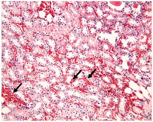
The proximal and distal tubules were observed to be natural in the Control Group (). On the other hand, tubular epithelial desquamation, tubular dilatation, and hydropic changes were observed in the I/R Group ( and ). It was observed that the tubular dilatation and epithelial desquamation regressed in the IR + HP Group when compared with the IR Group (). The most prominent finding in the IR + HP Group was the observing of obvious congestion areas among the tubules (). The histopathological score of kidney tissue among the groups is shown in .
Figure 1. Control group: proximal tubules, distal tubules and glomerular structures are observed in normal histological state, H–E; ×20.
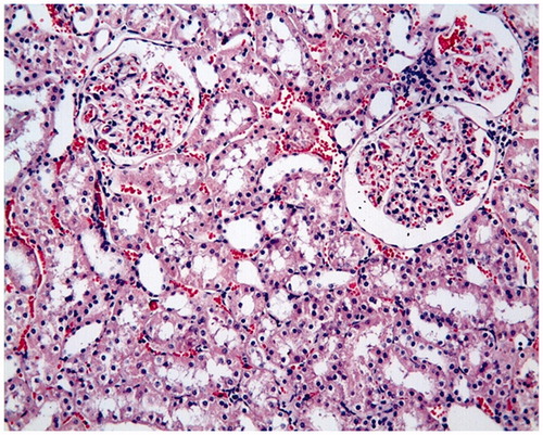
Figure 2. I/R group: degeneration of tubular epithelium (hydropic degeneration (small arrow) in the tubules and cell desquamation (great arrow), H–E; ×20.
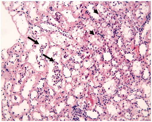
Figure 3. I/R group: tubular dilatation (*), H–E; ×20.
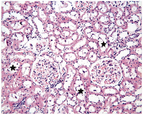
Figure 4. I/R + HP group: tubular dilatation (*) and tubular desquamation (arrow) are observed as being regressed when compared with the I/R group, H–E; ×20.
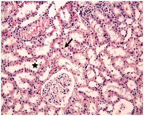
Figure 5. I/R + HP group: intertubular congestion obvious (arrow), H–E; ×20.
Discussion
The blood-flow may be cut off temporarily during urologic and vascular events like kidney transplantation and in operations in which the aorta or the renal pedicule is clamped.Citation22 Reestablishing the blood flow in the ischemic tissue is a situation that may increase the damage in the tissue, which is a paradoxical situation.Citation23 The kidney I/R is a dynamic process in which both the inflammation and some of the mediators that cause the inflammation influence each other. The inflammatory reactions happening during the I/R are among the causes of the oxidative stress and lipid peroxidation.Citation24 Endothelial damage, on the other hand, causes that the adhesion molecules increase. Then, leukocyte infiltration occurs towards the interstitial gap. Necrosis and fibrosis follow the polarity and brush border losses in the proximal tubules.Citation25 Vascular congestion, edema formation, diminished blood-flow have been showed in the corticomedullary junction of the kidney.Citation26 In empirical I/R damage studies conducted on rats, although different ischemia and reperfusion durations were applied, it was concluded in some studies that 45 min ischemia and 3 h reperfusion durations were suitable for the application of the empirical model.Citation3,Citation6
The reestablishment of oxygen to the ischemic kidney results in the production of free oxygen radicals. The free oxygen radicals cause lipid peroxidation and increase the damage in the kidney even more.Citation5 ROS triggers the cascade of cytokines and chemokines through NF-kB (Nuclear Factor kappa B) activation.Citation27 After I/R, the antioxidant defense system is disrupted. The level of the MDA, which is the product of lipid peroxidation, increases, while decreases are observed in SOD, CAT, and GSH-PX enzyme activations.Citation28 Similarly, in our study, while the SOD, CAT, and GSH-PX enzyme activation decreased in the I/R Group, the MDA level increased. In the rats in I/R + HP Group, the CAT, SOD, and GSH-PX enzyme activation increased, and the MDA level decreased. These findings show that HP decreases the oxidative stress developing due to I/R. In histological examination, tubular epithelial desquamation, tubular dilatation and hydropic changes were observed in the I/R Group. The tubular epithelial desquamation, tubular dilatation and hydropic changes decreased in the I/R + HP Group; however, tubular congestion was observed.
Hypericum extracts may be important for the treatment of many diseases that are related with free radical production with their antioxidant effects. In addition to its antidepressant activities, HP, in line with popular credence, also possesses anxiolytic, antiviral, wound-healing, antimicrobial, analgesic, and anti-inflammatory effectsCitation29 HP may be used alone or in combination with the substances that constitute it for treatment purposes.Citation10 HP contains hypericin, hyperphorin, flavonoids, and flavonoid derivatives, xanthone derivatives, and biapigenin. These active components show potential antioxidant activity by free radical scavenging, inhibition of lipid peroxidation, activation of signal transducers.Citation30,Citation31 It was determined that lipid peroxidation and cell-death were decreased in rat-cultured hippocampal neurons by ethanol extracts and the ones with flavonol glycosides, flavonol, and biflavone aglycone. The formation of amyloid-induced ROS were decreased by flavonoids in microgliaCitation32 Dinamarca et al. conducted a study and reported that the hyperphorinin decreased the Aβ-induced neurotoxicity and oxidative damage.Citation33
Benedi et al.Citation34 conducted another in vitro study and showed that the free radical production and lipid peroxidation formed by H2O2 decreased with HP extract application. In another in vitro study, it was reported that the HP application decreased the HO radical production.Citation35 The anti-inflammatory and antioxidant properties of HP resulted in its application in many in vivo study models. Mozaffari et al. conducted a study and found that HP extracts decreased inflammation and lipid peroxidation in rat irritable bowel syndrome model.Citation36 In another study, it was reported that HP application corrected the serum lipid level and oxidative damage in hyperlipidemic rats.Citation37
HP has been used in many I/R study models because of its antioxidant and anti-inflammatory effects. De Paola et al.Citation30 conducted a study in Splanchnic Artery Occlusion Model, and reported that HP extract decreased the lipid peroxidation and inflammation, and healed histopathological damage. Genovese et al.Citation38 conducted a study on rats and reported that applying HP on spinal cord injury decreased inflammation, apoptosis and histologic damage. In another study conducted in Hepatic I/R Model, it was demonstrated that applying HP decreased the inflammatory cytokine level, lipid peroxidation, and histopathological damage.Citation14 Bayramoglu et al.Citation39 conducted a study and applied 50 mg/kg intraperitonal single-dose HP, which is the same in our study. Similar to the findings obtained in our study, it was shown that HP application decreased MDA level and increased CAT, GSH-PX enzyme activity, thus decereasing liver I/R damage. In our study, we observed that the antioxidant property of HP is more obvious. HP and the compounds that constitute it may be used for treatment purposes because of their antioxidant properties in many diseases in which oxidative stress happens. HP, which has been used widely in the whole world for centuries, is a safe and cheap herbal medication. In this respect, it may be aplied both before and after I/R in clinics in a comfortable manner.
Table 2. The histopathological score of kidney tissue among the groups.
Conclusions
In this empirical study, we found that the HP treatment prevented the oxidative damage that occurred due to I/R in renal I/R model. The antioxidant effects of the HP observed in our study show similarities with those reported in the literature. We consider that the full healing of histological and biochemical symptoms being not observed stems from the shorter reperfusion time.
Disclosure statement
Authors declare no conflict of interest.
Funding
This study was supported by the Department of Scientific Research Projects of Inonu University (Project no: 2016/40).
References
- Bucuvic EM, Ponce D, Balbi AL. Risk factors for mortality in acute kidney injury. Rev Assoc Med Bras (1992). 2011;57:158–163.
- Hsu CY, McCulloch CE, Fan D, Ordonez JD, Chertow GM, Go AS. Community-based incidence of acute renal failure. Kidney Int. 2007;72:208–212.
- Ozer Sehirli A, Sener G, Ercan F. Protective effects of pycnogenol against ischemia reperfusion-induced oxidative renal injury in rats. Ren Fail. 2009;31:690–697.
- Wang ZS, Liu XH, Wang M, et al. Metformin attenuated the inflammation after renal ischemia/reperfusion and suppressed apoptosis of renal tubular epithelial cell in rats. Acta Cir Bras. 2015;30:617–623.
- Paller MS, Hoidal JR, Ferris TF. Oxygen free radicals in ischemic acute renal failure in the rat. J Clin Invest. 1984;74:1156–1164.
- Cakir M, Polat A, Tekin S, et al. The effect of dexmedetomidine against oxidative and tubular damage induced by renal ischemia reperfusion in rats. Ren Fail. 2015;37:704–708.
- Anaya-Prado R, Toledo-Pereyra LH. The molecular events underlying ischemia/reperfusion injury. Transplant Proc. 2002;34:2518–2519.
- Klemow KM, Bartlow A, Crawford J, Kocher N, Shah J, Ritsick M, Medical attributes of St. John's Wort (Hypericum perforatum). In: Benzie IFF, Wachtel-Galor S, eds. Herbal Medicine: Biomolecular and Clinical Aspects. Boca Raton, FL: Llc; 2011.
- Oliveira AI, Pinho C, Sarmento B, Dias AC. Neuroprotective activity of Hypericum perforatum and its major components. Front Plant Sci. 2016;7:1004.
- Lawvere S, Mahoney MC. St. John’s Wort. Am Fam Physician. 2005;72:2249–2254.
- Lu YH, Du CB, Liu JW, Hong W, Wei DZ. Neuroprotective effects of Hypericum perforatum on trauma induced by hydrogen peroxide in PC12 cells. Am J Chin Med. 2004;32:397–405.
- Tedeschi E, Menegazzi M, Margotto D, Suzuki H, Forstermann U, Kleinert H. Anti-inflammatory actions of St. John’s Wort: Inhibition of human inducible nitric-oxide synthase expression by down-regulating signal transducer and activator of transcription-1alpha (STAT-1alpha) activation. J Pharmacol Exp Ther. 2003;307:254–261.
- Zdunic G, Godevac D, Milenkovic M, et al. Evaluation of Hypericum perforatum oil extracts for an antiinflammatory and gastroprotective activity in rats. Phytother Res. 2009;23:1559–1564.
- Aydin A, Sakrak O, Yilmaz TU, Kerem M. The effects of Hypericum perforatum on hepatic ischemia-reperfusion injury in rats. Bratisl Lek Listy. 2014;115:209–215.
- Ocak T, Duran A, Ozyalvacli G, et al. Protective effects of montelukast and Hypericum perforatum against intestinal ischemia-reperfusion injury in hamsters. Turk J Med Sci. 2014;44:381–386.
- Ellman GL. Tissue sulfhydryl groups. Arch Biochem Biophys. 1959;82:70–77.
- Mihara M, Uchiyama M. Determination of malonaldehyde precursor in tissues by thiobarbituric acid test. Anal Biochem. 1978;86:271–278.
- Sun Y, Oberley LW, Li Y. A simple method for clinical assay of superoxide dismutase. Clin Chem. 1988;34:497–500.
- Aebi H. Catalase. In: Bergmeyer HU, ed. Methods of Enzymatic Analysis. New York, NY: Academic Press. 1974:673–677.
- Paglia DE, Valentine WN, Dahlgren JG. Effects of low-level lead exposure on pyrimidine 5′-nucleotidase and other erythrocyte enzymes. Possible role of pyrimidine 5′-nucleotidase in the pathogenesis of lead-induced anemia. J Clin Invest. 1975;56:1164–1169.
- Lowry OH, Rosebrough NJ, Farr AL, Randall RJ. Protein measurement with the Folin phenol reagent. J Biol Chem. 1951;193:265–275.
- Cologna AJ, Lima LV, Tucci S, Jr., et al. Cyclosporine action on kidneys of rats submitted to normothermic ischaemia and reperfusion. Acta Cir Bras. 2008;23:36–41. discussion 41.
- Koo DD, Welsh KI, West NE, et al. Endothelial cell protection against ischemia/reperfusion injury by lecithinized superoxide dismutase. Kidney Int. 2001;60:786–796.
- Malek M, Nematbakhsh M. Renal ischemia/reperfusion injury; from pathophysiology to treatment. J Renal Inj Prev. 2015;4:20–27.
- Bonventre JV, Yang L. Cellular pathophysiology of ischemic acute kidney injury. J Clin Invest. 2011;121:4210–4221.
- Molitoris BA, Sutton TA. Endothelial injury and dysfunction: Role in the extension phase of acute renal failure. Kidney Int. 2004;66:496–499.
- Hensley K, Robinson KA, Gabbita SP, Salsman S, Floyd RA. Reactive oxygen species, cell signaling, and cell injury. Free Rad Biol Med. 2000;28:1456–1462.
- Bircan B, Cakir M, Kirbag S, Gul HF. Effect of apelin hormone on renal ischemia/reperfusion induced oxidative damage in rats. Ren Fail. 2016;38:1122–1128.
- Flausino OA Jr, Zangrossi H Jr, Salgado JV, Viana MB. Effects of acute and chronic treatment with Hypericum perforatum L. (LI 160) on different anxiety-related responses in rats. Pharmacol Biochem Behav. 2002;71:251–257.
- De Paola R, Muia C, Mazzon E, et al. Effects of Hypericum perforatum extract in a rat model of ischemia and reperfusion injury. Shock. 2005;24:255–263.
- Saija A, Scalese M, Lanza M, Marzullo D, Bonina F, Castelli F. Flavonoids as antioxidant agents: Importance of their interaction with biomembranes. Free Radic Biol Med.1995;19:481–486.
- Kraus B, Wolff H, Heilmann J, Elstner EF. Influence of Hypericum perforatum extract and its single compounds on amyloid-beta mediated toxicity in microglial cells. Life Sci. 2007;81:884–894.
- Dinamarca MC, Cerpa W, Garrido J, Hancke JL, Inestrosa NC. Hyperforin prevents beta-amyloid neurotoxicity and spatial memory impairments by disaggregation of Alzheimer’s amyloid-beta-deposits. Mol Psychiatry. 2006;11:1032–1048.
- Benedi J, Arroyo R, Romero C, Martin-Aragon S, Villar AM. Antioxidant properties and protective effects of a standardized extract of Hypericum perforatum on hydrogen peroxide-induced oxidative damage in PC12 cells. Life Sci. 2004;75:1263–1276.
- Zou Y, Lu Y, Wei D. Antioxidant activity of a flavonoid-rich extract of Hypericum perforatum L. in vitro. J Agric Food Chem. 2004;52:5032–5039.
- Mozaffari S, Esmaily H, Rahimi R, et al. Effects of Hypericum perforatum extract on rat irritable bowel syndrome. Pharmacogn Mag. 2011;7:213–223.
- Ghosian Moghaddam MH, Roghani M, Maleki M. Effect of Hypericum perforatum aqueous extracts on serum lipids, aminotransferases, and lipid peroxidation in hyperlipidemic rats. Res Cardiovasc Med. 2016;5:e31326.
- Genovese T, Mazzon E, Menegazzi M, et al. Neuroprotection and enhanced recovery with hypericum perforatum extract after experimental spinal cord injury in mice. Shock. 2006;25:608–617.
- Bayramoglu G, Bayramoglu A, Engur S, Senturk H, Ozturk N, Colak S. The hepatoprotective effects of Hypericum perforatum L. on hepatic ischemia/reperfusion injury in rats. Cytotechnology. 2014;66:443–448.