Rubenstein DA, Venkitachalam SM, Zamfir D, Wang F, Lu H, Frame MD, Yin W. In vitro biocompatibility of sheath–core cellulose–acetate-based electrospun scaffolds towards endothelial cells and platelets. J. Biomater. Sci. Polym. Ed. 2010; 21:1713–1736. http://dx.doi.org/10.1163/092050609X12559317149363
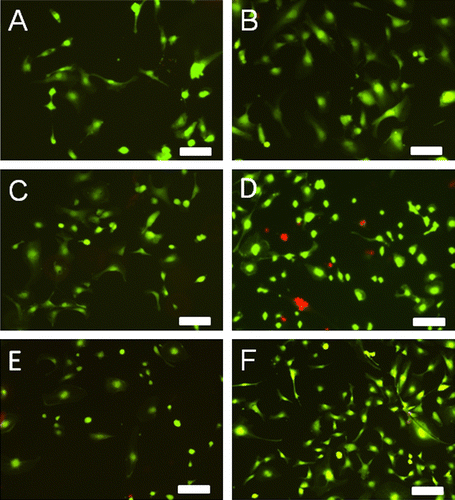
This Corrigendum is to correct a Figure and to clarify how some of the data was collected. When preparing Figure , we inadvertently duplicated Panels C and D. Panel C is the correct figure for the growth of endothelial cells on Chito-S/Chito-C for three days and the corresponding image for endothelial cell growth on Chito-S/Chito-C for five days (Panel D) was mistakenly not placed in the image. Additionally, Panels B and E were inadvertently duplicated during the image preparation. Panel B is the correct image, for endothelial cell growth on CA-S/CA-C for five days and Panel E has been replaced with the appropriate figure. We have prepared a new figure with a corrected Panel D and E. The quantitative analysis of our digital images, which was reported in Figure 4 and Table 3, remains the same. The corrected version of Figure is presented. All other figures in the original manuscript have been verified for accuracy. Additionally, it should be noted that some images were obtained using a Retiga Fast Cooled digital camera (Media Cybernetics, US), in lieu of the Coolsnap Fast Cooled digital camera (ES2), which was originally reported. When switching between these cameras, we were unable to document any differences in quantified data, magnification factors, etc. and thus, our quantitative data remains the same.
Figure 5 Digital images of HUVECs grown on cellulose–acetate-based dual-polymer electrospun scaffolds for 3 (A, C, E) or 5 (B, D, F) days. Scaffold formulations were CA-S/CA-C (A, B), Chito-S/Chito-C (C, D) or CA-S/Chito-C (E, F). Cells were stained with calcein (live, green) and ethidium (red, dead) as a live dead cell cytotoxicity assay. Scale bars are 100 μm. This figure is published in colour in the online edition of this journal that can be accessed via http://dx.doi.org/10.1080/09205063.2013.775835
