Article title: “AuNPs-PCL nanocomposite accelerated abdominal wound healing through photothermal effect and improving cell adhesion”
Authors: Cheng Zhao, Xiuwen Wu, Guopu Chen, Feng Wang, and Jianan Ren
Journal: Journal of Biomaterials Science, Polymer Edition
Bibliometrics: Volume 29, Number 16, pages 2035–2049.
DOI: 10.1080/09205063.2018.1526460
The original version of this article unfortunately contained a mistake. There was an error in . The correct can be found below. We apologize for the mistake.
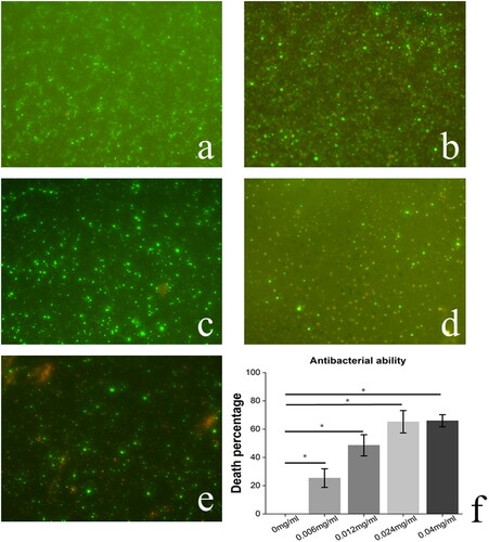
Figure 5. Bacteriostasis. Fluorescence microscope images of PCL scaffold and nanocomposites in resisting bacteria. (a) The pristine. (b) 200 μl 0.006 mg/ml AuNPs treated PCL scaffold. (c) 200 μl 0.012 mg/ml AuNPs treated PCL scaffold. (d) 200 μl 0.024 mg/ml AuNPs treated PCL scaffold. (e) 200 μl 0.04 mg/ml AuNPs treated PCL scaffold. (f) Quantification of the bacterial death percentage. *** means p < 0.001. ** means p < 0.01. * means p < 0.05.