
ABSTRACT
Purpose: To compare the clinical efficacies of 0.01% atropine and orthokeratology (OK) in slowing the development of myopia, and to evaluate their effects on basal tear secretion and tear film stability.
Methods: A prospectively, randomised study. A total of 120 children aged 8–14 years with myopia were included; of these, 60 participants were randomly assigned to use spectacles and 0.01% atropine (SA) and the remaining 60 wore OK lenses (OK). Comprehensive ophthalmologic examinations were performed before and after treatment every 3 months.
Results: The primary outcomes include the changes of spherical equivalent refraction (SE) and axial length (AL). After one-year, the SE and AL in participants aged ≤10 years were better controlled in the SA in low-myopia group (P < .05), whereas those aged ≥11 years were better controlled in the OK in high-myopia group (P < .05). Secondary outcomes include Schirmer’s test and tear film break-up time (TBuT). A statistically significant difference was found in TBuT between the SA and OK in the first 3 months and there was a statistically significant difference in TBuT in the OK between before and after 3 months; however, no statistically significant difference was observed after 6 months.
Conclusion: Both 0.01% atropine and OK lenses effectively retarded myopia progression; however, different effects were found in different ages and refractions. 0.01% atropine had no evident effect on Schirmer’s test and TBuT results. Furthermore, OK lenses had no effect on Schirmer’s test, whereas it had a significant effect on TBuT after only the first 3 months.
Introduction
The increasing incidence of myopia in children has attracted worldwide attention. High myopia in adults, which has been associated with hereditary factors, can be traced back to adolescence.Citation1 High myopia [≥6.00 diopters (D)] can cause vitreous opacity, macular degeneration, posterior sclera staphyloma, and choroidal neovascularization, which are closely related to cataract, glaucoma, retinal detachment, and even blindness.Citation2,Citation3 Therefore, it is important to control the occurrence and development of myopia in children. Many studies have shown that the use of 0.01% atropine and orthokeratology (OK) lenses can slow the progression of myopia and axial elongation, but the results vary greatly among individuals. Thus, it is essential to conduct additional clinical studies to compare the efficacies of 0.01% atropine and OK lenses. Moreover, whether the long-term use of 0.01% atropine or OK lenses can cause dry eye is a concern. In this study, we compared the clinical efficacies of 0.01% atropine and OK lenses in children of different ages and refractions and evaluated their effects on Schirmer’s test and the TBuT results to provide guidance for future clinical practice.
Subjects and methods
Patients
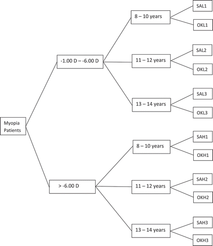
We observed 120 children aged 8–14 years with myopia from the Second Affiliated Hospital of Dalian Medical University between January 2019 and April 2020. According to the spherical equivalent refraction (SE) at the first visit, patients were randomly assigned in 1:1 ratio to low [1.00 diopters (D) < SE < 6.00 D] and high-myopia group (SE ≥ 6.00D) and then divided into age groups of 8–10, 11–12, and 13–14 years.
Groups are as follows:
Figure 1. Treatment and groups (SAL = spectacles + 0.01% atropine; OKL = orthokeratology; SAH = spectacles + 0.01% atropine; OKH = orthokeratology)
Inclusion criteria
The inclusion criteria were as follows: cycloplegic spherical equivalent refraction (SE) at least −1.00 diopters (D) and myopic astigmatism of ≤−1.00 DC and less than or equal to half the spherical diopter. All patients were randomly divided into the SA and OK groups. According to the SE of the first visit, they were divided into low- and high-myopia groups; and then divided based on age groups of 8–10 years, 11–12 years, and 13–14 years.
Exclusion criteria
The exclusion criteria were as follows: (1) wearing contact lenses within 3 days of the start of examination; (2) children with ocular disorders such as glaucoma, cataract, keratopathy, strabismus, and amblyopia and systemic disorders such as cardiac and respiratory illnesses; (3) anisometropia >1.5 D; (4) intraocular pressure (IOP) of >21 mmHg and difference between the eyes of >8 mmHg; (5) use of anticholinergic and cholinergic drugs that affect the evaluation of efficacy, such as atropine, pirenzepine, and pilocarpine, within the past 1 month; (6) use of other therapies that may affect the evaluation of efficacy within the past 3 months, such as wearing OK lenses and therapy of traditional Chinese medicine; (7) low birth weight (≤1500 g); and (8) history of hypersensitivity to atropine or anticholinergic drugs.
Before the experiment, the investigators explained the expected benefits and potential risks of using 0.01% atropine and OK lenses to the parents of children and also informed them about the details of the methods and precautions. Explanation to children was provided using an easy-to-understand method before obtaining informed consent.
Statement
All procedures performed in studies involving human participants were in accordance with the ethical standards of the Second Affiliated Hospital of Dalian Medical University (Dalian, China) and adhered to the tenets of the Declaration of Helsinki. Informed consent was obtained from all individual participants and their parents included in the study.
Methods
All subjects underwent comprehensive tests before treatment, including slit-lamp examination, distance and near visual acuity, noncycloplegic and cycloplegic autorefraction, IOP, AL, corneal topography, Schirmer’s test and TBuT. We applied 1% atropine eye gel for cycloplegic autorefraction after 3 days in children aged ≤10 years. The compound tropicamide was applied three times with an interval of 10 min to children aged >10 years, and we waited for 20 min for cycloplegic autorefraction.
Subjects were prescribed 0.01% atropine eye drops to be applied once per night before bedtime in both eyes in the SA groups. Subjects were required to wear OK lenses every night for at least eight consecutive hours in the OK groups. Spectacles were rematched when the SE dropped by 0.5 D and the vision of wearing glasses decreased. OK lenses were rematched when the naked vision during the day reached <0.5. Further, 0.01% atropine was procured from Shenyang Xingqi pharmaceutical company (Shenyang, China); it has a pH value between 3.5 and 4.0 and comprises no preservatives.
When the central corneal thickness stabilized after the first 1–2 months of OK treatment, the AL measurement was used as the baseline value after 3 months as in the studies conducted by Kakita et al.Citation4 The patients ceased wearing OK lenses and were examined weekly until the curvature regressed and the refractive error stabilized (difference between consecutive visits ≤0.25 D) during the follow-up visit. In this study, SE was measured using an autorefractor (KR-800, TOPCON, TOKYO, JAPAN). IOP was measured using an autotonometer (CT-IP, TOPCON, TOKYO, JAPAN). Corneal topography was evaluated using the OPD-Scan III (NIDEK, CO, LTD, TOKYO, JAPAN). AL was measured using the non-contact Biometer (lenstar LS 900, HAAG-STREIT AG, KOENIZ, SWITZERLAND). All experimental data were recorded using the average of five successive measurements for analysis.
Statistical methods
We applied SPSS 26.0 (IBM Corp., Armonk, NY) statistical analysis software for data analysis. The results of age, SE, AL, Schirmer’s test, and TBuT were described by mean (SD, standard deviation). Independent samples t-tests were used for intergroup comparisons and paired samples t-test was used for intragroup comparisons. Fisher’s exact test was used to analyze gender differences between the groups. A P value of <0.05 was considered statistically significant.
Results
A total of 240 eyes from 120 participants were included in this study. No statistically significant difference was found between the SAL1 and OKL1 groups, between the SAL2 and OKL2 groups, between the SAL3 and OKL3 groups, between the SAH1 and OKH1 groups, between the SAH2 and OKH2 groups, and between the SAH3 and OKH3 groups in terms of age, sex, SE, AL, Schirmer’s test, or TBuT results when they started treatment.
The primary outcomes included SE and AL. The increases in SE were −0.21 ± 0.05 D and −0.30 ± 0.07 D in the SAL1 group, and −0.26 ± 0.08 D and −0.41 ± 0.08 D in the OKL1 group (P < .05, independent samples t-test) over 6 and 12 months, respectively, see . The increases in SE were −0.28 ± 0.07 D and −0.41 ± 0.08 D in the SAH2 group, and −0.20 ± 0.09 D and −0.32 ± 0.09 D in the OKH2 group (P < .05, independent samples t-test) over 6 and 12 months, respectively, see . The increases in SE were −0.33 ± 0.07 D and −0.48 ± 0.07 D in the SAH3 group and −0.17 ± 0.08 D and −0.26 ± 0.11 D in the OKH3 group (P < .05, independent samples t-test) over 6 and 12 months, respectively, see . However, no statistically significant difference was found in the SE between the SAL2 and OKL2 groups, between the SAL3 and OKL3 groups, and between the SAH1 and OKH1 groups see . The increases in AL were 0.12 ± 0.04 mm and 0.24 ± 0.06 mm in the SAL1 group, and 0.18 ± 0.06 mm and 0.32 ± 0.07 mm in the OKL1 group (P < .05, independent samples t-test) over 6 and 12 months, respectively, see . The increases in AL were 0.18 ± 0.06 mm and 0.32 ± 0.07 mm in the SAH2 group, and 0.13 ± 0.05 mm and 0.24 ± 0.08 mm in the OKH2 group (P < .05, independent samples t-test) over 6 and 12 months, respectively, see . The increases in AL were 0.21 ± 0.05 mm and 0.36 ± 0.06 mm in the SAH3 group, and 0.11 ± 0.05 mm and 0.20 ± 0.18 mm in the OKH3 group (P < .05, independent samples t-test) over 6 and 12 months, respectively, see . However, no statistically significant difference was found in the AL between the SAL2 and OKL2 groups, between the SAL3 and OKL3 groups, and between the SAH1 and OKH1 groups, see .
Table 1. Alterations in SE and AL in the low-myopia group after treatment
Table 2. Alterations in SE and AL in the high-myopia group after treatment
Secondary outcomes presented in this article included Schirmer’s test and TBuT. No statistically significant difference was found in Schirmer’s test results between the SA and OK groups, see . A statistically significant difference was found in the TBuT results between the SA and OK groups in the first 3 months; however, no statistically significant difference was found after 6 months, see . No statistically significant difference was found in the Schirmer’s test results between the before treatment group and after 3, 6, and 12 months of treatment in the SA and OK groups, see . No statistically significant difference was found in the TBuT results between the before treatment group and after 3, 6, and 12 months of treatment in the SA groups, see . A statistically significant difference was found in the TBuT results between the before treatment group and after the first 3 months of treatment in the OK groups (both P < .05, paired samples t-test); however, the TBuT almost recovered to pretreatment levels after 6 months, see . The Schirmer’s test and TBuT results showed a tendency to be reduced in the SA groups even when all patients exhibited no symptoms and the data showed no statistical significance.
Table 3. Comparison of Schirmer’s test and TBuT between the SA and OK groups after treatment
Table 4. Comparison of Schirmer’s test and TBuT in the SA and OK groups between before treatment and after treatment
Discussion
With the increasing incidence of myopia in children in recent years, there has been an increasing attention toward controlling the development of myopia. Outdoor activities of 2–3 h every day have been demonstrated to exert protective effects on reducing the risk of myopia.Citation5 However, outdoor activities have proven difficult to practice because of the learning burden in children. Various methods have been used to impede the progression of myopia, including the use of atropine, pirenzepine, OK lenses, peripheral defocus modifying spectacle lenses, rigid gas-permeable contact lenses, and soft contact lenses.Citation6 The uses of 0.01% atropine and OK lenses are presently the most common and effective methods.
The mechanism underlying the development of myopia is still unknown, and how atropine and OK lenses reduce the progression of myopia is also unclear. Current theories have been proposed for atropine that is a nonselective mAChR antagonist; it blocks M1 and M4 receptors in the retina and sclera to restrict axial elongation and affect sclera remodeling.Citation7 Another theory is that atropine relieves near work-induced transient myopia (NITM). NITM is a transient myopia drift that resulted from near work for a period of time and is a crucial factor related to the development of permanent myopia.Citation8 Its mechanism may be related to the poor adjustment of the lens and may be associated with an accommodation lag phenomenon involving the interaction of the sympathetic and parasympathetic nervous systems.Citation8 A recent study has shown that 0.01% atropine has no clinical effect on cornea and lens; therefore, it has almost no side effect. Atropine mainly causes a reduction in AL elongation.Citation9 One studyCitation10 reported that adverse reactions were not significantly related to 0.01% atropine.
OK is an optical method that employs reverse-geometry-designed rigid oxygen-permeable contact lens, allowing corneal epithelial redistribution to correct refractive errors when worn at night.Citation11 Therefore, atropine and OK lenses control the progression of myopia through different mechanisms. Several studies have demonstrated the efficacies of atropine and OK lenses in slowing the progression of myopia and axial elongation in children; however, the results significantly vary among individuals.Citation12–15
In this study, patients with different ages and different refractions had different clinical efficacies when comparing the use of 0.01% atropine and OK lenses. For patients aged ≤10 years with an SE of <6.00 D, the clinical efficacy of 0.01% atropine on myopia progression and axial growth was better than that of OK lenses. Conversely, the clinical efficacy of OK lenses was better for patients aged ≥11 years having an SE of ≥6.00 D. For patients aged ≥11 years having an SE of <6.00 D and for those aged <11 years having an SE of ≥6.00 D, the results showed equal effects. Therefore, to slow the development of myopia, clinicians can select the most suitable method for children depending on their ages and refractions. Especially for children with an SE of ≥6.00 D, clinicians should first consider the use of OK lenses.
The results of this study showed that TBuT reduced after 3 months of wearing OK lenses. A possible mechanism was that OK lenses influenced the quality and stability of the tear film. Several clinical studies have reported that OK lenses affect the amount of tear secretion, causing reduced stability of the tear film.Citation16,Citation17 In this study, the TBuT results reduced in the first 3 months after wearing OK lenses and almost recovered to pretreatment levels after 6 months. Furthermore, the Schirmer’s test and TBuT results showed a tendency to be reduced after the use of 0.01% atropine. Atropine may have had an inhibitory effect on the tear gland secretions. None of the patients had dry eye symptoms during the follow-up; however, the specific effect of atropine and OK lenses on lacrimal secretion and stability of the tear film need further study.
There is currently no method available to stop the development of myopia. All methods of treatment only deal with the consequences of refractive error rather than its biological causes. More experiments are still needed to identify the fundamental mechanisms underlying the occurrence and development of myopia. In this study, the follow-up time was only 1 year. In future studies, therefore, we intend to continue to enroll additional subjects and conduct follow-up studies for a longer period of time.
Strengths and limitations
In this prospective randomized study, patients were grouped based on different ages and refractions. And we mainly compared the clinical efficacies of 0.01% atropine and orthokeratology (OK) in slowing the development of myopia and evaluated the effects of 0.01% atropine and OK on the Schirmer’s test and BuT results. However, our study also had a few limitations. First, the follow-up time of this study was only 1 year, a longer study period is required. Second, our study only used 0.01% atropine, although other concentrations such as 0.5%, 0.1%, 0.05%, and 0.025% are also used clinically. Further studies are required to determine the effect on the surface with different concentrations of atropine.
Disclosure statement
Author Qi Zhao and Qian Hao declare that they have no conflict of interest.
Additional information
Funding
References
- Xiang F, He M, Morgan IG, et al. The impact of parental myopia on myopia in Chinese children: population-based evidence. Optom Vis Sci. 2012;89(10):1487–1496. doi:https://doi.org/10.1097/OPX.0b013e31826912e0.
- Morgan IG, Ohno-Matsui K, Saw SM, et al. Myopia. Lancet. 2012;379:1739–1748. doi:https://doi.org/10.1016/S0140-6736(12)60272-4.
- ,Chen Z, Huangd S, Zhou J, et al. Adjunctive effect of orthokeratology and low dose atropine on axial elongation in fast-progressing myopic children—A preliminary retrospective study.
- Hiraoka T, Kakita T, Okamoto F, et al. Longterm effect of overnight orthokeratology on axial length elongation in childhood myopia: a 5-year follow-up study. Invest Ophthalmol Vis Sci. 2012;53:3913–3919. doi:https://doi.org/10.1167/iovs.11-8453.
- Sherwin JC, Reacher MH, Keogh RH, et al. The association between time spent outdoors and myopia in children and adolescents: a systematic review and meta-analysis. Ophthalmology. 2012;119:2141–2151. doi:https://doi.org/10.1016/j.ophtha.2012.04.020.
- Huang J, Wen D, Wang Q, et al. Efficacy comparison of 16 interventions for myopia control in children: a network metaanalysis. Ophthalmology. 2016;123:697–708. doi:https://doi.org/10.1016/j.ophtha.2015.11.010.
- Gallego P, Martinez-Garcia C, Perez-Merino P, et al. Scleral changes induced by atropine in chicks as an experimental model of myopia. Ophthalmic Physiol Opt. 2012;32(6):478–484. doi:https://doi.org/10.1111/j.1475-1313.2012.00940.x.
- Lin Z, Vasudevan B, Liang YB, et al. Near work induced transient myopia (NITM) in anisometropia. Ophthalmic Physiol Opt. 2013;33:311–317. doi:https://doi.org/10.1111/opo.12049.
- Li FF, Kam KW, Zhang Y, et al. Effects on ocular biometrics by 0.05%, 0.025%, and 0.01% atropine: low-concentration atropine for myopia progression (LAMP) study. Ophthalmology. 2020;10:1016.
- Yam JC, Jiang Y, Tang SM, et al. Low-concentration atropine for myopia progression (LAMP) study: a randomized, double-blinded, placebo-controlled trial of 0.05%, and 0.025%, and 0.01% atropine eye drops in myopia control. Ophthalmology. 2019;126(1):113–124. doi:https://doi.org/10.1016/j.ophtha.2018.05.029.
- Nichols JJ, Marsich MM, Nguyen M, et al. Bullimore, overnight orthokeratology. Optom Vis Sci. 2000;77(5):252–259. doi:https://doi.org/10.1097/00006324-200005000-00012.
- Cho P, Cheung SW. Retardation of myopia in Orthokeratology (ROMIO) study: a 2-year randomized clinical trial. Invest Ophthalmol Vis Sci. 2012;53:7077–7085. doi:https://doi.org/10.1167/iovs.12-10565.
- Cho P, Cheung SW, Edwards M. The longitudinal orthokeratology research in children (LORIC) in Hong Kong: a pilot study on refractive changes and myopic control. Curr Eye Res. 2005;30:71–80. doi:https://doi.org/10.1080/02713680590907256.
- Chia A, Chua WH, Wen L, et al. Atropine for the treatment of childhood myopia: changes after stopping atropine 0.01%, 0.1% and 0.5%. Am J Ophthalmol. 2014;157:451–457.e1. doi:https://doi.org/10.1016/j.ajo.2013.09.020.
- Fan DS, Lam DS, Chan CK, et al. Topical atropine in retarding myopic progression and axial length growth in children with moderate to severe myopia: A pilot study. Jpn J Ophthalmol. 2007;51:27–33. doi:https://doi.org/10.1007/s10384-006-0380-7.
- Kastelan S, Lukenda A, Salopek-Rabati, et al. Dry eye symptoms and signs in longterm contact lens wearers. Coll Antropol. 2013;37(1):199203.
- D H S, Iskander DR. Tear film dynamics on soft contact lenses. Optom Vis Sci. 2014;91:14061411.