Abstract
The SLC28 family of concentrative nucleoside transporter (CNT) proteins in mammalian cells contains members of two distinct phylogenic subfamilies. In humans, hCNT1 and hCNT2 belong to one subfamily, and hCNT3 to the other. All three CNTs mediate inwardly-directed Na+/nucleoside cotransport, and are either pyrimidine nucleoside-selective (hCNT1), purine nucleoside-selective (hCNT2), or broadly selective for both pyrimidine and purine nucleosides (hCNT3). While previous studies have characterized cation interactions with both hCNT1 and hCNT3, little is known about the corresponding properties of hCNT2. In the present study, heterologous expression in Xenopus oocytes in combination with radioisotope flux and electrophysiological techniques has allowed us to undertake a side-by-side comparison of hCNT2 with other hCNT family members. Apparent K50 values for Na+ activation were voltage-dependent, and similar in magnitude for all three transporters. Only hCNT3 was also able to couple transport of uridine to uptake of H+. The Na+/nucleoside stoichiometry of hCNT2, as determined from both Hill coefficients and direct charge/flux measurements, was 1:1. This result was the same as for hCNT1, but different from that of hCNT3 (2:1). The charge-to-22Na+ uptake stoichiometry was 1:1 for all three hCNTs. In parallel with their division into two separate CNT subfamilies, hCNT2 shares common cation specificity and coupling characteristics with hCNT1, which differ markedly from those of hCNT3.
Introduction
Physiological nucleosides and most therapeutic nucleoside analogs are hydrophilic molecules and require specialized plasma membrane nucleoside transport proteins (NTs) to cross cellular membranes (Cass [Citation1995], Griffith & Jarvis [Citation1996], Young et al. [Citation2000]). Two structurally unrelated NT protein families exist in mammalian cells: the SLC29 equilibrative nucleoside transporter (ENT) family, which is responsible for bidirectional nucleoside fluxes driven by nucleoside concentration gradients, and the SLC28 concentrative Na+-dependent nucleoside transporter (CNT) family, which couples nucleoside influx to movement of Na+ down its electrochemical gradient. NTs play important roles in salvage of precursors for nucleic acid biosynthesis, regulate adenosine availability to purinoreceptors, and permit cellular uptake of nucleoside drugs used in the treatment of cancer and viral infections (Young et al. [Citation2000], Damaraju et al. [Citation2003]).
In human (h) cells, three CNT isoforms (hCNT1-3) have been identified. Although both hCNT1 and hCNT2 transport uridine and adenosine, hCNT1 is otherwise pyrimidine nucleoside-selective and corresponds to functional nucleoside transport system cit (concentrative, insensitive to inhibition by nitrobenzylthioinosine (NBMPR), transports thymidine) while hCNT2 is purine nucleoside-selective and corresponds to functional nucleoside transport system cif (concentrative, insensitive to inhibition by NBMPR, transports formycin B) (Ritzel et al. [Citation1997], Wang et al. [Citation1997], Ritzel et al. [Citation1998]). hCNT1 and hCNT2 mediate inwardly-directed Na+/nucleoside cotransport, and are present primarily in intestinal and renal epithelia and other specialized cells. hCNT3 corresponds to functional nucleoside transport system cib (concentrative, insensitive to inhibition by NBMPR, broad selectivity), mediates Na+/nucleoside cotransport of both purine and pyrimidine nucleosides, and appears to have a wider tissue and cellular distribution than either hCNT1 or hCNT2 (Ritzel et al. [Citation2001]). Tissues containing hCNT3 transcripts include pancreas, bone marrow, trachea, mammary gland, liver, prostate, and regions of intestine, brain and heart (Ritzel et al. [Citation2001]). Mammalian hCNT1/2 and hCNT3 belong to two distinct CNT phylogenic subfamilies; the CNT3 subfamily also contains a second broad specificity cib-type CNT (hfCNT) from an ancient pre-vertebrate, the Pacific hagfish (Yao et al. [Citation2002]).
In addition to their well established differences in nucleoside preference, there is evidence that hCNT1, hCNT2 and hCNT3 may also differ with respect to cation interactions. For example, indirect Hill-type analyses of the relationship between nucleoside influx and Na+ concentration suggest Na+/nucleoside stoichiometries of 1:1 for cit (CNT1) and cif (CNT2) transport activities in different mammalian cells and tissues, compared to 2:1 for cib (CNT3) (Cass [Citation1995], Griffith & Jarvis [Citation1996]). Recently, a Na+/nucleoside coupling ratio of 2:1 has been confirmed for recombinant hCNT3 and hagfish hfCNT produced in Xenopus oocytes based upon direct charge versus radiolabelled uridine uptake measurements (Yao et al. [Citation2002], Smith et al. [Citation2005]). hCNT3 has also been shown to be H+-dependent, with a H+/nucleoside coupling ratio of 1:1 (Smith et al. [Citation2005]). In the case of recombinant hCNT1, however, corresponding analyses of oocyte charge vs. radiolabelled uridine uptake has provided conflicting Na+/nucleoside stoichiometries of either 1:1 (Smith et al. [Citation2004]) or 2:1 (Larráyoz et al. [Citation2004]). No corresponding data exists in the literature for recombinant hCNT2. To fill in this important gap with respect to hCNT2, and to address whether the Na+/nucleoside coupling ratio of hCNT1 is indeed different from that of hCNT3, we have used heterologous expression in Xenopus oocytes in combination with both radioisotope flux and electrophysiological techniques to undertake a systematic side-by-side comparison of cation interactions with all three human CNTs.
Materials and methods
In vitro transcription and expression in Xenopus oocytes
cDNAs encoding hCNT1/2/3 (GenBank™ accession numbers U62968, AF036109 and AF305210, respectively) in the enhanced Xenopus plasmid expression vector pGEM-HE with flanking 5′- and 3′-untranslated regions (UTRs) from the Xenopus β-globin gene were obtained as previously described (Ritzel et al. [Citation2001]). Plasmid DNA was linearized with Nhe1 (hCNT1/3) or Sph1 (hCNT2) and transcribed with T7 polymerase using the mMESSAGE mMACHINE™ (Ambion, Austin, TX, USA) transcription system. The remaining template was removed by digestion with RNase-free DNase1. Stage V-VI oocytes were isolated by collagenase treatment (2 mg ml−1 for 2 h) of ovarian lobes from female Xenopus laevis (Biological Sciences Vivarium, University of Alberta) that had been anaesthetized by immersion in 0.3% (w/v) tricaine methanesulphonate (pH 7.4). Frogs were humanely killed following collection of oocytes in compliance with guidelines approved by the Canadian Council on Animal Care. The remaining follicular layers were removed by phosphate treatment (100 mM K2PO4) and manual defolliculation. Oocytes were injected with either 10 nl of water containing 10 ng of RNA transcript encoding either hCNT1/2/3 or 10 nl of water alone. Injected oocytes were then incubated for either 4 days (radioisotope flux studies) or 4–7 days (electrophysiology) at 18°C in modified Barth's solution (changed daily) (88 mM NaCl, 1 mM KCl, 0.33 mM Ca(NO3)2, 0.41 mM CaCl2, 0.82 mM MgSO4, 2.4 mM NaHCO3, 10 mM Hepes, 2.5 mM sodium pyruvate, 0.1 mg ml−1 penicillin and 0.05 mg ml−1 gentamycin sulfate, pH 7.5) prior to the assay of nucleoside transport activity.
Transport media
Since steady-state current and radioisotope studies have shown that H+ ions are capable of driving the uptake of nucleoside substrate mediated by hCNT3 (Smith et al. [Citation2005]), all electrophysiological and radioisotope flux studies for hCNT1/2/3 in Na+-containing transport medium were performed at pH 8.5 to minimize the H+ inward driving force. Na+-containing transport medium was composed of 100 mM NaCl, 2 mM KCl, 1 mM CaCl2, 1 mM MgCl2 and 10 mM Hepes. In experiments examining the Na+-dependence of transport, Na+ in the transport medium was replaced by equimolar choline chloride (ChCl) to maintain isomolarity. Proton-dependence was tested in Na+-free choline-containing transport medium (100 mM ChCl) at pH values ranging from 5.5–8.5. In solutions with pH values ≤ pH 6.5, 10 mM Mes was used in place of Hepes.
Radioisotope flux studies
Radioisotope transport assays were performed as described previously (Ritzel et al. [Citation2001]) on groups of 10–12 oocytes at 20°C using [14C]-labelled uridine (1 µCi ml−1) in 200 µl of the appropriate transport medium. Nucleoside uptake was determined at a uridine concentration of 20 µM. Following incubation, seven rapid washes in ice-cold choline chloride transport medium (pH 7.5) removed extracellular label, and individual oocytes were dissolved in 1% (w/v) SDS for quantitation of cell-associated radioactivity by liquid scintillation counting (LS 6000 IC, Beckman, Fullerton, CA, USA). Uptake values represent initial rates of transport (1 min fluxes) and are presented as means±SEM. (standard error of the mean) of 10–12 oocytes from representative experiments. Individual experiments were performed on cells from single batches of oocytes used on the same day. Each experiment was repeated at least twice using oocytes from different frogs.
Electrophysiological studies
Nucleoside-evoked membrane currents were measured in CNT-producing oocytes at room temperature (20°C) using a GeneClamp 500B oocyte clamp (Axon Instruments Inc., Foster City, CA, USA) in the two-electrode, voltage-clamp mode. The GeneClamp 500B was interfaced to an IBM-compatible PC via a Digidata 1322A A/D converter and controlled by pCLAMP software (Version 9.0, Axon Instruments Inc.). The microelectrodes were filled with 3 M KCl and had resistances that ranged from 0.5–2.5 MΩ (megaohms). Oocytes were penetrated with the microelectrodes and their membrane potentials were monitored for periods of 10–15 min. Oocytes were discarded when membrane potentials were unstable, or more positive than −30 mV. For cation activation curves, the oocyte membrane potential was clamped at a holding potential (Vh) of −30 or −90 mV. Current signals were filtered at 20 Hz (four-pole Bessel filter) and sampled at a sampling interval of 20 ms.
Kinetic parameters
Kinetic parameters calculated from radioisotope flux and electrophysiological experiments were determined by least squares fits to the Hill equation,respectively, where V is the nucleoside-induced flux, Vmax is the predicted flux maximum, I is the nucleoside-induced current, Imax is the predicted current maximum, [Na+] is the concentration of sodium, K50 is the half-saturation constant for Na+ activation, and n is the Hill coefficient (Sigmaplot 9.0, Jandel Scientific Software, San Rafael, CA, USA). Kinetic parameters from radioisotope experiments were derived from curve fits to mediated averaged data from 10–12 oocytes and are presented as means±SE (standard error of the fitted estimate). Mediated data is determined as uptake in RNA-injected oocytes minus uptake in water-injected control oocytes. Kinetic parameters from electrophysiological experiments were determined from fits to data from individual oocytes normalized to the Imax value obtained for that oocyte and are presented as means±SEM of 4 or more cells.
Charge-to-nucleoside stoichiometry
The Na+/nucleoside coupling ratios for hCNT1, hCNT2 or hCNT3 were determined by simultaneously measuring Na+ currents and 14C-nucleoside (uridine or adenosine) (200 µM, 1 µCi ml−1) uptake under voltage-clamp conditions. In experiments measuring the Na+/nucleoside coupling ratio, 100 mM NaCl medium (pH 8.5) was used. Individual oocytes injected with RNA transcripts encoding hCNT1, hCNT2 or hCNT3 were voltage-clamped at Vh of −90 mV in a perfusion chamber and washed with nucleoside-free medium for a 5-min period to monitor baseline currents. When the baseline was stable, the perfusion was stopped and medium of the same composition containing unlabelled and radiolabelled nucleoside was manually added to the perfusion chamber. Current was measured for 2 min, and uptake of nucleoside was terminated by rapidly washing the oocyte with nucleoside-free medium until the current returned to baseline (<15 sec). The oocyte was then immediately transferred to a scintillation vial and solubilized with 1% (w/v) SDS for quantitation of oocyte-associated radioactivity. Nucleoside-induced current was obtained as the difference between baseline current and the inward nucleoside-induced current. The total charge translocated into the oocyte during the uptake period was calculated from the current-time integral and correlated with the measured radiolabelled flux for each oocyte to determine the charge/flux ratio. Basal 14C-nucleoside uptake was determined in control water-injected oocytes (from the same donor frog) under equivalent conditions and used to correct for endogenous non-mediated nucleoside uptake over the same incubation period. Coupling ratios (±SE) were calculated from slopes of least-squares fits of nucleoside-dependent charge versus nucleoside accumulation for 8 or more oocytes.
Charge-to-Na+ stoichiometry
CNT-mediated uptake of 22Na+ was optimized for specific activity and transport rate by using a saturating concentration of uridine (200 µM) and a 22Na+ concentration of 1 mM, a value close to the −90 mV Na+ apparent K50 for each transporter. Individual hCNT1-, hCNT2- or hCNT3-producing oocytes were voltage clamped at −90 mV and perfused with Na+-free medium (100 mM ChCl, pH 8.5) for a period of 5 min. A stable baseline current was recorded and perfusion was stopped. Medium containing 200 µM uridine, 1 mM 22Na+ (1 µCi ml−1) and 99 mM ChCl (pH 8.5) was manually added to the perfusion chamber. The uptake period was 2 min. The solution was then rapidly changed back to uridine- and Na+-free medium until the current returned to baseline (<15 sec). The oocyte was then transferred immediately to a scintillation vial and solubilized with 1% (w/v) SDS for quantitation of oocyte-associated radioactivity. Testing of each individual CNT-producing oocyte prior to addition of uridine and 22Na+ showed no shift in the baseline current when the composition of the bath solution was changed from 100 mM ChCl to 1 mM NaCl + 99 mM ChCl, indicating an absence of detectable CNT Na+ slippage under the experimental conditions used. Similarly, basal 22Na+ uptake in control water-injected oocytes (from the same donor frog) was determined under equivalent conditions over the same incubation period and used to correct for endogenous non-mediated Na+ uptake. For each CNT-producing oocyte, the total charge translocated during the uptake period was calculated from the current-time integral and correlated with the measured 22Na+ flux to determine the charge/flux ratio. The charge-to-Na+ stoichiometry (±SE) was calculated from the slope of a least-squares fit of uridine-dependent charge vs. 22Na+ accumulation for 5 or more oocytes.
Chemicals
Nucleosides and tricaine methanesulphonate were purchased from Sigma (Oakville, ON, Canada). [14C]-Labelled nucleosides and 22Na+ were purchased from Amersham Pharmacia Biotech (Piscataway, NJ, USA).
Results
Cation specificity
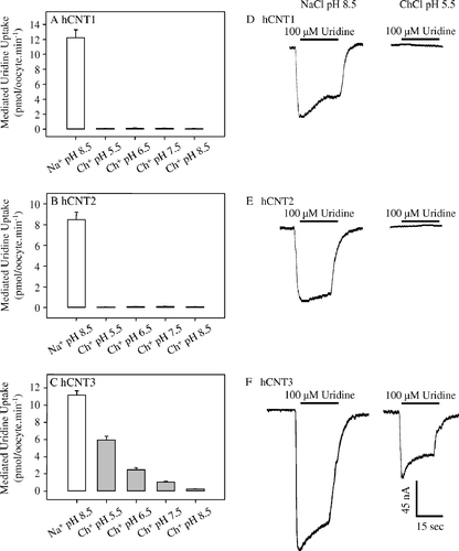
The experiment of directly compared the Na+- and H+-dependence of nucleoside transport mediated by hCNT1, hCNT2 and hCNT3. CNT-mediated uptake (influx) of [14C]-uridine (20 µM) in the presence of Na+ was measured in oocytes expressing hCNT1 (A), hCNT2 (B) or hCNT3 (C) in transport media containing 100 mM NaCl (pH 8.5). To examine CNT-mediated uptake of uridine in the presence of H+, Na+ in the transport medium was replaced by equimolar choline, and the pH varied from 5.5–8.5. Values were corrected for basal non-mediated uptake in control water-injected oocytes (<0.03 pmol/oocyte.min−1 under all conditions tested). All three recombinant transporters mediated similar levels of uridine influx in the presence of external Na+, but only hCNT3 demonstrated pH-dependent uridine influx. Previous studies have established that pH has minimal effect on hCNT3 Na+-mediated uridine binding affinity (Smith et al. [Citation2005]). The intracellular oocyte pH has been shown to be 7.3–7.6 (Burckhardt et al. [Citation1992]) and consistent with this, and in agreement with previous findings (Smith et al. [Citation2005]), H+-coupled uptake of uridine by hCNT3 was minimal at pH values of 7.5 or higher, but increased markedly as the external medium was acidified. In 100 mM ChCl at pH 5.5, hCNT3 mediated uridine influx was ∼60% of that obtained in the presence of 100 mM NaCl at pH 8.5. As the pH was raised from 5.5–8.5, uridine influx was reduced by 96%. Influx of uridine by hCNT2 and hCNT1 under the same conditions also exhibited marked Na+-dependence, but showed no indication of H+-coupling. This was confirmed by electrophysiology: all three transporters generated large inward Na+ currents in the presence of uridine, but only hCNT3 exhibited uridine-evoked H+ currents (D–1F). Subsequent experiments focused on the kinetics of Na+ activation and the determination of Na+/nucleoside coupling ratios.
Figure 1. Effects of Na+ and H+ on the transport activities of recombinant hCNT1, hCNT2 and hCNT3 produced in Xenopus oocytes. Radiolabelled fluxes of uridine (20 µM, 20°C, 1 min flux) in oocytes injected with RNA transcripts encoding hCNT1 (A), hCNT2 (B), or hCNT3 (C) were measured in transport media containing Na+ (open bar; 100 mM NaCl, pH 8.5) or choline (gray bars; 100 mM ChCl, pH 5.5–8.5). Values were corrected for basal non-mediated uptake in control water-injected oocytes and are means±SEM of 10–12 oocytes. The experiment was performed on a single batch of oocytes used on the same day. Representative cation/nucleoside current traces in single hCNT1- (D), hCNT2- (E), or hCNT3- (F) producing oocytes clamped at −50 mV in transport medium containing Na+ (100 mM NaCl, pH 8.5) or choline (100 mM ChCl, pH 5.5). The bar denotes addition of uridine (100 µM) to the bath. No currents were observed in control water-injected oocytes (traces not shown).
Na+ activation kinetics
Na+ activation curves measured by radioisotope nucleoside flux assays are presented in for oocytes producing either hCNT1, hCNT2 or hCNT3. Uptake was compared to that in control water-injected oocytes, and the kinetic parameters derived from these experiments for the CNT-mediated component of influx (uptake in RNA-injected oocytes minus uptake in water-injected oocytes) are presented in . For both hCNT1 and hCNT2, the relationship between uridine influx and Na+ concentration (0–100 mM NaCl, pH 8.5) was hyperbolic, with Hill coefficients consistent with an apparent Na+/nucleoside coupling stoichiometry of 1:1 (A, 2B). In marked contrast, the Na+ activation curve for hCNT3 was sigmoidal, with a Hill coefficient consistent with an apparent Na+/nucleoside coupling ratio of 2:1 (C). The three transporters (hCNT1/2/3) exhibited similar millimolar apparent affinities for Na+ and similar Vmax values ().
Figure 2. Cation activation kinetics of hCNT1, hCNT2 and hCNT3. Initial rates of 14C-uridine uptake (20 µM, 20°C, 1 min flux) were measured in Na+-containing (0–100 mM NaCl) transport media at pH 8.5 in oocytes injected either with water alone (open circles) or with water containing RNA transcripts encoding hCNT1 (A), hCNT2 (B) or hCNT3 (C) (solid circles). All of the fluxes were performed on the same batch of oocytes used on the same day. Kinetic parameters derived from these data for the hCNT-mediated component of transport (uptake in RNA transcript-injected oocytes minus uptake in water-injected oocytes) are presented in . The inset in each graph shows the Hill plot for the data.
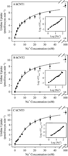
Table I. Na+ activation kinetics of uridine uptake by hCNT1, hCNT2 and hCNT3. Apparent affinities (K50), predicted maximum flux values (Vmax) and Hill coefficients (n) were determined from Na+ concentration response curves (0–100 mM NaCl, pH 8.5) in oocytes producing hCNT1, hCNT2 or hCNT3 measured at a 14C-uridine concentration of 20 µM (A–2C). Values were obtained from curve fits to mediated averaged data from 10–12 oocytes, and are presented as means±SE (standard error of the fitted estimate).
The effects of membrane potential on CNT cation activation kinetics were examined electrophysiologically by measuring hCNT1/2/3 apparent affinities (K50 values) for Na+ and corresponding Hill coefficients (n) at two different holding potentials (Vh= − 30 and −90 mV). The kinetic parameters for Na+ activation were determined at the same external uridine concentration used in the radioisotope studies (20 µM). The relationship between CNT-mediated uridine-evoked currents and Na+ concentration (pH 8.5) is illustrated in for single representative oocytes measured at −30 and −90 mV. Mean kinetic parameters for hCNT1, hCNT2 and hCNT3 calculated from four or more individual oocytes are presented in . In agreement with the 14C-uridine influx experiments (B), hCNT2 Na+ activation curves were hyperbolic, with Hill coefficients consistent with an apparent Na+/nucleoside coupling stoichiometry of 1:1. The Hill coefficient was independent of membrane potential, while the Na+ apparent K50 value decreased from 16–2.8 mM as the membrane potential was made more negative. Similar to hCNT2, the Hill coefficient for hCNT1 was ∼1 and independent of membrane potential; the Na+ apparent K50 value decreased from 11–1.9 mM as the membrane potential was changed from −30 to −90 mV (). In marked contrast to hCNT1/2, but again in agreement with the 14C-radioisotope flux experiments (C), hCNT3 Na+ activation curves were sigmoidal, with Hill coefficients of ∼2, suggesting an apparent Na+/nucleoside coupling stoichiometry of 2:1 (). Similar to hCNT1/2, however, the Hill coefficient was independent of membrane potential, while the Na+ apparent K50 value decreased from 4.7–1.7 mM as the membrane potential was made more negative.
Figure 3. Voltage-dependence of hCNT1, hCNT2 and hCNT3 cation activation kinetics. Na+ concentration-response curves (pH 8.5) measured in single representative hCNT1- (A, B), hCNT2- (C, D) and hCNT3- (E, F) producing oocytes at membrane potentials of −30 (A, C, E) and −90 (B, D, F) mV (20 µM uridine). Currents at each Na+ concentration were normalized to the fitted Imax value for that oocyte. Imax values ranged from 45–101 nA. No currents were observed in control water-injected oocytes. Mean Na+-activation data for hCNT1, hCNT2, and hCNT3 is summarized in . The insets are Hill plots of the data.
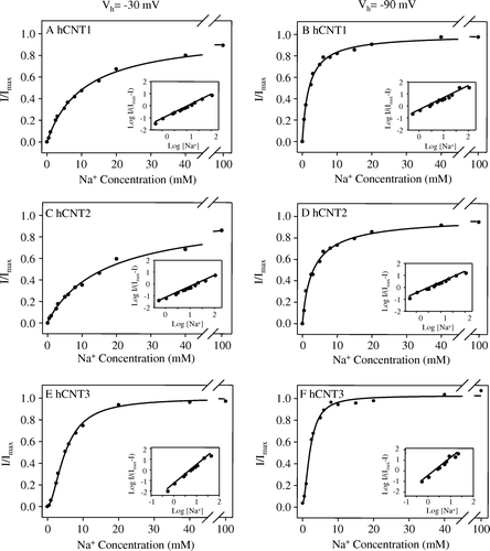
Table II. Voltage-dependence of Na+ activation kinetics of hCNT1, hCNT2 and hCNT3. Apparent affinities (K50) and Hill coefficients (n) for Na+ were determined from Na+ concentration response curves (0–100 mM NaCl, pH 8.5) in oocytes producing hCNT1, hCNT2 or hCNT3 measured at a uridine concentration of 20 µM and membrane potentials of −30 and −90 mV (see for representative hCNT1-, hCNT2-, or hCNT3-producing oocytes). Values were obtained from fits to data from individual oocytes normalized to the fitted Imax value obtained for that cell, and are presented as means±SEM. The numbers in parentheses denote the number of oocytes.
Na+/nucleoside coupling ratios
The Na+/nucleoside stoichiometries of hCNT1, hCNT2 and hCNT3 were directly determined by simultaneously measuring nucleoside-evoked currents and 14C-nucleoside uptake under voltage-clamp conditions. Data for the Na+/uridine stoichiometry of hCNT2 is compared with those of hCNT1 and hCNT3 in . To further characterize the stoichiometries of human CNTs, the coupling ratios for hCNT2 and hCNT3 were also measured with adenosine as permeant (), in transport medium containing 1 µM deoxycoformycin to inhibit adenosine deaminase activity (Yao et al. [Citation1996]). Due to the small magnitudes of adenosine-evoked hCNT1 Na+ currents (≤3 nA; Smith et al. [Citation2004]), a corresponding hCNT1 Na+/adenosine coupling ratio could not be measured. Each data point in and represents a single oocyte and the Na+/nucleoside coupling ratio is given by the slope of the linear fit of charge (pmol) versus uptake (pmol) ().
Figure 4. Uridine coupling ratios of hCNT1, hCNT2 and hCNT3. (A) Representative example of the current generated during application of 200 µM 14C-uridine to an hCNT2-producing oocyte in Na+-containing transport medium (100 mM NaCl, pH 8.5) at a membrane potential of −90 mV. Integration of the uridine-evoked current over the uptake period (2 min) yielded the charge moved which was converted to pmol and plotted against radiolabeled uridine uptake (pmol) in the same oocyte. The experiment was repeated in 12 different hCNT2-producing oocytes (C) Corresponding charge-to-14C-uridine uptake ratio plots are also shown for hCNT1 (B, n=11) and hCNT3 (D, n=12) (100 mM NaCl, pH 8.5; Vh= − 90 mV). Linear regression analysis of the data for each plot is indicated by the solid line. The dashed line indicates a theoretical 1:1 charge/flux ratio in (B and C) and a 2:1 charge/flux ratio in (D). Lines were fitted through the origin. Stoichiometries (±SE) obtained from these data are given in .
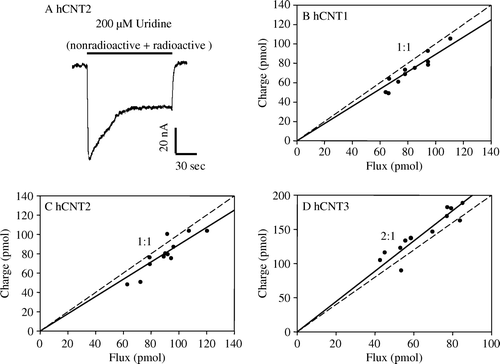
Figure 5. Adenosine coupling ratios of hCNT2 and hCNT3. Charge to 14C-adenosine uptake ratio plots were generated at a membrane potential of −90 mV in Na+-containing transport media (100 mM NaCl, pH 8.5) in hCNT2- (A, n=8) and hCNT3- (B, n=10) producing oocytes. The time of exposure of oocytes to 14C-adenosine (200 µM) was 2 min. Integration of the adenosine-evoked current was used to calculate the net cation influx (charge) and was correlated to the net 14C-adenosine influx (flux). Linear regression analysis of the data for each plot is indicated by the solid line. The dashed line indicates a theoretical 1:1 charge/flux ratio in (A) and a 2:1 charge/flux ratio in (B). Lines were fitted through the origin. Stoichoimetries (±SE) obtained from these data are indicated in .
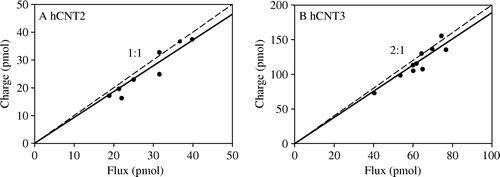
Table III. Stoichiometry of hCNT1, hCNT2 and hCNT3. Charge to 14C-nucleoside ( and ; 100 mM NaCl, pH 8.5) and charge to 22Na+ (data not shown; 1 mM NaCl, pH 8.5) uptake ratio plots were generated at a membrane potential of −90 mV in hCNT1-, hCNT2- and hCNT3-producing oocytes. The numbers in parentheses denote the number of oocytes.
Stoichiometries were measured at a nucleoside concentration of 200 µM in Na+-containing transport medium (100 mM) at pH 8.5 and at a holding potential of −90 mV. A is a representative uridine-dependent current recording in an hCNT2-producing oocyte. As previously observed for several other cotransporters, including hCNT1 and hCNT3 (Smith et al. [Citation2004], [Citation2005]), current reached an initial maximal value and then progressively decreased, returning to baseline upon removal of nucleoside. The linear correlations between integrated uridine-dependent charge and [14C]-uridine accumulation measured in Na+-containing transport medium (100 mM) gave calculated stoichiometries of 0.89 for hCNT2 (C) vs. 0.89 for hCNT1 (B) and 2.1 for hCNT3 (D) (). Similarly, the linear correlation between adenosine-dependent charge and adenosine accumulation (100 mM NaCl, pH 8.5) gave calculated stoichiometries of 0.93 for hCNT2 (A) and 1.9 for hCNT3 (B) ().
Charge-to-Na+ stoichiometry
The relationship between uridine-evoked charge influx (pmol) and 22Na+ uptake (pmol) was measured in oocytes producing hCNT1, hCNT2 or hCNT3 at a holding potential of −90 mV. A linear fit of the data gave regression lines with slopes of 0.93 for hCNT2 vs. 0.90 for hCNT1 and 0.90 for hCNT3, indicating that for all three transporters 1 net inward positive charge was transported for every Na+ ion cotransported with uridine into the cell ().
Discussion
The cation-coupled concentrative nucleoside transport processes cit, cif and cib are mediated by isoforms of the CNT (SLC28) transporter family, designated in humans as hCNT1, hCNT2 and hCNT3, respectively. All three proteins transport uridine and adenosine (although hCNT1-mediated adenosine transport activity is low), but are otherwise pyrimidine nucleoside-selective (hCNT1), purine nucleoside-selective (hCNT2), or broadly selective for both pyrimidine and purine nucleosides (hCNT3). Conducted under identical experimental conditions, the present study utilized heterologous expression in Xenopus oocytes in combination with radioisotope flux experiments and electrophysiological measurements to demonstrate that hCNT2 has cation coupling characteristics similar to those of subfamily member hCNT1, and markedly different from those of hCNT3.
Like hCNT1, hCNT2 was Na+-specific, while hCNT3 was able to utilize both Na+ and H+ to drive permeant uptake into cells. Dual Na+/H+-coupling is not, however, a general characteristic of the CNT3/hfCNT subfamily: hagfish hfCNT has been shown to be Na+- but not H+-dependent (Yao et al. [Citation2002]). CNTs from other species that exclusively use H+ as the coupling cation include CeCNT3 from Caenorhabditis elegans (Xiao et al. [Citation2001]), CaCNT from Candida albicans (Loewen et al. [Citation2003]) and NupC from Escherichia coli (Craig et al. [Citation1994], Loewen et al. [Citation2004]). hCNT3 is closer evolutionarily to these proteins than hCNT1/2 (Ritzel et al. [Citation2001]). H+-coupling of hCNT3 may be physiologically and pharmacologically important in tissues such as the duodenum, proximal jejunum and kidney proximal tubule where apical contents can be relatively acidic. The CNT protein family is therefore similar to other cotransporter families in which members are able to utilize Na+ and/or H+ to drive permeant uptake into cells (Pourcher et al. [Citation1990], Mackenzie et al. [Citation1996], Wright et al. [Citation2004]). Since hCNT2 was not H+-coupled, subsequent analyses focused on a comparison of hCNT2 and hCNT1/3 interactions with Na+.
Na+-concentration dependence experiments directly compared the apparent affinities (K50) of all three hCNTs for Na+. [14C]-Uridine influx studies revealed that hCNT1/2/3 exhibited apparent K50 values for Na+ activation in the range of 10–12 mM, and similar results were seen by electrophysiology (apparent K50 values ranging from 5–16 mM at Vh= −30 mV). Similar to hCNT1 and hCNT3, and in agreement with results reported previously for hCNT3 (Smith et al. [Citation2005]), the Na+ binding affinity of hCNT2 increased as the membrane potential was made more negative.
Previously published studies suggest that differences in Na+/H+ specificity between hCNT1/2 and hCNT3 may also extend to differences in cation-coupling ratios. Hill-type analyses of the relationships between nucleoside influx and Na+ concentration, for example, suggest a 1:1 Na+/nucleoside coupling stoichiometry for (i) various cit (CNT1) and cif (CNT2) transport activities in different mammalian cells and tissues (Plagemann et al. [Citation1990], Dagnino et al. [Citation1991], Plagemann [Citation1991], Williams & Jarvis [Citation1991], Che et al. [Citation1992], Griffith & Jarvis [Citation1996]), (ii) recombinant rCNT1 transport of both adenosine and uridine (Yao et al. [Citation1996]), and (iii) recombinant rCNT2 transport of adenosine and 2’,3’-dideoxyinosine (Li et al. [Citation2001a],Citation[b]). In marked contrast, Hill coefficients consistent with a 2:1 Na+/nucleoside coupling stoichiometry have been reported for system cib in choroid plexus (Wu et al. [Citation1992]) and microglia (Hong et al. [Citation2000]), and for recombinant mouse CNT3 (mCNT3), hCNT3 and hfCNT transport of uridine (Ritzel et al. [Citation2001], Yao et al. [Citation2002]). Direct determination of coupling stoichiometries by simultaneous measurement of radiolabelled nucleoside influx and cation current has yielded Na+/uridine coupling ratios of of either 1:1 (Smith et al. [Citation2004]) or 2:1 for hCNT1 (Larráyoz et al. [Citation2004]), and 2:1 for hCNT3 (Smith et al. [Citation2005]). The equivalent H+/uridine coupling ratio for hCNT3 was 1:1 (Smith et al. [Citation2005]). No corresponding published data are available for recombinant hCNT2.
In the present study, we utilized both Hill analyses of Na+ activation curves and charge/flux studies to determine the Na+/nucleoside coupling ratio of hCNT2, and to compare results for this transporter with those for hCNT1 and hCNT3 determined under identical experimental conditions and in the same batches of oocytes. Na+ activation curves derived from radioisotope flux experiments for both hCNT1 and hCNT2 were hyperbolic, with Hill coefficients consistent with a Na+/nucleoside coupling ratio of 1:1. In contrast, there was a sigmoidal relationship between nucleoside flux and Na+ concentration for hCNT3, with a Hill coefficient consistent with a Na+/nucleoside coupling ratio of 2:1. Parallel electrophysiology experiments measuring the relationship between uridine-evoked inward current and Na+ concentration confirmed these results. Similar to members of the SGLT protein family (Díez-Sampedro et al. [Citation2001]), Hill coefficients for hCNT2 and the other hCNTs were independent of membrane potential.
Hill coefficients reflect the number of ions interacting with the transporter rather than the actual number of ions entering the cell as a result of transport activity, and therefore provide only indirect estimates of transporter coupling ratios (Weiss [Citation1997]). We therefore also undertook charge vs. nucleoside and charge vs. Na+ uptake experiments under voltage-clamp conditions to determine hCNT2 stoichiometry directly and to compare that stoichiometry with hCNT1 and hCNT3. The ratio of charge to uridine uptake determined for both hCNT2 and hCNT1 yielded a Na+/uridine coupling ratio of 1:1. In contrast, the Na+/uridine coupling ratio of hCNT3 was 2:1. Na+/nucleoside coupling ratios for hCNT2 and hCNT3 were also determined with the purine nucleoside adenosine as permeant and yielded results identical to those seen with the pyrimidine nucleoside uridine. Contrary to a report by Larráyoz et al. ([Citation2004]), adenosine is also transporterd by the CNT1 isoform (Huang et al. [Citation1994], Fang et al. [Citation1996], Yao et al. [Citation1996], Ritzel et al. [Citation1997], Loewen et al. [Citation1999], Gray et al. [Citation2004], Smith et al. [Citation2004]). Kinetically, adenosine functions as a high-affinity, but low-capacity CNT1 permeant, precluding studies of hCNT1 Na+/adenosine coupling stoichiometry. Measurements correlating 22Na+ uptake to the translocated charge indicated that 1 net positive charge was transported for every Na+ ion cotransported, verifying that, like hCNT1 and hCNT3, hCNT2 nucleoside-dependent currents are carried by Na+ ions.
Both direct and indirect methods therefore give Na+/nucleoside coupling ratios of 1:1 for hCNT1 and hCNT2, and 2:1 for hCNT3. The Na+/nucleoside stoichiometry determined for hCNT1 agrees with our previously published results (Smith et al. [Citation2004]), and contradicts the 2:1 coupling ratio reported by Larráyoz et al. ([Citation2004]). The corresponding 1:1 stoichiometry for recombinant hCNT2 is the first report of this transporter's Na+/nucleoside coupling ratio, while the contrasting characteristics of hCNT3 (2:1 coupling ratio) also confirm our previous findings (Smith et al. [Citation2005]). A 2:1 Na+/nucleoside stoichiometry for hCNT3 also mirrors that for hfCNT (Yao et al. [Citation2002]). CNTs therefore resemble other transporter families in which individual members have different cation-coupling ratios. For example, members of the SGLT family have been described with 1:1 and 2:1 Na+/glucose coupling ratios (1:1 for SGLT2 and 2:1 for SGLT1/3) (Chen et al. [Citation1995], Mackenzie et al. [Citation1996], [Citation1998], Díez-Sampedro et al. [Citation2001]). Similarly, the PepT1 and PepT2 proton-linked peptide transporters have 1:1 and 2:1 H+/peptide coupling ratios, respectively (Chen et al. [Citation1999]). Energetically, a 2:1 Na+/nucleoside coupling stoichiometry for hCNT3 versus a 1:1 coupling ratio for hCNT1/2 implies a greater ability of hCNT3 to transport permeants (including nucleoside drugs) against their concentration gradient and, hence, achieve higher levels within the cell.
Conclusion
In conclusion, the cation coupling characteristics of hCNT2 are similar to those of subfamily member hCNT1, and different from those of hCNT3. Our results therefore validate and expand upon previously reported differences in cation coupling between human members of the CNT1/2 and CNT3/hfCNT subfamilies of CNT proteins. The observed functional differences in Na+/H+ specificity and Na+ coupling that we have described, in addition to being of potential physiologic and therapeutic importance, will facilitate future structure/function analyses to identify protein structural domains and specific amino acid residues responsible for CNT cation recognition and coupling.
This paper was first published online on prEview on 29 September 2006.
This work was supported by the National Cancer Institute of Canada, with funds from the Canadian Cancer Society, the Alberta Cancer Board, the Heart and Stroke Foundation, Canada, and the Medical Research Council, UK. JDY is a Heritage Scientist of the Alberta Heritage Foundation for Medical Research. CEC is a Canada Research Chair in Oncology at the University of Alberta. MDS is funded by a Studentship from the Alberta Heritage Foundation for Medical Research. KMS and MDS contributed equally to this work.
References
- Burckhardt BC, Kroll B, Frömter E. Proton transport mechanism in the cell membrane of Xenopus laevis oocytes. Pflugers Archiv 1992; 420: 78–82
- Cass CE. Drug transport in antimicrobial and anticancer chemotherapy, NH Georgopapadakou. Marcel Dekker, New York 1995
- Che M, Nishida T, Gatmaitan Z, Arias IM. A nucleoside transporter is functionally linked to ectonucleotidases in rat liver canalicular membrane. J Biol Chem 1992; 267: 9684–9688
- Chen X-Z, Coady MJ, Jackson F, Berteloot A, Lapointe JY. Thermodynamic determination of the Na+: glucose coupling ratio for the human SGLT1 cotransporter. Biophys J 1995; 69: 2405–2414
- Chen X-Z, Zhu T, Smith DE, Hediger MA. Stoichiometry and kinetics of the high-affinity H+-coupled peptide transporter PepT2. J Biol Chem 1999; 274: 2773–2779
- Craig JE, Zhang Y, Gallagher MP. Cloning of the nupC gene of Escherichia coli encoding a nucleoside transport system, and identification of an adjacent insertion element, IS 186. Mol Microbiol 1994; 11: 1159–1168
- Damaraju VL, Damaraju S, Young JD, Baldwin SA, Mackey J, Sawyer MB, Cass CE. Nucleoside anticancer drugs: the role of nucleoside transporters in resistance to cancer chemotherapy. Oncogene 2003; 22: 7524–7536
- Dagnino L, Bennett LL, Jr, Paterson AR. Substrate specificity, kinetics, and stoichiometry of sodium-dependent adenosine transport in L1210/AM mouse leukemia cells. J Biol Chem 1991; 266: 6312–6317
- Díez-Sampedro A, Eskandari S, Wright EM, Hirayama BA. Na+-to-sugar stoichiometry of SGLT3. Am J Physiol Renal Physiol 2001; 280: F278–F282
- Fang X, Parkinson FE, Mowles DA, Young JD, Cass CE. Functional characterization of a recombinant sodium-dependent nucleoside transporter with selectivity for pyrimidine nucleosides (cNT1rat) by transient expresssion in cultured mammalian cells. Biochem J 1996; 317: 457–465
- Griffith DA, Jarvis SM. Nucleoside and nucleobase transport systems of mammalian cells. Biochem Biophys Acta 1996; 1286: 153–181
- Gray JH, Owen RP, Giacomini KS. The concentrative nucleoside family, SLC28. Pflugers Archiv 2004; 447: 728–734
- Hong M, Schlichter L, Bendayan R. A Na+-dependent nucleoside transporter in microglia. J Pharmacol Exp Ther 2000; 292: 366–374
- Huang Q-Q, Yao SYM, Ritzel MWL, Paterson ARP, Cass CE, Young JD. Cloning and functional expression of a complementary DNA encoding a mammalian nucleoside transport protein. J Biol Chem 1994; 269: 17757–17760
- Larráyoz IM, Casado FJ, Pastor-Anglada M, Lostao MP. Electrophysiological characterization of the human Na+/nucleoside cotransporter 1 (hCNT1) and role of adenosine on hCNT1 function. J Biol Chem 2004; 279: 8999–9007
- Li JY, Boado RJ, Pardridge WM. Differential kinetics of transport of 2′,3′-dideoxyinosine and adenosine via concentrative Na+ nucleoside transporter CNT2 cloned from rat blood-brain barrier. J Pharmacol Exp Ther 2001a; 299: 735–740
- Li JY, Boado RJ, Pardridge WM. Cloned blood-brain barrier adenosine transporter is identical to the rat concentrative Na+ nucleoside cotransporter CNT2. J Cereb Blood Flow Metab 2001b; 21: 929–936
- Loewen SK, Ng AML, Mohabir NN, Baldwin SA, Cass CE, Young JD. Functional characterization of a H+/nucleoside co-transporter (CaCNT) from Candida albicans, a fungal member of the concentrative nucleoside transporter (CNT) family of membrane proteins. Yeast 2003; 20: 661–675
- Loewen SK, Ng AM, Yao SY, Cass CE, Baldwin SA, Young JD. Identification of amino acid residues responsible for the pyrimidine and purine nucleoside specificities of human concentrative Na+ nucleoside cotransporters hCNT1 and hCNT2. J Biol Chem 1999; 274: 24475–24484
- Loewen SK, Yao SY, Slugoski MD, Mohabir NN, Turner RJ, Mackey JR, Weiner JH, Gallagher MP, Henderson PJ, Baldwin SA, Cass CE, Young JD. Transport of physiological nucleosides and anti-viral and anti-neoplastic nucleoside drugs by recombinant Escherichia coli nucleoside-H(+) cotransporter (NupC) produced in Xenopus laevis oocytes. Mol Membr Biol 2004; 21: 1–10
- Mackenzie BM, Loo DDF, Panayotova-Heiermann M, Wright EM. Biophysical characteristics of the pig kidney Na+/glucose cotransporter SGLT2 reveal a common mechanism for SGLT1 and SGLT2. J Biol Chem 1996; 271: 32678–32683
- Mackenzie B, Loo DD, Wright EM. Relationships between Na+/glucose cotransporter (SGLT1) currents and fluxes. J Membr Biol 1998; 162: 101–106
- Plagemann PG. Na(+)-dependent, concentrative nucleoside transport in rat macrophages. Specificity for natural nucleosides and nucleoside analogs, including dideoxynucleosides, and comparison of nucleoside transport in rat, mouse and human macrophages. Biochem Pharmacol 1991; 42: 247–252
- Plagemann PG, Aran JM, Woffendin C. Na(+)-dependent, active and Na(+)-independent, facilitated transport of formycin B in mouse spleen lymphocytes. Biochem Biophys Acta 1990; 1022: 93–102
- Pourcher T, Bassilana M, Sarkar HK, Kaback HR, Leblanc G. The melibiose/Na+ symporter of Escherichia coli: kinetic and molecular properties. Philos Trans R Soc Lond B Biol Sci 1990; 326: 411–423
- Ritzel MW, Ng AM, Yao SY, Graham K, Loewen SK, Smith KM, Ritzel RG, Mowles DA, Carpenter P, Chen XZ, Karpinski E, Hyde RJ, Baldwin SA, Cass CE, Young JD. Molecular identification and characterization of novel human and mouse concentrative Na+-nucleoside cotransporter proteins (hCNT3 and mCNT3) broadly selective for purine and pyrimidine nucleosides (system cib). J Biol Chem 2001; 276: 2914–2927
- Ritzel MW, Yao SY, Huang M-Y, Elliot JF, Cass CE, Young JD. Molecular cloning and functional expression of cDNAs encoding a human Na+-nucleoside cotransporter (hCNT1). Am J Physiol Cell Physiol 1997; 272: C707–C714
- Ritzel MW, Yao SY, Ng AM, Mackey JR, Cass CE, Young JD. Molecular cloning, functional expression and chromosomal localization of a cDNA encoding a human Na+/nucleoside cotransporter (hCNT2) selective for purine nucleosides and uridine. Mol Membr Biol 1998; 15: 203–211
- Smith KM, Ng AML, Yao SY, Labedz KA, Knaus EE, Wiebe LI, Cass CE, Baldwin SA, Chen XZ, Karpinski E, Young JD. Electrophysiological characterization of a recombinant human Na+-coupled nucleoside transporter (hCNT1) produced in Xenopus oocytes. J Physiol 2004; 558: 807–823
- Smith KM, Slugoski MD, Loewen SK, Ng AML, Yao SYM, Chen XZ, Karpinski E, Cass CE, Baldwin SA, Young JD. The broadly selective human Na+/nucleoside cotransporter (hCNT3) exhibits novel cation-coupled nucleoside transport characteristics. J Biol Chem 2005; 280: 25436–25449
- Wang J, Su S-F, Dresser MJ, Schaner ME, Washington CB, Giacomini KM. Na+-dependent purine nucleoside transporter from human kidney: cloning and functional characterization. Am J Physiol Renal Physiol 1997; 273: F1058–F1065
- Weiss JN. The Hill equation revisited: uses and misuses. FASEB J 1997; 11: 835–841
- Williams TC, Jarvis SM. Multiple sodium-dependent nucleoside transport systems in bovine renal brush-border membrane vesicles. Biochem J 1991; 274: 27–33
- Wright EM, Loo DD, Hirayama BA, Turk E. Suprising versatility of Na+-glucose cotransporters: SLC5. Physiol 2004; 19: 370–376
- Wu X, Yuan G, Brett CM, Hui AC, Giacomini KM. Sodium-dependent nucleoside transport in choroid plexus from rabbit. Evidence for a single transporter for purine and pyrimidine nucleosides. J Biol Chem 1992; 267: 8813–8818
- Xiao G, Wang J, Tangen T, Giacomini KM. A novel proton-dependent nucleoside transporter, CeCNT3, from Caenorhabditis elegans. Mol Pharmacol 2001; 59: 339–348
- Yao SYM, Ng AML, Loewen SK, Cass CE, Baldwin SA, Young JD. An ancient prevertebrate Na+-nucleoside cotransporter (hfCNT) from the Pacific hagfish (Eptatretus stouti). Am J Physiol Cell Physiol 2002; 283: C155–C168
- Yao SY, Ng AM, Ritzel MW, Gati WP, Cass CE, Young JD. Transport of adenosine by recombinant purine- and pyrimidine-selective sodium/nucleoside cotransporters from rat jejunum expressed in Xenopus laevis oocytes. Mol Pharmacol 1996; 50: 1529–1535
- Young JD, Cheeseman CI, Mackey JR, Cass CE, Baldwin SA. Molecular mechanisms of nucleoside and nucleoside drug transport. Current Top Membr 50, KE Barrett, M Donowitz. Academic Press, San Diego 2000; 329–378