
ABSTRACT
Fur ranching has been a prosperous industry for decades. Despite its worldwide distribution, little published information is available regarding the importance of the various causes of death in chinchilla (Chinchilla lanigera). In the period 1999–2013, 698 captive chinchillas from different commercial ranches were presented for necropsy at the Pathology Department (UNLP). Two-hundred and forty-four animals (35.0%) had classical enteritis, 198 (28.4%) had pneumonia, 63 (9.0%) had other infections, 40 (5.7%) had traumatic injuries, 109 (15.6%) had miscellaneous conditions, meanwhile 44 (6.3%) had no significant lesions. Although some disease processes may be underrepresented (i.e. heat stroke and the shock syndrome), the data were collected from a field wide enough and over a sufficient period to give a reliable overview of the fatal problems of this rodent in captivity.
1. Introduction
Chinchilla lanigera is a small South American rodent belonging to the family Chinchillidae, its fur is possibly the thickest and warmest of any animals (Vietmeyer Citation1991; Spotorno et al. Citation2004). Once found throughout the Andes mountains, chinchilla remains highly endangered in the wild (IUCN Citation2014). It has been successfully raised for commercial markets for decades and this global farming industry represents a highly specialized and costly business (Tremblay Citation2000; Martin Citation2012).
The main difficulty is the multi-factorial nature of problems arising from the production of this animal in captivity (Norton & Reynolds Citation2012). Due to the scarce reference studies, essential knowledge about the fundamental biology for many fur-bearing animals including the chinchilla is lacking or limited (Eidmann Citation1995; Fehr Citation2008). After the historical publications from the 1950s and 1960s describing systematic post-mortem examinations (Bowden Citation1959; Dall Citation1963), case studies on particular diseases have been mostly reported with some frequency in this species since then. The purpose of the present survey was to describe in brief the findings of chinchilla necropsies and to determine the probable cause of death based on systematical diagnostic studies.
2. Materials and methods
Six-hundred and ninety-eight chinchilla necropsies were performed at the Pathology Department (UNLP) between 1999 and 2013. The age of chinchillas was determined primarily from dentition and the appearance of organs. Thus, all chinchillas were divided into two possible groups: young (under one year of age) and adult (over one year).
A complete necropsy was performed and testing was conducted to determine the cause of the disease, including routine tests for specific bacterial and fungal diseases, protozoa, toxins and blood analyses. Histological examination was restricted to major organs (i.e. brain, lungs, liver, intestines, kidneys) and obvious abnormal tissues.
Bacteria and fungi were identified by means of standard procedures (Carter & Carter Citation1990) and by the use of the API 20E System (API® 20E, Biomerieux, France). Toxin production and other virulence genes of diarrheagenic Escherichia coli were assayed by enzyme-linked immunosorbent assay, the polymerase chain reaction multiplex (PCR multiplex) and a further subtypification by pulse-field gel electrophoresis (XbaI-PFGE) (Beutin et al. Citation1997). E. coli isolates containing a virulence factor were also serotyped using available antisera. The presence of Salmonella spp. in collected samples was assessed by performing the pre-enrichment culture, followed by multiplex PCR assay (12).
Giardia spp. cysts and trophozoites were detected on faecal and intestinal samples by microscopy of wet mounts and by a commercial immunofluorescence assay (Merifluor® Giardia, Meridian, USA) (Bautista Citation2009).
Listeria monocytogenes isolates were confirmed with a real-time PCR assay (O’Gradya et al. Citation2008); meanwhile listeria antigens were identified on paraffin-embedded brain and intestine sections with immunohistochemistry.
Attempts to demonstrate viral particles in tissues from suspected lesions were done by direct-contrast electron microscopy or by inoculation onto cell cultures. Toxicological studies were based on case history and the analytical determination of the specific toxic compound in food (Fehr Citation2008; Greco et al. Citation2012).
Cause of death, determined by interpreting the combination of lesions found in each animal, was recorded into one of the following six categories: classical enteritis, pneumonia, other infections, trauma, miscellanea and an undetermined group. Judgements as to what factors were primary and what were contributory were based mainly on the necropsy findings, but other factors were also taken into account (i.e. sanitary status of the farms, diet, management).
Sex and age frequencies of diagnoses were statistically evaluated for temporal differences using a Chi square test with the SAS 8.0 statistical package (SAS® Institute Inc., Cary, NC, USA).
3. Results
presents the occurrence of the main necropsy and the distribution of recorded causes.
Table 1. Diagnostic findings in 698 chinchilla mortalities by cause of death, sex and age, from 1999 to 2013.
The age and sex distribution was similar among the mortality categories, with only the significantly highest incidence in young females of the enteritis group (p < .05, relative risk (rr) = 1.03, 95% confidence limits (cl) 0.89–1.33) and in adult females of the miscellaneous group (p < .05, rr = 0.89, 95% cl 0.63–1.27).
Overall, infectious agents associated with the diseases seen (enteritis, pneumonia and other infections) were identified in 72.4% of the necropsies, significantly outnumbering the rest of the non-infectious entities (trauma, miscellanea and undetermined) (p < .01, rr = 1.12, 95% cl 1.00–1.72). Moreover, enteritis and pneumonia cases, altogether accounted for the 63% of the total investigated accessions (p < .01, rr = 1.37, 95% cl 0.88–1.61).
In the present survey, most cases of these six categories showed concurrently non-fatal entities or secondary conditions, like dermatomycoses, teeth malocclusion, fur-chewing, facial abscesses, alopecia, keratoconjunctivitis and footpad ulcerations.
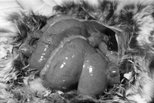
The most common casualties were due by localized classical enteritis (i.e. catharral, fibrino-haemorrhagic or necrotic enteritis, tiflocolitis) (). This group also includes those cases that showed other simultaneous digestive tract lesions, like meteorism or hepatic necrosis. The bulk of the animals were in poor body condition because of persistent diarrhoea. E. coli, Salmonella enterica subsp. typhimurium, arizonae and enteritidis, Pseudomonas aeruginosa, Yersinia pseudotuberculosis, Yersinia enterocolitica, Klebsiella pneumoniae and Clostridium perfringens were identified as the principal agents recovered. Among them, haemolytic E. coli was the most common bacterial pathogen significantly isolated (117 out of the 244 cases, p < .05, rr = 1.16, 95% cl 0.77–1.46) and mostly in pure culture (98 out of the 117 strains). Some of these strains were classified as enteropathogenic E. coli (eaea+ enteroadherent) and as enterotoxigenic E. coli. The bulk of these strains could not be grouped with H-antigen, but with the O-antigen (i.e. O2, O7, O45 and O117). XbaI-pulse-field gel electrophoresis revealed that at least 64 strains isolated from different ranches were distinguishable, sharing a 91.5–94% similarity among them. Six cases with E. coli infection represented severe outbreaks which devastated three ranches in the winter of 2008 and with mortalities exceeding 70%. In addition, Giardia spp. cysts and trophozoites were often identified among these cases (84 out of 244), although not significantly associated (p > 0.05, rr = 2.73, 95% cl 1.68–3.03). Presence of Coccidiae (Eimeria spp.), Tricostrongylus sp., Trichuris sp., Cryptosporidium spp. and Saccharomyces gluturatus was recorded but they were not present in sufficient numbers to have caused ill health or death.
Figure 1. A severe case of tiflocolitis with meteorism by E. coli on a silver 11-month-old female.
Pneumonia was the second most frequently diagnosed disease (i.e. interstitial pneumonia, bronchopneumonia), disregarding the age or sex groups. Pasteurella multocida, Bordetella bronchiseptica, Staphylococcus aureus, P. aeruginosa, Streptococcus sp., K. pneumoniae and L. monocytogenes were the most commonly isolated agents. Positive bacterial cultures in 48% of these infections yielded at least two of these agents combined. One adult with pasteurellosis also had the presence of calicivirus in the lungs although the association is not clear. Pasteurellosis accounted for 72 autopsies (53 of them came from one farm, in which the 71% of the chinchillas died within 14 days). Pneumonia was sometimes seen concurrently with pleuritis, myocarditis and metritis, from which those bacteria were also yielded.
Deaths by other infections were represented by diverse bacterial infections: 31 cases of septicaemia by Y. enterocolitica, P. aeruginosa, S. aureus, Enterobacter sp., E. coli or Proteus spp., 12 cases of peritonitis by C. perfringens and S. aureus, 6 cases of omphalitis by S. aureus, 6 cases of metritis by P. multocida, Corynebacterium pyogenes and E. coli, 5 cases of nephritis by Salmonella spp. and Leptospira interrogans serovars ictero-haemorrhagiae and pomona, and single cases of myocarditis (S. aureus), meningoencephalitis (L. monocytogenes) and rodenciosis (Y. pseudotuberculosis).
In the trauma group, 17 cases (mostly young females) violently died from fatal injuries, apparently attacked by adult breeders or rats, meanwhile 23 animals had either fractures (i.e. on the spine, ribs, limbs and mandibles) or/and ruptured livers and internal haemorrhage from suspected mismanagement of handlers or from getting caught on passageways of cages. The remaining two cases were kits which presented signs of suffocation with congestion and frothy oedema in the respiratory tree after being inadvertently crushed by their genitors. The bulk of the animals that died dramatically were in good body condition.
The miscellaneous category had a vast range of pathologies. Thirty-nine animals died by suffering adverse environmental factors such as heat stroke (n = 27) or hypothermia by extreme temperatures (n = 12). Nineteen suspected intoxications accounted by mycotoxins (n = 12), the ionophore narasin (n = 4) and the organophosphate dieldrin (n = 3). In the first case, aflatoxins and zearalenone were found at 19.8 and 162.2 ppb in the pelleted feed, respectively; the animals came from a single farm and died with suspected hepatic lesions (i.e. mottling and discolouration of the liver, with replication of ducts on the histopathology study). Regarding the acute poisoning by dieldrin and narasin, these episodes were part of unusual clusters involving several simultaneous deaths in one farm each, and they were attributed to careless disposal of these products on the feed recipients. Dieldrin was detected at high levels not only in the feed (79 ppm) but also on the livers of the animals (up to 15 ppm of liver dry matter), while gross necropsies revealed fatty livers, haemorrhagic kidneys and gastric ulcers. The outbreak was reported by the owner to be more severe in adults than in young animals. On the other hand, intoxicated animals with narasin acutely died after having fed fodder containing 88 ppm of this toxin. At post-mortem examination, these animals revealed myocardial and renal degeneration. This category also includes 16 cases of shock syndrome (with exhausted adrenals and collapsed lungs), 14 cases of nutritional problems which lead to starvation (i.e. by food shortage or absence of milk), 6 kits drowned in water containers, intestinal intussusception in 4 animals, malformations in 3 newborn kits (i.e. hydrocephalia), neoplasia in 4 aged females with lymphosarcoma of extensive involvement, 3 cases of gastrointestinal obstruction by foreign bodies and a single case of periportal fibrosis on liver. The diagnosis of nutritional deficiencies was confirmed by noting the response of the rest of the animals to an improved diet or a proper supply.
Undetermined cases included those accessions that generally appeared in good bodily condition but showed no obvious underlying disease at the time of examination. Laboratory investigations failed to reveal any aetiological factor.
4. Discussion
This paper describes in brief the retrospective study of necropsies performed on chinchilla in captivity. Sometimes, the interpretation of necropsy findings was limited by two factors: the lack of adequate ante-mortem data, and the delay between death and necropsy examination (Bautista et al. Citation2007). Thus, the results in this survey may suffer from a degree of bias. In addition, the idea of the severity of the outbreaks could not be properly assessed. For example, heat stroke accounted for only 27 cases, but they came mostly from 2 ranches which had lost almost the entire stock of hundreds of animals.
There is an increasing awareness to seek veterinary attention, but most farmers do not utilize or have poor access to veterinarians.
Despite the potential unknown factors regarding case submissions, infections were clearly considered responsible for more than 70% of the total accessions. Most diseases are caused by poor management, and unsanitary conditions increase the susceptibility (Dall Citation1963; Bautista Citation2009). Domesticated chinchilla’s high susceptibility of organism to stress in breeding colonies plays a substantial role, leading particularly to infectious diseases (Norton & Reynolds Citation2012).
Enteritis and pneumonia, two fatal disorders to which the species is especially prone, accounted here for the bulk of the casualties (Bowden Citation1959; Schaffer & Donnelly Citation1997; Lucena et al. Citation2012). Herein, the most frequent bacterial agents of the enteritis cases are comparable with those found by other workers (Dall Citation1963; Bartoszcze et al. Citation1990; Novak et al. Citation1994). From observations at several farms during the epizootic periods, it was concluded that sporadic outbreaks may occur at any time of the year and young animals were particularly susceptible. In a recent survey over 202 post-mortem examinations in Brazil, enteric and respiratory infections were also the most prevalent causes of death (25.7% of the total losses), followed by intoxications and physical injuries (22% and 10%, respectively) (Lucena et al. Citation2012). There was a frequent presence of Giardia in our cases of enteritis. It is well known that giardiasis is endemic in many chinchilla colonies and concomitant bacterial infections may occur complicating both diagnosis and treatment (Schönball Citation1992; Bautista Citation2009). Although Giardia spp. populate the intestinal tracts of almost every group of vertebrates, molecular characterization is still lacking to support that chinchilla is really infected with this flagellate (Eidmann Citation1995; Sulaiman et al. Citation2003).
Spontaneous fatal outbreaks of pneumonia with significant morbidity and mortality was recorded, with the same pathological pattern and bacterial agents described elsewhere (Dall Citation1963; Lucena et al. Citation2012). Stress brought on by high temperatures, high humidity and wet conditions can lead to respiratory disorders and even sudden death (Alworth & Harvey Citation2012). Although the horizontal mode of transmission of these bacterial agents (i.e. P. multocida, B. bronchiseptica, P. aeruginosa) is relatively well understood, the factors involved in the progression from commensal infection to clinically significant disease are far from clear in chinchillas (Fehr Citation2008).
In the category of trauma, several causes were involved, but holding the animals incorrectly by unexperienced handlers is, unfortunately, a common factor.
Among the miscellaneous conditions, heat stroke posed an important problem in the case of chinchilla (Fehr Citation2008). Dead animals came from areas where temperatures can reach more than 30°C, and did not have air-conditioning units. Hot humid weather and environmental temperatures over 27°C are liable to cause heat exhaustion in chinchilla which can die very easily (Building Citation1977).
The unusual intoxications by narasin and dieldrin can be considered as sporadic incidents, but mycotoxicosis is a common problem among Argentinian farms (González Pereyra et al. Citation2008; Greco et al. Citation2012). These reports demonstrated that fungi and mycotoxins were usually present in chinchilla feed, with fungal contamination exceeding the limits (up to 4.5 × 10⁴ CFU g−1) and the co-occurrence of the five most important mycotoxigenic mould genera recovered at high concentrations. Exposure to these toxic substances may lead to impairment of immune function resulting in increased susceptibility to infectious agents (Shareef Citation2010). Though the synergic effects of mycotoxins on health and productivity of other animal species such as poultry have been well documented, more studies are needed for chinchillas.
Chinchillas are very prone to die suddenly without symptoms (Kraft Citation1994; Alworth & Harvey Citation2012). Herein, there were many historical cases of animals which have shown no actual clinical symptoms of illness but found dead a few hours later. This is the case of the shock syndrome, which has been referred to as one of the most serious facing the world’s chinchilla industry (Höefler Citation1994; Lucena et al. Citation2012). The shock syndrome is underrepresented here as only 16 cases were recorded. These deaths are undoubtedly due to the cumulative effect of various stress factors and only a careful examination of the carcasses might suggest the syndrome (Eidmann Citation1995).
One accession of pasteurellosis observed a concomitant presence of calicivirus during electron microscopy examination, meanwhile no virus was identified in the remaining cases. There are no reports in the literature documenting identified fatal calicivirus infections in chinchilla. In fact, virologic disease appears to be unimportant in this rodent, as spontaneous viral episodes are rarely reported in the literature (Höefler Citation1994; Norton & Reynolds Citation2012).
Major conclusions cannot be statistically extracted from the age-sex distribution among the death categories. Young female were particularly exposed to enteritis in this survey, while the adult females were significantly represented in the miscellaneous group. In the rest of the categories, an even distribution is observed. It is not possible to even speculate on the present population sizes or densities because formal systematic publications on post-mortem findings are rarely reported in this species. In commercial farms, the number of males is normally restricted compared to the females, with the most frequent ratio being between 1 male to 6–8 females. In spite of the rapid worldwide increase of the chinchilla farming, its nutritional requirements and disease profile are poorly understood by comparison with other fur-bearing animals (Tremblay Citation2000; Wolf et al. Citation2003).
Further research is needed in this field to investigate a number of problems. Overall, taking into account that present data are coming from several ranches with a meaningful number of cases, and during a long period of investigations, some valuable information has been gathered regarding the diagnosis of many fatal entities which may be useful to the area of chinchilla pathology.
Disclosure statement
No potential conflict of interest was reported by the authors.
Additional information
Funding
Related Research Data
References
- Alworth LC, Harvey SB. 2012. Chinchillas. Anatomy, physiology and behaviour. In: Suckow MA, Stevens KA, Wilson RP, editors. The laboratory rabbit, guinea pig, hamster, and other rodents. 1st ed. American College of Laboratory Animal Medicine Series. San Diego (CA): Academic Press (Elsevier); p. 955–965.
- Bartoszcze M, Nowakoski J, Roskowskin J, Matras J, Palec S, Wystupe E. 1990. Chinchilla deaths due to Clostridium perfringes A enterotoxin. Vet Rec. 126:341–342.
- Bautista EL. 2009. Epidemiological study on giardiasis among the chinchillas farms of La Plata City [doctoral thesis]. Argentina: Veterinary College, La Plata University; 94 pp.
- Bautista EL, Martino PE, Manacorda A, Cossu ME, Stanchi N. 2007. Spontaneous Proteus mirabilis and Enterobacter aerogenes infection in chinchilla (Chinchilla lanigera). Scientifur. 31:27–30.
- Beutin L, Horbach I, Zimmermann S, Gleier K. 1997. Comparative evaluation of different diagnostic methods for the detection of verotoxin (shiga-toxin) producing strains of Escherichia coli (VTEC) in human clinical stool specimens. J Lab Med. 21:537–546.
- Bowden RS. 1959. Diseases of chinchillas. Vet Rec. 71:33–35.
- Building JC. 1977. Raising chinchillas. Diseases and disorders, love printing service. Ottawa: Canada Department of Agriculture.
- Carter G, Carter JR. 1990. Diagnostic procedures in veterinary bacteriology and mycology. 5th ed. New York: Academic Press, Inc.
- Dall J. 1963. Diseases of the chinchilla. J Small Anim Pract. 4:207–212. doi: 10.1111/j.1748-5827.1963.tb01849.x
- Eidmann S. 1995. Untersuchüngen zur Ätiologie und Pathogenese von Fellscaden beim Chinchilla [doctoral thesis]. Med. Vet., Tierärztliche Hochschule Hannover; 163 pp.
- Fehr M. 2008. Chinchilla. In: Gabrisch K, Zwart P, editors. Krankheiten der Heimtiere. Hannover: Schlütersche Verlagsgesellschaft mbH & Co KG; p. 103–205.
- González Pereyra M, Carvalho E, Tissera J, Keller K, Magnoli C, Rosa C, Dalcero A, Cavaglieri L. 2008. An outbreak of acute aflatoxicosis on a chinchilla (Chinchilla lanigera) farm in Argentina. J Vet Diagn Invest. 6:853–856. doi: 10.1177/104063870802000629
- Greco M, Pardo G, Ludemann V, Martino PE, Pose GN. 2012. Mycoflora and natural incidence of selected mycotoxins in rabbit and chinchilla feeds. Scientific World J. 2012:1–6. doi: 10.1100/2012/956056
- Höefler HL. 1994. Chinchillas. Exotic pet medicine. Vet Clin North Am Small Anim Pract. 24:102–111.
- Kraft H. 1994. Krankheiten der Chinchillas. Stuttgart: Ferdinand Enke.
- Lucena R, Giaretta P, Tessele B, Fighera R, Kommers G, Irigoyen L, Barros C. 2012. Doenças de chinchilas (Chinchilla lanigera). Pesq Vet Bras. 32:529–535. doi: 10.1590/S0100-736X2012000600010
- Martin BJ. 2012. Chinchillas. Taxonomy and history. In: Suckow MA, Stevens KA, Wilson RP, editors. The laboratory rabbit, guinea pig, hamster, and other rodents. 1st ed. American College of Laboratory Animal Medicine Series. San Diego (CA): Academic Press (Elsevier); p. 949–952.
- Norton JN, Reynolds RP. 2012. Chinchillas. Diseases and veterinary care. In: Suckow MA, Stevens KA, Wilson RP, editors. The laboratory rabbit, guinea pig, hamster, and other rodents. American College of Laboratory Animal Medicine Series. San Diego (CA): Academic Press (Elsevier); p. 993–1026.
- Novak S, Ruttkay D, Solar I. 1994. Results of screening for bacterial diseases in large scale chinchilla (Chinchilla lanigera). Slovak Vet J. 19:19–21.
- O’Gradya J, Sedano S, Maherb M, Smith T, Barrya T. 2008. Rapid real-time PCR detection of Listeria monocytogenes in enriched food samples based on the ssrA gene, a novel diagnostic target. Food Microbiol. 25:75–84. doi: 10.1016/j.fm.2007.07.007
- Schaffer DO, Donnelly TM. 1997. Disease problems of guinea pigs and chinchillas. In: Hillyer EV, Quesenberry KE, editors. Ferrets, rabbits and rodents. Philadelphia: W.B. Saunders; p. 260–281.
- Schönball U. 1992. Fallbericht: Giardieninfektion bei einem Chinchilla – mögliche Infektionsquelle für den Menschen. Kleintierpraxis. 37:785–788.
- Shareef AM. 2010. Moulds and mycotoxins in poultry feeds from farms of potential mycotoxicosis. Iraqi J Vet Sci. 24:17–25.
- Spotorno AE, Zuleta CA, Valladares JP, Deane AL, Jiménez, JE. 2004. Chinchilla lanigera. Mamm Species. 758:1–9. doi: 10.1644/758
- Sulaiman IM, Fayer R, Bern C, Gilman RH, Trout JM, Schartz PM, Das P, Lal AA, Xiao L. 2003. Triosephosphate isomerase gene characterization and potential zoonotic transmission of Giardia duodenalis. Emerg Inf Dis. 9:1444–1452. doi: 10.3201/eid0911.030084
- The IUCN Red List of Threatened Species. 2014. [ Downloaded 2015 Mar 23]. Available from: http://www.iucnredlist.org.ar.
- Tremblay M. 2000. Le Chinchilla. Le Jour Éditeur, France; p. 3–17.
- Vietmeyer ND. 1991. Chinchilla. Microlivestock: little-known small animals with a promising economic future. Washington (DC): National Academy Press; p. 277.
- Wolf P, Schröder A, Wenger A, Kamphues J. 2003. The nutrition of the chinchilla as a companion animal. Basic data, influences and dependences. J Anim Physiol Anim Nutr. 87:129–133. doi: 10.1046/j.1439-0396.2003.00425.x