
Abstract
Distal hair segments collected at delivery may allow for the assessment of maternal cortisol secretion in early pregnancy, an important time window for fetal development. Therefore, an investigation of the validity of distal hair cortisol concentrations is warranted. We examined the concordance between proximal and distal hair cortisol concentrations (HCC), both representing the first trimester of pregnancy. The study population was comprised of a random sample of 97 women participating in the Pregnancy Outcomes Maternal and Infant Study, a prospective cohort study of pregnant women attending prenatal clinics in Lima, Peru. Each participant provided two hair samples: once at enrollment [mean gestational age (GA) = 13.1 weeks] and again at full-term delivery (mean GA = 39.0 weeks). Hair segments reflecting the first trimester were: 3 cm hair segments closest to the scalp on the first hair sample (proximal) and 6–9 cm from the scalp on the second hair sample (distal). HCC was determined using Luminescence Immunoassay. A subset (N = 28) had both hair segments additionally analyzed using liquid chromatography tandem mass spectrometry (LC-MS/MS). HCC values were log-transformed (logHCC), and proximal–distal differences tested using paired sample t-tests. Concordance was evaluated within and across assay types. LogHCC, measured using immunoassay, in distal hair segments was lower compared to proximal hair segments (1.35 versus 1.64 respectively; p = .02). No difference was observed using LC-MS/MS (1.99 versus 1.83, respectively; p=.33). Proximal–distal concordance was low within assay (immunoassay: Pearson = 0.27 and κ = 0.10; LC-MS/MS: Pearson = 0.37 and κ = 0.07). High correlation was observed across assays for both distal (Pearson = 0.78, p < .001; κ = 0.64) and proximal segments (Pearson = 0.96, p < .001; κ = 0.75). In conclusion, distal first-trimester hair segments collected at delivery have lower absolute HCC compared to HCC in proximal first trimester hair segments collected in early pregnancy, and are poorly concordant with HCC in proximal segments. Findings may inform the design of future studies.
1. Introduction
Cortisol, a glucocorticoid hormone released by the hypothalamic–pituitary–adrenal (HPA) axis, plays a key role in maintaining homeostatic conditions and aids in the functioning of the metabolic, immune, and neurologic systems (McEwen & Seeman, Citation1999; Smith & Cidlowski, Citation2010; Van Londen et al., Citation1998). Chronically dysregulated cortisol concentrations have been associated with early miscarriage (Nepomnaschy et al., Citation2006), low birth weight delivery (Bolten et al., Citation2011), and depression in the perinatal period (Bjelanovic et al., Citation2015; Diego et al., Citation2009; Field et al., Citation2009; Hoffman, Mazzoni, Wagner, Laudenslager, & Ross, Citation2016; Lommatzsch et al., Citation2006; Murphy et al., Citation2015; O’Connor et al., Citation2014; O’Keane et al., Citation2011; Peer, Soares, Levitan, Streiner, & Steiner, Citation2013; Voegtline et al., Citation2013). Traditional cortisol monitoring techniques, such as saliva, serum and urine, have been useful in assessing how deviations in diurnal cortisol profiles are associated with such outcomes. However, these techniques are limited in their ability to monitor long-term cortisol secretion due to their reflection of cortisol concentrations in the past 1–24 h, thereby requiring the collection of multiple hourly or daily samples over the course of many days.
Hair has emerged as a relatively noninvasive, stable, and easily stored biospecimen that represents a retrospective measure of integrated cortisol concentrations spanning months (Cirimele, Kintz, Dumestre, Goullé, & Ludes, Citation2000; D’Anna-Hernandez, Ross, Natvig, & Laudenslager, Citation2011; Davenport, Tiefenbacher, Lutz, Novak, & Meyer, Citation2006; Gow, Thomson, Rieder, Van Uum, & Koren, Citation2010; Kirschbaum, Tietze, Skoluda, & Dettenborn, Citation2009; Raul, Cirimele, Ludes, & Kintz, Citation2004; Sauve, Koren, Walsh, Tokmakejian, & Van Uum, Citation2007; Stalder & Kirschbaum, Citation2012; Wosu, Valdimarsdottir, Shields, Williams, & Williams, Citation2013). Cortisol concentrations in hair are thought to result from the passive diffusion of unbound circulating cortisol from nearby blood vessels, sweat and sebaceous glands (Stalder & Kirschbaum, Citation2012). Hair cortisol concentrations (HCC) increase during pregnancy and correlate with salivary cortisol concentrations during pregnancy (D’Anna-Hernandez et al., Citation2011). The average hair growth in humans is ∼1 cm per month (or 1.1 ± 0.2 cm) (Barman, Astore, & Pecoraro, Citation1965; Barth, Citation1986; Loussouarn, El Rawadi, & Genain, Citation2005; Pragst & Balikova, Citation2006). Despite increases in the percentage of scalp hairs in active growth during pregnancy (Conrad & Paus, Citation2004; Lynfield, Citation1960; Pecoraro, Astore, & Barman, Citation1967) and increases to scalp hair diameter during pregnancy (Nissimov & Elchalal, Citation2003), similar rates are observed in scalp and pubic hair (Astore, Pecoraro, & Pecoraro, Citation1979; Pecoraro et al., Citation1967). Therefore, researchers interested in assessing long-term cortisol secretion and release during the pregnancy period have collected hair samples of ≥9 cm at delivery. However, due to washout of cortisol from distal hair segments over time, such samples may be limited in their ability to reflect early pregnancy cortisol synthesis and release. For researchers interested in early pregnancy cortisol secretion, and its role in fetal development and maternal health, an evaluation of the validity of distal segments collected at delivery is warranted.
Previous studies have observed increases in HCC during pregnancy when using 9 cm hair samples collected at delivery, with segments representing the first trimester (6–9 cm from the scalp), second trimester (3–6 cm from the scalp), and third trimester (0–3 cm nearest the scalp) (D’Anna-Hernandez et al., Citation2011; Kirschbaum et al., Citation2009). While this is consistent with observed increases in cortisol during pregnancy in saliva (Braithwaite, Murphy, & Ramchandani, Citation2016; Davis et al., Citation2007; Giesbrecht, Campbell, Letourneau, Kooistra, & Kaplan, Citation2012; Lachelin, Citation2013), plasma (Burke & Roulet, Citation1970), and urine (Burke & Roulet, Citation1970), the lower cortisol concentrations in hair further from the scalp may be influenced by degradation or “washout effects” from prolonged environmental exposures (Hamel et al., Citation2011; Li et al., Citation2012). These exposures may lead to artificially low levels in hair segments beyond 6 cm from the scalp, a commonly cited methodological limitation of long-term cortisol monitoring in hair (D’Anna-Hernandez et al., Citation2011; Hamel et al., Citation2011; Kirschbaum et al., Citation2009; Russell, Koren, Rieder, & Van Uum, Citation2012). However, the magnitude of such “washout effects” over time, and the extent to which distal segments are concordant with proximal segments presumed to reflect the same time period requires further investigation. Furthermore, assessing the concordance across laboratory methods of cortisol determination separately for proximal and distal hair segments would benefit researchers deciding between the two laboratory approaches. Combined, such findings could help inform the design of future studies, and aid in the interpretation of existing studies that utilize HCC. Therefore, using hair samples collected from a cohort of pregnant women in Lima, Peru, we evaluated the concordance between HCC in proximal hair segments collected in early pregnancy with HCC in distal hair segments collected at delivery, both presumed to reflect the first trimester of pregnancy ().
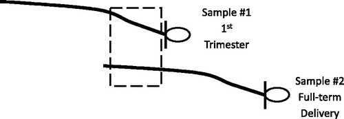
Figure 1. Diagram showing the proximal and distal first-trimester hair segments for comparison. The dashed box indicates segments on each hair sample collection used for comparisons: 0–3 cm from the scalp on hair sample 1 collected in the first trimester (proximal) and 6–9 cm from the scalp on hair sample 2 collected at delivery (distal).
2. Materials and methods
2.1. Study participants and procedures
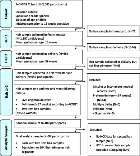
Data were gathered as part of the Pregnancy Outcomes, Maternal, and Infant Study (PrOMIS), a prospective cohort study consisting of pregnant women attending prenatal clinics at the Instituto Nacional Materno Perinatal (INMP) in Lima, Peru, the primary reference establishment for maternal and perinatal care operated by the Ministry of Health of the Peruvian government (Barrios et al., Citation2015). The institutional review boards of the Harvard T.H. Chan School of Public Health and INMP approved this study, and written informed consent was obtained from all participants. Recruitment for the PrOMIS study began in February of 2012, and scalp hair samples were collected from participants enrolled in the cohort during the period of October 2014 to November 2015. Participants who were ≥18 years of age, were able to speak and read in Spanish, and initiated prenatal care in early pregnancy were invited to participate [mean gestational age = 13.1 weeks, standard deviation (SD) = 3.9]. Recruited participants were then followed from early pregnancy to delivery. Among enrolled PrOMIS cohort participants, 96% provided a first hair sample at enrollment in early pregnancy, 32% of which contributed a second hair sample at full-term delivery (mean gestational age 39.0 weeks, SD = 1.0). Since it was neither necessary nor financially feasible to conduct biochemical analyses of all participants, we randomly selected 100 women from all eligible women with two hair samples. Selected women did not differ from non-selected women. The selection process for the present analysis is provided in . Briefly, women were excluded if they failed to meet the following criteria: live singleton delivery, full-term delivery (≥37 weeks), and two hair sample collections. Three women were excluded from the 100 randomly selected participants due to either undetectable cortisol values, or cortisol values that exceeded 100 pg/mg (>4 SDs from the mean). Our final analytic sample consists of 97 participants, each providing two first-trimester hair segments: the proximal 3 cm segment measured as 0–3 cm from the scalp collected in early pregnancy, and the distal 3 cm segment measured as 6–9 cm from the scalp collected at full-term delivery (). Assuming a two-tailed α of 0.05, a sample size of 100 participants had 80% power to detect a correlation of 0.28. The power for our sample size of 97 participants was 79.7%.
Figure 2. Flow chart showing selection into study. *ACOG: American Congress of Obstetricians and Gynecologists.
Hair collection procedures were similar to those described elsewhere (Gao et al., Citation2013). In brief, trained research staff collected two hair samples from the posterior vertex region of the scalp as close to the scalp as possible twice during the perinatal period, first at enrollment and again at full-term delivery. Collected hair samples were then wrapped in aluminum foil, and stored in manila envelopes away from light and at room temperature using desiccants. Prior to assay, women’s hair samples were randomly ordered, and samples from the same woman assayed in the same immunoassay batch. From this sample of 97 women, approximately one-third (N = 28) were randomly selected for a sub-study validation using liquid chromatography tandem mass spectrometry (LC-MS/MS). Assuming a two-tailed α of 0.05, a sample size of 28 participants was 80% powered to detect a correlation of 0.50.
2.2. Laboratory analysis
Hair processing procedures for cortisol were as described in previous studies (Albar et al., Citation2013). First, both 9 cm hair samples from each participant were segmented into three 3 cm hair segments. This analysis was restricted to values from the first-trimester hair segments only (). Lab personnel used 7.5 mg of whole non-pulverized hair per segment for analysis with the Cortisol Saliva Luminescence Immunoassay, IBL International® (Hamburg, Germany). One immunoassay batch was defined as one 96-well plate, and all hair segments from the same woman were analyzed together in the same batch, thereby reducing the influence of variability across batches (Tworoger & Hankinson, Citation2006). For our sub-study validation, a subset of 28 randomly selected participants had both first trimester hair segments additionally analyzed using LC-MS/MS, where samples were run consecutively rather than in batches. The same lot of reagents was used for all samples. Cortisol units of both techniques were reported in picograms per milligram (pg/mg), and the lower limit of detection was 0.1 pg/mg (Gao, Kirschbaum, Grass, & Stalder, Citation2016; Gao et al., Citation2013). Six blinded quality control (QC) samples were randomly dispersed to assess variability (Tworoger & Hankinson, Citation2006). For the immunoassay, inter-assay and intra-assay coefficients of variation (CV) were 19.4 and 11.9%, respectively. For the LC-MS/MS, the inter-assay CV was 8.1%. Since LC-MS/MS analyses did not employ batches, intra-assay CV’s are not reported. CV’s up to 20% are regarded as acceptable (Tworoger & Hankinson, Citation2006). In sensitivity analyses, immunoassay-derived cortisol concentrations were batch-corrected using the Rosner method (Rosner, Cook, Portman, Daniels, & Falkner, Citation2008) to determine if batch-corrected agreement findings differed from original findings.
2.3. Participant characteristics
At enrollment, structured interviewer-administered questionnaires were used to collect information on participants’ hair and sociodemographic characteristics, anthropometrics, and medical and reproductive history. Hair characteristics included: natural hair color, hair structure, hair washing frequency, shampoo and conditioner use, chemical hair treatment use, and hair cutting frequency. Sociodemographic characteristics included: age, educational attainment, smoking status prior to the study pregnancy, alcohol consumption prior to the study pregnancy, ethnicity, marital status, employment during the study pregnancy, and difficulty paying for basics. Early pregnancy body mass index (BMI) (kg/m2) was measured using the participants’ weight to the nearest 0.1 kg and height to the nearest 0.1 cm. Medical and reproductive history questions assessed whether the study pregnancy was planned, parity, gestational age at enrollment and full-term delivery using last menstrual period (in weeks), and asthma diagnosis before the study pregnancy. Psychological measures such as perceived stress, generalized anxiety disorder, depression, and post-traumatic stress disorder were assessed at enrollment using the 14-item Perceived Stress Scale (Cohen, Kamarck, & Mermelstein, Citation1983), the 7-item Generalized Anxiety Disorder scale (Zhong et al., Citation2015), the 9-item Patient Health Questionnaire (Zhong et al., Citation2014), and the Post-Traumatic Stress Disorder Checklist-Civilian version (Blanchard, Jones-Alexander, Buckley, & Forneris, Citation1996; Gelaye et al., Citation2017), respectively. Since cortisol concentrations in hair may be influenced by ultraviolet light (UV) exposure (Hansen, Garde, Skovgaard, & Christensen, Citation2001; Li et al., Citation2012; Stalder et al., Citation2012), categories of UV exposure during the 3-month time period of hair growth were estimated using meteorological UV levels. Hair growth occurring exclusively during the high UV Peru summer months of December to April were defined as “high” (N = 16), and hair growth occurring exclusively during the low UV Peru non-summer months of May to November were defined as “low” (N = 20). Hair growth occurring during both high UV summer months and low UV non-summer months was defined as “intermediate” (N = 61). Lastly, in order to assess whether differences in the time between the two hair sample collections (at enrollment and full-term delivery) influenced concordance, categories distinguishing participants whose hair samples were collected 5–6 months apart (N = 58) versus not (N = 39) were created. If time between hair samples played a role, we hypothesized that participants whose hair samples were collected 5–6 months apart would be most concordant due to the average time between hair collections.
2.4. Statistical analysis
The Shapiro–Wilk test statistic was used to test for skewness in HCC. Based on findings of right-skewness, HCC values were transformed on the natural logarithm scale (logHCC) to approximate normality. To facilitate comparison of HCC with other studies, we report geometric mean HCC and standard deviations (SD) for proximal and distal hair segments for all participants and according to participant characteristics. Student’s t-tests or analysis of variance (ANOVA) were used to evaluate differences in logHCC across maternal characteristics for proximal and distal hair segments. We then used paired sample t-tests to evaluate the mean difference between proximal and distal logHCC. Linear regression models were used to determine whether proximal–distal differences (deltas) varied according to maternal characteristics at enrollment. Bland–Altman plots were used to evaluate for evidence of systematic bias in differences. Concordance was further evaluated using scatter plots, Pearson correlation coefficients, Cohen’s weighted kappa test statistics using logHCC tertiles, and intra-class correlation coefficients (ICCs). In the subset of women whose first-trimester hair segments were analyzed using both laboratory methods (N = 28), concordance across laboratory methods was evaluated separately for distal and proximal segments. All statistical analyses used SAS® version 9.4 software (SAS Institute, Inc., Cary, NC) and p values are two-sided at the α = 0.05 level.
3. Results
Participant ages ranged from 18 to 44 years (mean = 26.5 years, SD = 5.8), and the mean gestational ages at the two hair collections (i.e. early pregnancy and delivery) were 13.1 weeks (SD = 3.9) and 39.0 weeks (SD = 1.0), respectively. Participant sociodemographic characteristics, anthropometrics, medical history, and hair characteristics are described in . Participants reported no medication use or diabetes at time of interview. Differences in proximal HCC were observed across categories of education, asthma, generalized anxiety disorder, hair structure, and UV light exposure. Differences in distal HCC were observed in the similar directions as proximal HCC, albeit only statistically significantly for education and alcohol use.
Table 1. Hair cortisol concentrations according to study population characteristics (N = 97 participants, each with one distal and one proximal hair segment).
Mean logHCC, measured using immunoassay, in proximal hair segments were higher compared with distal segments, 1.64 versus 1.35, respectively (p = .02) (). Bland–Altman plots showed no evidence of systematic bias in difference estimates (). No statistical difference was observed using LC-MS/MS (1.99 versus 1.83, p = .33). The magnitudes of the delta values were investigated according to maternal characteristics in . Delta values statistically differed according to the following maternal characteristics at enrollment: employment status, UV exposure, and asthma diagnosis (Supplementary Table S1). Specifically, larger differences were observed among participants who were unemployed versus employed, among participants exposed to higher UV in early pregnancy versus low UV, and among participants with a history of asthma versus participants without a history of asthma.
Figure 3. Bland–Altman plots comparing first trimester proximal (0–3 cm hair sample 1) and distal (6–9 cm on hair sample 2) log-transformed hair cortisol concentrations (logHCC), by laboratory method of detection, immunoassay (N = 97) and liquid chromatography tandem mass spectrometry (LC-MS/MS) (N = 28).
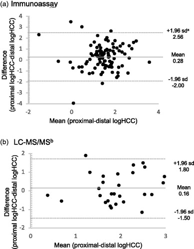
Table 2. Comparisons of proximal and distal first-trimester hair cortisol concentrations by laboratory method of detection.
The Pearson correlation between proximal and distal first-trimester hair segments measured using immunoassay was 0.27 (p = .008) (). Correlation coefficients were highest among the subset of participants whose hair samples were collected 5–6 months apart (N = 58, r = 0.36, p = .006) as compared to participants whose hair samples were not collected 5–6 months apart (N = 39, r = 0.14, p = .39). The Pearson correlation between proximal and distal first-trimester hair segments measured using LC-MS/MS was 0.37 (p = .05) (). The proportion of hair samples concordantly ranked as either low, middle, or high according to HCC tertiles was low comparing proximal and distal rankings (immunoassay weighted κ = 0.10, LC-MS/MS weighted κ = 0.07) (). Intra-class correlation (ICC) values were also low (immunoassay ICC: 0.46, LC-MS/MS ICC: 0.34) (). In the subset of participants whose proximal and distal hair samples were analyzed using both immunoassay and LC-MS/MS, concordance was high among distal segments (r = 0.78, p < .001; weighted κ = 0.64) and high among proximal segments (r = 0.96, p < .001; weighted κ = 0.75) (). Batch-corrected immunoassay agreement measures did not substantially differ from the immunoassay agreement measures provided (Supplementary Table S2).
Figure 4. Scatterplots comparing proximal and distal log-transformed hair cortisol concentrations (logHCC) by laboratory method of detection. ar: Pearson correlation coefficient, bLC-MS/MS: liquid chromatography tandem mass spectrometry.
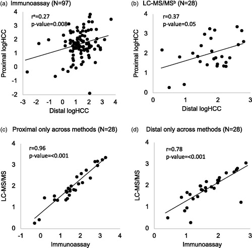
Table 3. Agreement between proximal and distal first trimester hair cortisol concentrations according to method of cortisol analysis.
4. Discussion
On average, HCC measured in distal hair segments collected at delivery were lower and had poor agreement with HCC measured in proximal hair segments collected in early pregnancy. This suggests that distal hair segments collected at the time of delivery cannot be used to reflect cortisol concentrations in the first trimester of pregnancy, and more generally that distal hair segments beyond 6 cm from the scalp may not be appropriate for use in studies aiming to assess HCC over a 9-month period. Our findings also show that despite lower absolute measures of HCC using immunoassay as compared to LC-MS/MS, the two laboratory methods strongly agree and preserve relative rankings, as previously shown in an inter-laboratory round robin (Russell et al., Citation2015).
Reasons for why distal segments beyond 6 cm from the scalp may not reflect cortisol concentrations in the presumed time period have been hypothesized to be due to “washout effects” over time (Russell et al., Citation2012). Successive laboratory washes of hair samples have been shown to result in the decreases in HCC due to potential leaching of cortisol from the hair (Davenport et al., Citation2006), mainly from water exposure rather than shampoo treatment (Hamel et al., Citation2011; Li et al., Citation2012). In our study, we observed lower HCC with increased self-reported hair washing frequency in early pregnancy; however, these differences were not statistically significant. One study among non-pregnant women determined that HCC naturally declines by 30–40% as one moves from proximal to distal hair segments on the same hair sample (Kirschbaum et al., Citation2009). In comparison, we observed a smaller magnitude of difference comparing proximal and distal hair segments across different hair samples (mean logHCC values of 1.64 and 1.35, a 32% difference in HCC on the original scale).
We are aware of only one other study that compared early pregnancy cortisol concentrations in proximal and distal hair segments (D’Anna-Hernandez et al., Citation2011). In their study of 14 women recruited prior to 17 weeks gestational age with non-complicated pregnancies, D’Anna-Hernandez et al. (Citation2011) reported no agreement in HCC proximal and distal segments reflecting early pregnancy (Pearson correlation coefficient = −0.2, p = .29). Both our study and D’Anna-Hernandez et al. (Citation2011) sampled hair from the posterior vertex of the scalp at two times (first in early pregnancy and again at delivery), used similar laboratory methods, recruited participants of comparable maternal age and at comparable times in pregnancy, and included participants with no medication use. Studies differed in sample size (14 versus 97), region (USA versus Peru), in exclusion criteria [our study included smokers (14.5%) and those who used hair treatments (39.2%)], and magnitude of correlation (−0.2, p = .29 versus 0.27, p = .008). Despite differences in study design, geographic region, and behavioral characteristics, our present findings and that of D’Anna-Hernandez et al. (Citation2011) indicate that distal hair segments collected at delivery are in poor agreement with proximal hair segments collected in early pregnancy.
Our study also builds upon previous findings by suggesting that differences in proximal and distal HCC values may vary according to participant characteristics. Specifically, we observed larger differences among participants who were unemployed compared to participants who were employed, among participants exposed to higher UV compared to participants exposed to low UV, and among participants with a history of asthma compared to participants without a history of asthma. Reasons for a larger difference among the non-employed as compared to the employed are unclear, and differences according to asthma status were based on a small sample of asthmatics (10.3%, N = 10) requiring replication. However, given that asthma is commonly treated with corticosteroids, researchers interested in early pregnancy maternal cortisol levels should take this into consideration. Despite the fact that we observed higher HCC values in proximal hair segments that grew during seasons of higher UV, we observed larger proximal–distal differences at higher UV indices. This is plausible given that high UV irradiation is an environmental factor believed to facilitate washout of cortisol from hair over time (Dettenborn, Tietze, Kirschbaum, & Stalder, Citation2012). If such differences across subgroups hold, differential measurement error of HCC in distal segments may result in biased estimates. Therefore, researchers interested in utilizing distal hair segments may need to consider how they will account for such differences in the study design and analytic phases.
Our findings of strong agreement across laboratory methods of cortisol analysis are consistent with earlier reports. A recent international inter-laboratory round robin of 15 hair samples showed excellent agreement comparing immunoassay and LC-MS/MS analyzed HCC levels (r’s ranged from 0.89 to 0.98) (Russell et al., Citation2015), though it is unclear if the hair samples used were proximal or distal segments. We show that correlations comparing proximal segments were stronger than correlations comparing distal segments (r’s = 0.96 versus 0.78), and that relative HCC rankings are preserved, albeit to a stronger extent in proximal segments compared to distal segments. Furthermore, ICC values indicated lower between-person and total variation using LC-MS/MS values compared to immunoassay values, a difference potentially due to the smaller sample size of the LC-MS/MS values. This information may be of importance for researchers deciding between the two laboratory methods.
In our investigation of the concordance between HCC in proximal and distal hair segments, both presumed to reflect the first trimester of pregnancy, our study had some limitations. First, our study assessed all maternal characteristics once at enrollment. Therefore, any changes that occurred later in pregnancy, such as hair washing frequency and chemical treatment, were not accounted for. Second, it is possible that morphological changes in hair (such as increased thickness) may have occurred during the second and third trimesters of pregnancy, potentially affecting porosity and leaching of cortisol from hair segments. However, the impact of these changes on distal first trimester hair segments is likely minimal given that first trimester hair growth was already complete. Third, the timing between hair collections was not exactly 6 months for all participants. According to the average hair growth (1 cm/month), a 6-month window between the two hair collections would have resulted in optimal overlap of the 3 cm proximal and distal hair segments. Therefore, differences in time between hair collections may have impacted our findings. However, the mean timing between hair collections was 5.8 months (SD = 1.1), close to this optimal window of overlap. To investigate the extent to which timing between hair collections impacted our findings, we evaluated concordance among a subset collected 5–6 months apart (N = 58). In doing so, we observed stronger correlations (r = 0.37). Therefore, the timing between hair collections may have had some impact on our findings. However, the magnitude of this influence does not appear substantial (r = 0.27 among all participants versus r = 0.37 among this subset). Interestingly, slower underarm hair growth has been observed with pregnancy progression (Pecoraro, Astore, & Barman, Citation1971). While this is not a major concern given our focus on scalp hair, we provide it as potential evidence for the impacts of pregnancy on general hair growth. Fourth, our CV’s are on the higher end of the acceptable range. However, batch-corrected findings were very similar. Lastly, we cannot extrapolate our findings of poor concordance of distal and proximal segments to non-pregnant populations.
Despite these limitations our study had many strengths. First, our study used standardized hair collection and extraction procedures. For example, the posterior vertex region of the scalp has the lowest intra-individual variation of HCC compared to samples obtained from other areas of the scalp (16 versus 31%) (Sauve et al., Citation2007), and strong agreement has been observed comparing HCC across the posterior vertex region of the scalp during pregnancy (Pearson correlation = 0.85, p < .001) (D’Anna-Hernandez et al., Citation2011). Second, our study assessed concordance using two laboratory methods of cortisol analysis, immunoassay and LC-MS/MS. Third, our study assessed multiple concordance measures, which yield similar conclusions. Fourth, our sample size of 97 participants, each of whom provided one proximal and one distal hair segment, exceeds the sample size of the only other known comparable study, although, an evaluation of concordance using a larger sample of participants or in non-pregnant populations is warranted. Lastly, our restriction to full-term deliveries helped to ensure that distal and proximal hair segments represented similar times during pregnancy.
5. Conclusion
HCC in distal hair segments collected at delivery do not appear to reflect HCC in proximal hair segments collected in early pregnancy. Therefore, in accordance with previous suggestions, we suggest that investigators interested in long-term maternal cortisol secretion in early pregnancy restrict analyses to proximal hair segments collected in early pregnancy, or if possible, perform a validation sub-study similar to our own study to determine the extent of cortisol degradation if any.
Supplemental_Table_2.docx
Download MS Word (12.1 KB)Supplemental_Table_1.docx
Download MS Word (12.8 KB)Disclosure statement
No potential conflict of interest was reported by the authors.
Additional information
Funding
Related Research Data
References
- Albar, W.F., Russell, E.W., Koren, G., Rieder, M.J., & van Umm, S.H. (2013). Human hair cortisol analysis: comparison of the internationally-reported ELISA methods. Clinical and Investigative Medicine, 36, E312–E316. doi:10.25011/cim.v36i6.20629
- Astore, I.P., Pecoraro, V., & Pecoraro, E.G. (1979). The normal trichogram of pubic hair. British Journal of Dermatology, 101, 441–445. doi:10.1111/j.1365-2133.1979.tb00023.x
- Barman, J.M., Astore, I., & Pecoraro, V. (1965). The normal trichogram of the adult. Journal of Investigative Dermatology, 44, 233–236. doi:10.1038/jid.1965.42
- Barrios, Y.V., Gelaye, B., Zhong, Q., Nicolaidis, C., Rondon, M.B., Garcia, P.J., … Williams, M.A. (2015). Association of childhood physical and sexual abuse with intimate partner violence, poor general health and depressive symptoms among pregnant women. PloS One, 10, e0116609. doi:10.1371/journal.pone.0116609
- Barth, J.H. (1986). Measurement of hair growth. Clinical and Experimental Dermatology, 11, 127–138. doi:10.1111/j.1365-2230.1986.tb00437.x
- Bjelanovic, V., Babic, D., Hodzic, D., Bjelanovic, A., Kresic, T., Dugandzic-Simic, A., & Oreskovic, S. (2015). Correlation of psychological symptoms with cortisol and CRP levels in pregnant women with metabolic syndrome. Psychiatria Danubina, 27(Suppl. 2), 578–585.
- Blanchard, E.B., Jones-Alexander, J., Buckley, T.C., & Forneris, C.A. (1996). Psychometric properties of the PTSD Checklist (PCL). Behaviour Research and Therapy, 34, 669–673. doi:10.1016/0005-7967(96)00033-2
- Bolten, M.I., Wurmser, H., Buske-Kirschbaum, A., Papoušek, M., Pirke, K.M., & Hellhammer, D. (2011). Cortisol levels in pregnancy as a psychobiological predictor for birth weight. Archives of Women's Mental Health, 14, 33–41. doi:10.1007/s00737-010-0183-1
- Braithwaite, E.C., Murphy, S.E., & Ramchandani, P.G. (2016). Effects of prenatal depressive symptoms on maternal and infant cortisol reactivity. Archives of Women's Mental Health, 19, 581–590. doi:10.1007/s00737-016-0611-y
- Burke, C.W., & Roulet, F. (1970). Increased exposure of tissues to cortisol in late pregnancy. British Medical Journal, 1, 657–659. doi:10.1136/bmj.1.5697.657
- Cirimele, V., Kintz, P., Dumestre, V., Goullé, J.P., & Ludes, B. (2000). Identification of ten corticosteroids in human hair by liquid chromatography-ionspray mass spectrometry. Forensic Science International, 107, 381–388. doi:10.1016/S0379-0738(99)00180-2
- Cohen, S., Kamarck, T., & Mermelstein, R. (1983). A global measure of perceived stress. Journal of Health and Social Behavior, 24, 385–396. doi:10.2307/2136404
- Conrad, F., & Paus, R. (2004). Estrogens and the hair follicle. Journal of the German Society of Dermatology, 2, 412–423. doi:10.1046/j.1439-0353.2004.04037.x
- D’Anna-Hernandez, K.L., Ross, R.G., Natvig, C.L., & Laudenslager, M.L. (2011). Hair cortisol levels as a retrospective marker of hypothalamic-pituitary axis activity throughout pregnancy: comparison to salivary cortisol. Physiology & Behavior, 104, 348–353. doi:10.1016/j.physbeh.2011.02.041
- Davenport, M.D., Tiefenbacher, S., Lutz, C.K., Novak, M.A., & Meyer, J.S. (2006). Analysis of endogenous cortisol concentrations in the hair of rhesus macaques. General and Comparative Endocrinology, 147, 255–261. doi:10.1016/j.ygcen.2006.01.005
- Davis, E.P., Glynn, L.M., Schetter, C.D., Hobel, C., Chicz-Demet, A., & Sandman, C.A. (2007). Prenatal exposure to maternal depression and cortisol influences infant temperament. Journal of the American Academy of Child and Adolescent Psychiatry, 46, 737–746. doi:10.1097/chi.0b013e318047b775
- Dettenborn, L., Tietze, A., Kirschbaum, C., & Stalder, T. (2012). The assessment of cortisol in human hair: associations with sociodemographic variables and potential confounders. Stress, 15, 578–588. doi:10.3109/10253890.2012.654479
- Diego, M.A., Field, T., Hernandez-Reif, M., Schanberg, S., Kuhn, C., & Gonzalez-Quintero, V.H. (2009). Prenatal depression restricts fetal growth. Early Human Development, 85, 65–70. doi:10.1016/j.earlhumdev.2008.07.002
- Field, T., Diego, M., Hernandez-Reif, M., Deeds, O., Holder, V., Schanberg, S., & Kuhn, C. (2009). Depressed pregnant black women have a greater incidence of prematurity and low birthweight outcomes. Infant Behavior & Development, 32, 10–16. doi:10.1016/j.infbeh.2008.09.005
- Gao, W., Kirschbaum, C., Grass, J., & Stalder, T. (2016). LC-MS based analysis of endogenous steroid hormones in human hair. The Journal of Steroid Biochemistry and Molecular Biology, 162, 92–99. doi:10.1016/j.jsbmb.2015.12.022
- Gao, W., Stalder, T., Foley, P., Rauh, M., Deng, H., & Kirschbaum, C. (2013). Quantitative analysis of steroid hormones in human hair using a column-switching LC-APCI-MS/MS assay. Journal of Chromatography. B, Analytical Technologies in the Biomedical and Life Sciences. 928, 1–8. doi:10.1016/j.jchromb.2013.03.008
- Gelaye, B., Zheng, Y., Medina-Mora, M.E., Rondon, M.B., Sánchez, S.E., & Williams, M.A. (2017). Validity of the posttraumatic stress disorders (PTSD) checklist in pregnant women. BMC Psychiatry, 17, 179. doi:10.1186/s12888-017-1304-4
- Giesbrecht, G.F., Campbell, T., Letourneau, N., Kooistra, L., & Kaplan, B. (2012). Psychological distress and salivary cortisol covary within persons during pregnancy. Psychoneuroendocrinology, 37, 270–279. doi:10.1016/j.psyneuen.2011.06.011
- Gow, R., Thomson, S., Rieder, M., Van Uum, S., & Koren, G. (2010). An assessment of cortisol analysis in hair and its clinical applications. Forensic Science International, 196, 32–37. doi:10.1016/j.forsciint.2009.12.040
- Hamel, A.F., Meyer, J.S., Henchey, E., Dettmer, A.M., Suomi, S.J., & Novak, M.A. (2011). Effects of shampoo and water washing on hair cortisol concentrations. International Journal of Clinical Chemistry, 412, 382–385. doi:10.1016/j.cca.2010.10.019
- Hansen, A.M., Garde, A.H., Skovgaard, L.T., & Christensen, J.M. (2001). Seasonal and biological variation of urinary epinephrine, norepinephrine, and cortisol in healthy women. International Journal of Clinical Chemistry, 309, 25–35. doi:10.1016/S0009-8981(01)00493-4
- Hoffman, M.C., Mazzoni, S.E., Wagner, B.D., Laudenslager, M.L., & Ross, R.G. (2016). Measures of maternal stress and mood in relation to preterm birth. Obstetrics and Gynecology, 127, 545–552. doi:10.1097/AOG.0000000000001287
- Kirschbaum, C., Tietze, A., Skoluda, N., & Dettenborn, L. (2009). Hair as a retrospective calendar of cortisol production-Increased cortisol incorporation into hair in the third trimester of pregnancy. Psychoneuroendocrinology, 34, 32–37. doi:10.1016/j.psyneuen.2008.08.024
- Lachelin, G.C. 2013. Introduction to clinical reproductive endocrinology. Oxford, UK: Butterworth-Heinemann.
- Li, J., Xie, Q., Gao, W., Xu, Y., Wang, S., Deng, H., & Lu, Z. (2012). Time course of cortisol loss in hair segments under immersion in hot water. International Journal of Clinical Chemistry, 413, 434–440. doi:10.1016/j.cca.2011.10.024
- Lommatzsch, M., Hornych, K., Zingler, C., Schuff-Werner, P., Hoppner, J., & Virchow, J.C. (2006). Maternal serum concentrations of BDNF and depression in the perinatal period. Psychoneuroendocrinology, 31, 388–394. doi:10.1016/j.psyneuen.2005.09.003
- Loussouarn, G., El Rawadi, C., & Genain, G. (2005). Diversity of hair growth profiles. International Journal of Dermatology, 44(Suppl. 1), 6–9. doi:10.1111/j.1365-4632.2005.02800.x
- Lynfield, Y.L. (1960). Effect of pregnancy on the human hair cycle. Journal of Investigative Dermatology, 35, 323–327. doi:10.1038/jid.1960.127
- McEwen, B.S., & Seeman, T. (1999). Protective and damaging effects of mediators of stress. Elaborating and testing the concepts of allostasis and allostatic load. Annals of the New York Academy of Sciences, 896, 30–47. doi:10.1111/j.1749-6632.1999.tb08103.x
- Murphy, S.E., Braithwaite, E.C., Hubbard, I., Williams, K.V., Tindall, E., Holmes, E.A., & Ramchandani, P.G. (2015). Salivary cortisol response to infant distress in pregnant women with depressive symptoms. Archives of Women’s Mental Health, 18, 247–253. doi:10.1007/s00737-014-0473-0
- Nepomnaschy, P.A., Welch, K.B., McConnell, D.S., Low, B.S., Strassmann, B.I., & England, B.G. (2006). Cortisol levels and very early pregnancy loss in humans. Proceedings of the National Academy of Sciences of the United States of America, 103, 3938–3942. doi:10.1073/pnas.0511183103
- Nissimov, J., & Elchalal, U. (2003). Scalp hair diameter increases during pregnancy. Clinical and Experimental Dermatology, 28, 525–530. doi:10.1046/j.1365-2230.2003.01331.x
- O’Connor, T.G., Tang, W., Gilchrist, M.A., Moynihan, J.A., Pressman, E.K., & Blackmore, E.R. (2014). Diurnal cortisol patterns and psychiatric symptoms in pregnancy: short-term longitudinal study. Biological Psychology, 96, 35–41. doi:10.1016/j.biopsycho.2013.11.002
- O’Keane, V., Lightman, S., Marsh, M., Pawlby, S., Papadopoulos, A.S., Taylor, A., … Patrick, K. (2011). Increased pituitary-adrenal activation and shortened gestation in a sample of depressed pregnant women: a pilot study. Journal of Affective Disorders, 130, 300–305. doi:10.1016/j.jad.2010.10.004
- Pecoraro, V., Astore, I., & Barman, J.M. (1971). Growth rate and hair density of the human axilla. A Comparative study of normal males and females and pregnant and post-partum females. Journal of Investigative Dermatology, 56, 362–365. doi:10.1111/1523-1747.ep12261236
- Pecoraro, V., Astore, I.P.L., & Barman, J.M. (1967). The normal trichogram of pregnant women. In W. Montagna & J.M. Dobson (Eds.), Advances in biology of the skin (Vol. 9). New York: Pergamon Press.
- Peer, M., Soares, C.N., Levitan, R.D., Streiner, D.L., & Steiner, M. (2013). Antenatal depression in a multi-ethnic, community sample of Canadian immigrants: psychosocial correlates and hypothalamic-pituitary-adrenal axis function. Canadian Journal of Psychiatry, 58, 579–587. doi:10.1177/070674371305801007
- Pragst, F., & Balikova, M.A. (2006). State of the art in hair analysis for detection of drug and alcohol abuse. International Journal of Clinical Chemistry, 370, 17–49. doi:10.1016/j.cca.2006.02.019
- Raul, J.S., Cirimele, V., Ludes, B., & Kintz, P. (2004). Detection of physiological concentrations of cortisol and cortisone in human hair. Clinical Biochemistry, 37, 1105–1111. doi:10.1016/j.clinbiochem.2004.02.010
- Rosner, B., Cook, N., Portman, R., Daniels, S., & Falkner, B. (2008). Determination of blood pressure percentiles in normal-weight children: some methodological issues. American Journal of Epidemiology, 167, 653–666. doi:10.1093/aje/kwm348
- Russell, E., Kirschbaum, C., Laudenslager, M.L., Stalder, T., de Rijke, Y., van Rossum, E.F., … Koren, G. (2015). Toward standardization of hair cortisol measurement: results of the first international interlaboratory round robin. Therapeutic Drug Monitoring, 37, 71–75. doi:10.1097/FTD.0000000000000148
- Russell, E., Koren, G., Rieder, M., & Van Uum, S. (2012). Hair cortisol as a biological marker of chronic stress: current status, future directions and unanswered questions. Psychoneuroendocrinology, 37, 589–601. doi:10.1016/j.psyneuen.2011.09.009
- Sauve, B., Koren, G., Walsh, G., Tokmakejian, S., & Van Uum, S.H. (2007). Measurement of cortisol in human hair as a biomarker of systemic exposure. Clinical and investigative medicine. Medecine Clinique Et Experimentale, 30, E183–E191. doi:10.25011/cim.v30i5.2894
- Smith, L.K., & Cidlowski, J.A. (2010). Glucocorticoid-induced apoptosis of healthy and malignant lymphocytes. Progress in Brain Research, 182, 1–30. doi:10.1016/S0079-6123(10)82001-1
- Stalder, T., & Kirschbaum, C. (2012). Analysis of cortisol in hair–state of the art and future directions. Brain, Behavior, and Immunity, 26, 1019–1029. doi:10.1016/j.bbi.2012.02.002
- Stalder, T., Steudte, S., Miller, R., Skoluda, N., Dettenborn, L., & Kirschbaum, C. (2012). Intraindividual stability of hair cortisol concentrations. Psychoneuroendocrinology, 37, 602–610. doi:10.1016/j.psyneuen.2011.08.007
- Tworoger, S.S., & Hankinson, S.E. (2006). Use of biomarkers in epidemiologic studies: minimizing the influence of measurement error in the study design and analysis. Cancer Causes & Control, 17, 889–899. doi:10.1007/s10552-006-0035-5
- Van Londen, L., Goekoop, J.G., Zwinderman, A.H., Lanser, J.B., Wiegant, V.M., & De Wied, D. (1998). Neuropsychological performance and plasma cortisol, arginine vasopressin and oxytocin in patients with major depression. Psychological Medicine, 28, 275–284. doi:10.1017/S0033291797006284
- Voegtline, K.M., Costigan, K.A., Kivlighan, K.T., Laudenslager, M.L., Henderson, J.L., & DiPietro, J.A. (2013). Concurrent levels of maternal salivary cortisol are unrelated to self-reported psychological measures in low-risk pregnant women. Archives of Women’s Mental Health, 16, 101–108. doi:10.1007/s00737-012-0321-z
- Wosu, A.C., Valdimarsdottir, U., Shields, A.E., Williams, D.R., & Williams, M.A. (2013). Correlates of cortisol in human hair: implications for epidemiologic studies on health effects of chronic stress. Annals of Epidemiology, 23, 797–811.e792. doi:10.1016/j.annepidem.2013.09.006
- Zhong, Q., Gelaye, B., Rondon, M., Sanchez, S.E., Garcia, P.J., Sanchez, E., … Williams, M.A. (2014). Comparative performance of Patient Health Questionnaire-9 and Edinburgh Postnatal Depression Scale for screening antepartum depression. Journal of Affective Disorders, 162, 1–7. doi:10.1016/j.jad.2014.03.028
- Zhong, Q.Y., Gelaye, B., Zaslavsky, A.M., Fann, J.R., Rondon, M.B., Sanchez, S.E., & Williams, M.A. (2015). Diagnostic Validity of the Generalized Anxiety Disorder-7 (GAD-7) among pregnant women. PLoS One, 10, e0125096. doi:10.1371/journal.pone.0125096