
 ?Mathematical formulae have been encoded as MathML and are displayed in this HTML version using MathJax in order to improve their display. Uncheck the box to turn MathJax off. This feature requires Javascript. Click on a formula to zoom.
1. Introduction
The measurement of Fractional Flow Reserve (FFR) becomes essential for an interventional cardiologist to decide whether or not an angioplasty or stenting procedure needs to be performed on his patient. The FFR is an invasive technique used in clinical routine for evaluating stenosis severity and functional significance of the coronary artery disease. In practice, FFR is calculated as the ratio of the distal trans-stenotic pressure to the proximal coronary or aortic pressure during induced hyperemia.
Several theoretical, experimental, animal and clinical studies have demonstrated the usefulness of measuring FFR to assess the hemodynamic significance of two serial stenoses (CitationPijls et al. 2000; CitationD’Souza et al. 2014). However, the effects of the distance separating the two lesions (Dss) and the combined severity of the two serial stenoses (S1, S2) on the global and intermediate FFR amplitudes remain unclear. Therefore, the aim of this study was to develop an experimental hemodynamic bench to elucidate the influences of such geometrical parameters on the FFR assessment. As a first approach, the present work was focused on the influence of the severity of S1 and S2.
2. Materials and Methods
2.1 Experimental bench
A straight tube vessel phantom, made of silicone with an internal diameter (ID) of 4 mm and a wall thickness of 0.5 mm was used to conduct the experiments. The measured elastic tangent moduli for the material was about 1600 KPa. Two stenosis models were built with silicone: the first one with an ID of approximately 2.8 mm (i.e. 30% of diameter reduction) and the second one with an ID of 1.6 mm (i.e. 60% of diameter reduction). The distance Dss between the two stenosis was fixed to 20 mm for all the experiments.
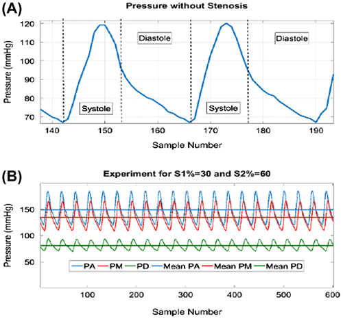
A circulatory hemodynamic bench was built (see Figure ). A pulsatile pump, compliance chambers and resistive elements were incorporated to mimic the human physiological coronary flow conditions. Beats per minute were set to 65. Periodic systolic-dominant pulses with 120 mmHg and 70 mmHg for systolic and diastolic pressures respectively were obtained for no stenosis conditions (see Figure A). An absolute flow Q = 300 ml/min was generated in order to simulate the hyperemic conditions of the major coronary vessels. Water was used as working fluid. Pressure measurements were performed via 1mm-diameter liquid-filled catheter connected to a pressure transducer.
Figure 1 Cartoons of the experimental hemodynamic bench.
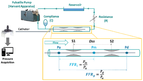
Figure 2 Example of pulsatile pressures recorded for no stenosis and serial stenoses
2.2 Pressure-derived FFR measurements
For single stenosis experiments, mean pressure amplitude Pa was recorded first at location A (before lesion). Then, the catheter passed through the stenosis (S1) to location D (after lesion) where mean pressure Pd was recorded. Global FFR (FFRg) was defined as:(1)
(1)
In the serial stenoses experiments, mean pressure amplitude was measured at locations A, M and D (see Figure , insert). Two FFRs were acquired, the global FFRg (given by Equation 1) and the intermediate FFR (FFRi) estimated as:(2)
(2)
Before each experiment the catheter was re-filled with water in order to avoid bubbles formation. The mean pressures Pa, Pm and Pd were recorded through n=35 pressure cycles only after the pulses stabilized and became periodic.
3. Results
Figure B illustrates a time-pressure variation recorded at 3 locations (A, M and D) when the catheter went through two serial stenoses with S1=30% and S2=60%.
The single stenosis FFR measurements agree with those published in the literature and measured on patients (CitationTakagi et al. 1999). The results presented in Table (Mean ± SD for n>5) clearly show that the presence of a second stenosis affects the amplitudes of the two fractional flow reserves FFRi and FFRg. While a significant decrease of the global FFRg is observed when introducing a second stenosis, the intermediate FFRi tends to increase slightly.
Table 1 Measurements of FFRg and FFRi for 4 experimental protocols
4. Conclusions
These preliminary results are promising and validate the hemodynamic bench which will be used in the near future: 1) to highlight the existing relationships between the FFR amplitudes and geometrical parameters S1, S2 and Dss, 2) to mimic complex situations of coronary branches with several stenosis, and, 3) to test the hemodynamic perturbations induced by the presence of stents.
The perspective for the future work is to improve the hemodynamic bench by: 1) including a phantom with an elastic behaviour more similar to the one of an artery wall, 2) using a working fluid with the same density and viscosity as blood and 3) changing the 1mm catheter for a 0.35 mm one with the aim of decreasing the effect of catheter size on FFR measurements. This will allow to evaluate worse lesions up to 80% of diameter reduction.
Acknowledgements
Ricardo Coppel and Armida L. Gomez express their gratitude to the Mexican National Council for Science and Technology (CONACYT) and the Nuevo Leon Institute of Innovation and Technology Transfer (I2T2).
References
- D’Souza GA, Peelukhana SV, Banerjee RK. 2014. Diagnostic Uncertainties During Assessment of Serial Coronary Stenoses: An In Vitro Study. J Biomech Eng. 136(2):021026.
- Pijls NH, De Bruyne B, Bech GJ, Liistro F, Heyndrickx GR, Bonnier HJ, Koolen JJ. 2000. Coronary Pressure Measurement to Assess the Hemodynamic Significance of Serial Stenoses Within One Coronary Artery: Validation in Humans. Circulation. 102(19):2371–2377.
- Takagi A, Tsurumi Y, Ishii Y, Suzuki K, Kawana M, Kasanuki H. 1999. Clinical Potential of Intravascular Ultrasound for Physiological Assessment of Coronary Stenosis: Relationship Between Quantitative Ultrasound Tomography and Pressure-Derived Fractional Flow Reserve. Circulation. 100:250–255.