
Keywords:
1. Introduction
Intensive tennis practice, with its unilateral solicitation, leads to sport-specific adaptations particularly in shoulder range of motion and muscular strength at the dominant shoulder (Gillet et al., Citation2017). However, excessive or imbalance changes are known to increase the risk of shoulder injuries (Chorley et al., Citation2017). Shoulder injuries can be evaluated and prevented by monitoring the scapulohumeral rhythm (SHR). The SHR assess the contribution of the humerus and the scapula during the arm elevation. Commonly, a 2:1 ratio is observed and its alterations are related to shoulder injuries (Hosseinimehr et al., Citation2015). However, asymptomatic overhead adult athlete presents a lower SHR ratio in the dominant side compared with the nondominant one (Hosseinimehr et al., Citation2015). To our knowledge, the SHR in young competitive tennis players has not been characterized yet. A better description of the SHR in such a population may help to improve the shoulder injury prevention.
This study aimed to describe the SHR of young competitive tennis players. We hypothesized that the SHR in the dominant side should be lower than the nondominant one.
2. Methods
Twenty-seven young competitive players (age: 11.4 ± 1.1 years; height: 149 ± 7 cm; mass: 38.2 ± 5.8 kg, tennis weekly exposure: 8 ± 2 h; years of practice: 6 ± 2 years) were volunteered to participate in this study (IRB 00009118). A written consent was signed by the legal guardian.
Players performed bilaterally two arm elevations with the thumb pointing up in the scapular plan. i.e. 30° anterior to the frontal plan. During this motion, the scapular and humeral kinematic data were collected using an electromagnetic device (100 Hz; Trackstar, Ascension Technology Corporation, Chicago, Ill), with six sensors of six degrees-of-freedom each. Five sensors were located on incisura jugularis, flat portion of the right and left acromion, and distal portion of the right and left humerus. A sixth sensor within a stylus was used to digitalize bony landmarks (T8, processus xiphoideus, C7, incisura jugularis, scapular angulus acromialis, trigonum scapulae, angulus inferior, humeral medial and lateral epicondyle) (Wu et al., Citation2005). These bony landmarks were then used to convert the sensor system of coordinates to an anatomic one. The glenohumeral joint center was estimated with functional method during circumvolutions (Wu et al., Citation2005). Raw kinematic data were filtered with a low-pass 4th-order Butterworth filter at 10 Hz. With the Euler angle decompositions, the humerus relative to the scapula rotation sequence (glenohumeral elevation angle, plane of glenohumeral elevation and glenohumeral internal/external rotation) was calculated with the YXY rotation sequence, while the scapular orientation relative to the thorax was determined with the YX’Z’’ rotation sequence. The scapulohumeral rhythm was the ratio between the glenohumeral elevation and the upward scapular rotation. The SHR was calculated at 30°, 60°, 90°, 120°, during rising of the arm; and at 120°, 90°, 60°, and 30° during lowering of the arm. At each position, the SHR of the dominant and the nondominant sides were compared with a paired t-test using SPSS 11.0. (SPSS Science Inc., Chicago, IL). After the application of the Bonferroni correction, the level of significance was set at p ≤ 0.006 (p = 0.05/8).
3. Results and discussion
In the literature, the scapulohumeral rhythm varies between 1.25:1 and 7.9:1 (Hosseinimehr et al., Citation2015; Robert-Lachaine et al., Citation2015). The SHR calculated in our population was in this range except at the 30° of elevation in lowering of the arm. However, a large inter-subject variability was found at this low angle (Robert-Lachaine et al., Citation2015). In our population of young competitive tennis players, no significant difference was found for the SHR between the dominant and the nondominant sides (Figure) whatever the arm elevation. In contrast, for the asymptomatic adult overhead athletes, the SHR was found lower in the dominant than the nondominant arm due to an increased scapular upward rotation during the arm elevation (Hosseinimehr et al., Citation2015).
Figure 1. Mean (± standard errors) glenohumeral elevation, scapular upward rotation (degrees) and scapulohumeral rhythm during rising and lowering of the arm in the scapular plane.
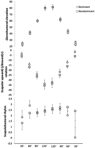
In contrast, for the asymptomatic adult overhead athletes, the SHR was found lower in the dominant than the nondominant arm due to an increased scapular upward rotation during the arm elevation (Hosseinimehr et al., Citation2015). Scapular and humeral positions during arm motion are the result of muscular actions. Particularly, the scapular position results of coaction of the mobilizer and stabilizer muscles. To evaluate the scapular functions, strength ratios between mobilizer and stabilizer muscles are calculated. Such ratios were similar between the dominant and the nondominant side in young asymptomatic competitive tennis players (Gillet et al., Citation2017) that could explain the absence of difference in SHR in our study. Asymptomatic young tennis players seem then to present a similar contribution of the humerus and scapula to raise and lower their arm in both sides. The SHR asymmetry could be a sport-related adaptation occurring latter in the practice. This study presents some limitations. First, by including only young boys, our results cannot be generalized to
players of different age or sex. Moreover, players included in this study were 10 to 14 years that may introduce variablity due to the growth process. It may then explain the lack of significant difference in SHR. Furthermore, although the 2D-method is reliable to calculate the SHR, other methods (3D) are more accurate (Robert-Lachaine et al., Citation2015). Nevertheless, this study is the first to analyse the scapulohumeral rhythm in young competitive tennis players and showed that the dominant and nondominant arm present similar characteristics during rising and lowering of the arm. Futures studies should investigate the relationship between pain and the SHR in such populations.
4. Conclusions
This study focused on the scapulohumeral rhythm in young competitive tennis players and showed that the dominant and the nondominant arm present the same coordination during caption. This result brings new knowledge on the shoulder of young overhead athletes and might help coaches and clinicians in the shoulder health monitoring in such a population.
Acknowledgements
We thank players and coaches of the Ligue du Lyonnais de Tennis for their participation at this study. We also thank the region of Auvergne-Rhone-Alpes and Mitacs for their financial support.
References
- Chorley J, Eccles RE, Scurfield A. 2017. Care of Shoulder Pain in the Overhead Athlete. Pediatric Annals. 46:e112–e113.10.3928/19382359-20170216-01
- Gillet B, Begon M, Sevrez V, Berger-Vachon C, Rogowski I. 2017. Adaptive Alterations in Shoulder Range of Motion and Strength in Young Tennis Players. J Athl Train. 52:137–144.10.4085/1062-6050.52.1.10
- Hosseinimehr SH, Anbarian M, Norasteh AA, Fardmal J, Khosravi MT. 2015. The comparison of scapular upward rotation and scapulohumeral rhythm between dominant and non-dominant shoulder in male overhead athletes and non-athletes. Manual Therapy.20:758–762.10.1016/j.math.2015.02.010
- Robert-Lachaine X, Marion P, Godbout V, Bleau J, Begon M. 2015. Elucidating the scapulo-humeral rhythm calculation: 3D joint contribution method. Computer Methods in Biomechanics and Biomedical Engineering.18:249–258.10.1080/10255842.2013.792810
- Wu G, van der Helm FC, Veeger HD, Makhsous M, Van Roy P, Anglin C, Nagels J, Karduna AR, McQuade K, Wang X. 2005. ISB recommendation on definitions of joint coordinate systems of various joints for the reporting of human joint motion – Part II: shoulder, elbow, wrist and hand. Journal of Biomechanics.38:981–992.10.1016/j.jbiomech.2004.05.042