1. Introduction
Electromyography (EMG) has already been investigated to provide insights into the changes of the functional abilities of the masticatory system related to temporomandibular disorders (TMD) or changes of the dental occlusion (Castroflorio et al. Citation2012; Mappelli et al., Citation2016). However, the clinical applications are limited by the various approaches to measure and post-process the EMG signals. The present study proposes a method to measure jaw muscle activity during a sub-maximal and controlled biting task.
2. Methods
2.1 Subjects
One asymptomatic subject (male, 27 years old) and one subject with moderate TMD (female, 23 years old) gave their consent to participate to the study. Both subjects had a complete denture, without current or recent orthodontic treatment and no pain or discomfort. The identified moderate TMD was a clicking joint without pain or limitation of the mouth opening.
2.2 Electrode positioning
The skin was cleaned with an exfoliating gel and scrubbed with alcohol soaked gauzes. The man had a clean-shaven skin. Paired pre-gelled electrodes (electrode distance: 2 cm, sensor area: 1cm of diameter) were placed on the skin at the muscle belly and along the muscular fibres. The location and orientation of each electrode were conscientiously defined and measured using home-made soft tools placed on the face to ensure a reproducible positioning. These locations and orientations were chosen to provide maximal amplitudes of muscle activity, following a preliminary experimental design.
2.3 EMG recordings
The left and right masseter (M), anterior temporalis (AT) and sternocleidomastoid (SCM) muscles were investigated. EMG activity was recorded using a wireless device (WinJaw EMG, Zebris Medical GmbH, Germany). The analog signals were pre-amplified and digitized (gain = 1000, CMRR = 110 dB in the range 4 - 310 Hz, input impedance = 120 GΩ, noise = 0.28 μV, sampling frequency = 800 Hz).
The subjects sat on a chair with the feet on the floor, the thigh perpendicular to the shank, in a natural erected position with closed eyes. They were asked to perform three repetitions of maximal voluntary contraction (MVC) held 5 seconds, after a complete rest of 10 seconds. MVC of the SCM was obtained by turning the head at its maximum towards the contralateral side. Biting on 12mm-thick cotton rolls placed on the left and right premolars and molars provided MVC of the M and AT muscles. The third MVC was held 5 seconds more (total of 10 seconds) to assess the muscular fatigue. The subjects trained themselves once before the first acquisition and they were invited to relax for about one minute between each record. At the end, the subjects bit five times on a calibrated dynamometer placed between the incisors. The biting force globally delivered at each time step was assessed from the recording of the dynamometer stretch using the JMA motion capture system (WinJaw JMA, Zebris Medical GmbH, Germany – sampling frequency 75 Hz). The resulting biting force varied from 0 to 25 N.
2.4 Post-processing
Raw signals were high-pass filtered (Butterworth filter, cutting frequency = 10 Hz). The SCM signals were additionally high-pass filtered at 30 Hz to reduce ECG artifacts. The filtered signals were rectified and then smoothed by computing the root mean square (RMS) on a moving time window of 100 ms without overlapping. The rest muscular activity was computed as the RMS value on a 10 s window corresponding to the rest at the beginning of each acquisition. An averaged value was then computed from the three resulting values for each subject. An averaged value of the MVC was similarly computed as the RMS value on a 3 s interval where the signal was maximal and the most constant. Since the muscular fatigue is partly due to a decrease of the motor unit firing rates during a long time of maximal contractions, the density spectrums of the first and the last five seconds of the MVC signal were estimated using the Welch’s method (Hamming window, 50% of overlap, Giannasi et al. Citation2014) and their mean power frequencies (MPF) were computed and compared. Finally, the average value of the muscle activities at maximal force of biting was computed.
3. Results and discussion
The mean rest activities were lower than 5 μV for all the muscles and quite reproducible (Table ). An asymmetric MVC pattern was observed: the position of the mandible was stabilized by a higher contraction of the AT of one side and the contralateral M. The activities of the M muscles during the MVC exercises were significantly higher for the subject with moderate TMD than for the asymptomatic subject. Moreover, a shift of the MPF was observed for the M and AT muscles for the subject with moderate TMD, which is a myoelectric manifestation of fatigue. However, these patterns may not be directly due to the TMD since it seemed that the asymptomatic subject did not will to produce maximal clenching during the MVC exercises. This problem was earlier reported and is partly due to the sensitivity of the teeth even with soft cottons in between (Castroflorio et al. Citation2012; Crawford et al., Citation2015).
Table 1 Muscle activities (RMS values in μV): comparison between an asymptomatic subject and a subject having moderate TMD
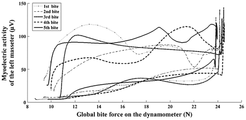
In order to compare the activities of the two subjects, they were asked to bite on a calibrated dynamometer placed between the incisors. The global force to deliver at the maximal stretch was equal to 25 N. The muscle activity was reproducible during biting (Figure ): it was first quite constantly low and suddenly increased at the maximal stretch of the dynamometer. While returning at the initial state, the muscle activity was higher than during biting. Sudden picks occurred underlying the difficulty to control the opening movement. The area between the curves was nearly the same whatever the cycle showing that the muscular demand was nearly the same during the five successive exercises. The maximal activity was in the same order of magnitude for the two subjects and the curve profiles were similar. These patterns were similar for the AT and M muscles. The activity of the SCM muscles was constantly low (about 10 μV, Table ) while the mouth was open. It is consistent with the function of the SCM aimed to stabilize the mandible when open.
4. Conclusions
The present study underlined the difficulty to obtain willing maximal clench. In this context, a method to measure jaw muscle activity during a sub-maximal and controlled biting task is proposed. The evolution of the myoelectric activities as a function of the global force delivered on the dynamometer provides new insights of the behavior of the masticatory muscles. These results give also promising data to improve the subject-specific numerical models of the cranio-maxillofacial area, either as inputs or as validation data.
References
- Castroflorio T, Falla D, Tartaglia GM, Sforza C, Deregibus A. 2012. Myoelectric manifestations of jaw elevator muscle fatigue and recovery in healthy and TMD subjects. JOR. 39:648–658.
- Crawford SR, Burden AM, Yates JM, Zioupos P, Winwood K. 2015. Can masticatory electromyography be normalised to submaximal bite force? JOR. 42:323–330.
- Giannasi LC, Matsui MY, Politti F, Batista SRF, Caldas BF, Amorim JBO, De Oliveira LVF, Santos Oliveira C, Fernandes Gomes M. 2014. Test–retest reliability of electromyographic variables of masseter and temporal muscles in patients with cerebral palsy. Arch Oral Biol. 59:1352–1358.
- Mappelli A, Zanandréa Machado BC, Dantas Giglio L, Sforza C, de Felicio CM. 2016. Reorganization of muscle activity in patients with chronic temporo mandibular disorders. Arch Oral Biol. 72:164–171.

