
1. Introduction
One of the crucial tasks in experimental biomechanics is the measurement of displacement and strain fields in the bone tissues. With the advancements of the computational methods, Digital Volume Correlation (DVC) has emerged as an innovative and elite method for full volume displacement and strain measurements in the biomechanics field (Palanca et al., Citation2015). However, bone tissue remains a heterogeneous material and has complex behaviour due to its microstructure and density characteristics variation even for a simple loading state like a compression test. The discontinuities or crack initiation appears under loading which can lead to damage or fracture of the specimen. We need to improve the knowledge of crack apparition and behaviour but classical DVC approaches are not able to retrieve mechanical fields close to the discontinuity.
Considering this, in the present work, we developed a Heaviside based DVC method to identify the discontinuity fields and the presence of cracks using the X-ray tomography images of the cancellous bone tissues under compression testing. The work presented here aims to address the objectives of (a) application of Heaviside based DVC for identifying the local displacement fields in cancellous bone tissues. (b) to evaluate equivalent strain fields and to compute fracture volume (FV/TV) parameters using classical DVC methods and H-DVC methods for comparison.
2. Methods
A bovine shinbone chosen with slaughter age of about six months. The bone was cleaned exhaustively with water and excess flesh and tissues were removed. The samples first roughly cut with a handsaw. Then a cancellous bone specimen extracted from the proximal epiphysis region of the bovine tibia with dimensions of 9.4*8.6*10.5mm3. Samples were stored at −20 °C until the chemical cleaning and testing.
2.1. Heaviside digital volume correlation (H-DVC)
Heaviside DVC (Valle et al. Citation2018) was adapted to measure displacement fields and to detect the presence of cracks in the cancellous bone. To ensure the fast computation of the volumes, the process has been implemented in Massively Parallelized Computation (Valle et al. Citation2018) giving 100,000 data to be analysed at the same time.
2.2. Experimental procedure
The X-ray tomography was carried out in Pprime Institute with an Ultratom scanner (RX Solutions, France) with a Hamatsu X-ray source (150 kV, 75 W) and a Paxscan Varian at panel imager (1920*1536 pixels with a pixel size 127 µm). The scans were performed at a tube voltage of 45 kV and a current of 222 µA, with a frame rate of eight images per second and an averaging with ten images for each projection. A total time of 45-50 minutes needed for acquiring the images for each scan stage.
To perform mechanical testing, we used a specific compression testing setup () built with a PMMA tube and instrumented with load sensors at the bottom to monitor the load variations. For each loading step, a set of 1155 projections are recorded during rotation of the sample, step by step, over 360°. The rotation was under loading without removing the load for relaxation.
A small preload (10 N) was first applied to ensure good end contact prior to testing and the initial scan was then step wise compressed at six loading stages until its ultimate apparent strength and final fracture state respectively. The 3 D reconstruction of the samples performed with 20 µm voxel size ().
Figure 1. X-ray tomography device with in situ custom made compression testing rig setup with the cancellous bone specimen. Arrow (red) indicates the direction of load application.
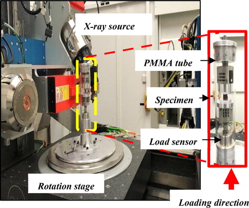
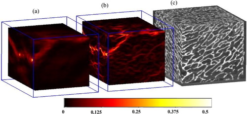
Figure 2. Equivalent strain distribution results of the cancellous bone sample (for final loading stage). (a) using classical DVC, (b) using H DVC, (c) 3 D reconstruction of the sample. White arrows indicate the micro-cracks.
3. Results and discussion
Displacement fields (u,v,w) and equivalent strain distribution (Valle et al. Citation2018) were calculated at different loading conditions. All the calculations were made with a subset size of 48*48*48 voxels. The computation used one data for every four voxels given that the final resolution of 1.8 million data for each volume. represents the results for the equivalent strain map for the final load state (step-6) to illustrate the discontinuity and strain localization obtained using classical DVC and H-DVC. The discontinuities (indicated by white arrows in ) were observed both near and far from the crack with variation in the gradient values throughout the boundaries in all directions using H-DVC. However, when a classical DVC used, only the major crack is visible but not the micro cracks.
From the strain maps, we evaluated the fracture volume to total volume ratio (FV/TV) for a threshold of 10percent of equivalent strain, which corresponds to 16 µm jump value as shown in . For the same specimen with the same voxel size, H-DVC revealed a fracture volume two times better than that obtained from DVC method (which was observed for different threshold levels of equivalent strain).
Table 1. Fracture parameter (FV/TV) evaluated using DVC and H-DVC.
Owing to the above results, the present study has a few limitations. Only one sample, with six loading steps, was tested in the present study. We need to perform more experiments to better understand the fracture mechanisms and to correlate the fracture parameters with the bone porosity and morphology.
4. Conclusions
The H-DVC approach was applied successfully for the measurement of local strain and localization of micro cracks in bone tissues. These results have confirmed better performances of the proposed H-DVC method and it could precisely detect discontinuities in all directions. H-DVC retrieved the fracture volume (FV/TV) better than compared to classical DVC. With this approach, it is now possible to localize the initiation of micro-cracks and measure strain fields until the final fracture. Future work is mandatory to distinctly, understand in situ fracture mechanics in bone tissue at µ-scale and to establish a relation between density and fracture.
References
- Palanca M, Tozzi G, Cristofolini L, Viceconti M, Dall’Ara E. 2015. Three-dimensional local measurements of bone strain and displacement: comparison of three digital volume correlation approaches. J Biomech Eng. 137:071006.
- Valle V, Bokam P, Germaneau A, Hedan S. 2018. New development of digital volume correlation for the study of fractured materials. Exp Mech. 59:1–15.