
1. Introduction
Most of ray-finned fish (Actinopterygii) use suction feeding to capture their prey. The rapid expansion of their mouth generates a high velocity water flow (e.g. Muller and Osse Citation1984), which leads the prey inside the buccal cavity. In spite of the prominent role of hydrodynamics to catch food successfully in suction feeding (Day et al. Citation2015) difficult optical access to the buccal cavity makes it still poorly understood. To go beyond computational simulations (Provini and Van Wassenbergh Citation2018) and overcome the technical constraints faced in situations without optical access, we combined two X-ray based methods to study suction feeding in Carp (Cyprinus carpio).
We used a biplanar X-ray system to obtain a 3D animation of the head bones, using X-Ray Reconstruction of Moving Morphology (XROMM) (Brainerd et al. Citation2010). Simultaneously, we optimised the technique of 3D X-ray particle tracking (Seeger et al. Citation2001), to visualize intra-oral flow dynamics using neutrally-buoyant radio-opaque particles. The combination of these two techniques allowed us to quantify the water flow involved during suction feeding and to correlate it to the 3D kinematics of the head bones in vivo.
2. Methods
2.1. X-Ray reconstruction of moving morphology
We performed the experiment at the University of Antwerp, where two synchronized and calibrated X-ray sources were associated with high-speed video cameras to film two individuals of Carps during suction feeding (N = 4 sequences for each individual). Prior to the experiment, the carps were implanted with 0.35 mm diameter radio-opaque markers on the upper jaw, lower jaw, hyoid, suspensorium, branchial arches, operculum, neurocranium and cleithrum. The tracking of these markers on each frame of each video, associated with the 3D model of the implanted bones, provided an animation (in Autodesk Maya) of the implanted bones and the six-degree of freedom kinematic data.
The experiments were ethically approved by the University of Antwerp (ECD-2017-22).
2.2. 3D X-ray particle tracking (3D XPT)
During the X-ray video acquisition, the food was surrounded by radio-opaque particles. We specifically designed small, approximately neutrally-buoyant radio-opaque particles composed of 1.4 mm diameter polystyrene foam spheres with an insert of an X-ray absorbing metal marker. A food item was also implanted with radio-opaque metal markers.
The tracking of these markers allowed us to reconstruct the 3D trajectory of the water tracers and the food particle outside and inside the buccal cavity.
3. Results and discussion
For the first time we were able to visualize and quantify the 3D intra-oral water flow () associated with the motions of the head bones during suction feeding ().
Figure 1. Trajectory of the water and food particles on a lateral view (a) and a dorsoventral view (b) for a representative sequence.
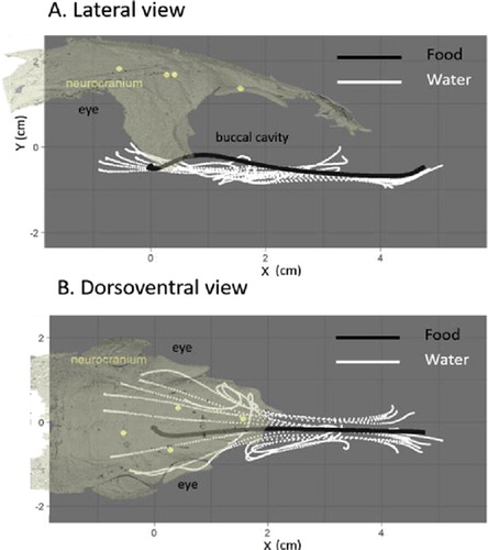
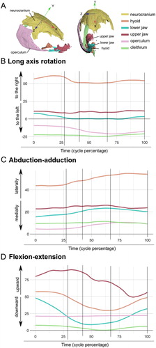
Figure 2. Location of the bones and neurocranium axis (a), Rotations in degrees of the bones along the X, Y, and Z axes (b, c, d) for a representative sequence.
During the first strike, we found the same pattern in each trial. The flow of water entering the mouth follows a different path in comparison to the food item trajectory.
On the lateral view, the water tracers display a homogenous linear ventral trajectory (). In contrast, the food particles show a dorsal trajectory, especially at the back of the buccal cavity. On the dorsoventral view, the water flow divides into two symetrical paths, from the entrance of the mouth to the location of the gill rackers. The food particles, however, keep a straight trajectory () from the entrance of the mouth to the location of the pharyngeal jaws, corresponding to the entrance of the oesophagus.
The 3D kinematics analysis of the head bones relative to the neurocranium, revealed an increase of the volume of the buccal cavity during the first strike of suction feeding, mostly due to the opening of the mouth (upward motion of the upper jaw and downward motion of the lower jaw, ). The downward motion of the hyoid participates to guiding the water and the food in a ventral direction. The opening of the opercula (), contributes to the lateral motion of the particles after they entered the buccal cavity.
4. Conclusions
For the first time, we were able to image the 3D trajectory of water inside the buccal cavity in vivo. Being able to visualize and quantify these previously concealed motions will open new questions and help explain the morphology, behavior, and thus the evolution of ray-finned fish, one of the most extraordinary animal radiations on earth.
Acknowledgements
We thank Peter Aerts for the access to the facility at the University of Antwerp, Jonathan Brecko for the CT-scan acquisition at the Royal Belgian Institute of Natural Science, as well as Anthony Herrel for helpful discussions.
References
- Brainerd EL, Baier DB, Gatesy SM, Hedrick TL, Metzger KA, Gilbert SL, Crisco JJ. 2010. X-ray reconstruction of moving morphology (XROMM): precision, accuracy and applications in comparative biomechanics research. J Exp Zool Part A. 313A:262–279.
- Day SW, Higham TE, Holzman R, Van Wassenbergh S. 2015. Morphology, kinematics, and dynamics: the mechanics of suction feeding in fishes. Integr Comp Biol. 55(1):21–35.
- Muller M, Osse J. 1984. Hydrodynamics of suction feeding in fish. Trans Zool Soc Lond. 37(2):51–135.
- Provini P, Van Wassenbergh S. 2018. Hydrodynamic performance of suction feeding is virtually unaffected by variation in the shape of the posterior region of the pharynx in fish. R Soc Open Sci. 5(9):181249.
- Seeger A, Affeld K, Goubergrits L, Kertzscher U, Wellnhofer E. 2001. X-ray-based assessment of the three-dimensional velocity of the liquid phase in a bubble column. Exp Fluids. 31(2):193–201.