
Keywords:
1. Introduction
Cranial cruciate ligament (CCL) disease is the most common cause of pelvic limb lameness in dogs. Complete rupture must be surgically treated.
In the past, intra-articular ligament prostheses have been implanted in dogs with mitigate results. Prosthesis tended to fail upon time (Legnani et al. Citation2010) and to increase progression of osteoarthritis. Up to date surgical techniques rely mostly on tibial osteotomies. They focus on modifying the biomechanical function of the knee to avoid cranial tibial thrust instead of replacing the injured ligament (Cabrera et al. Citation2008).
In human surgery intra-articular techniques are actually a gold standard (Hospodar and Miller Citation2009) because of the spreading of a mini-invasive approach with arthroscopy for implantation of graft or new type of ligaments (Purchase et al. Citation2007; Fedorová et al. Citation2015). It appears that the weakest link of the intra-articular CCL repair is the bone fixation of the ligament. Interference screws allow the best fixation. In veterinary medicine, this technique is new and not well known. The aim of this study was to test biomechanical properties (Kim et al. Citation2012) of a repair of CCL rupture with an ultra-high molecular weight polyethylene (UHMWPE) prosthesis (Fmax 8000 N) (Novalig, Novetech, Monaco) fixed with titanium interference screws on ex-vivo dog knee joints. We hypothesized that the resistance to failure of theses prosthesis would be superior to normal walking and trotting conditions during immediate postoperative period. The purpose of this specific study was to assess the number of screws needed to achieve this resistance.
2. Methods
17 hindlimbs from seven adult dogs between 25 and 35 kg were taken. Dogs were of similar size and died from reasons unrelated to this study. Knees were dissected to let intact only tibia, CCL and femur. Each extremity of the bones was resin-sealed in two supports. Tensile tests of the CCL were performed using a traction system (AGS-X Shimadzu, Japan) with in a pretest of 20 mm/s traction until the strength reached 10 N, straightening the system. The final test consisted in a 1 mm/sec traction to failure.
Failure was considered if the strength dropped to a value less than 10% of maximum strength or if displacement reached 25 mm and initial failure was considered at the first decrease of tensile strength during the test.
Once CCL ruptured, prosthetic ligament was implanted and fixed with 2 interference screws (1 tibial oblique and 1 femoral oblique) and tested in the same way to failure. The screws were 4.5 mm wide and the tunnel diameter was 3,6mm wide. Each limb was re-implanted and re-tested with a new prosthetic ligament fixed with 3 interference screws (1 tibial oblique + 1 tibial transverse and 1 femoral oblique). The 4.5 mm screw was removed and replaced by a 5 mm screw in the same tunnel to avoid weakening from the previous test.
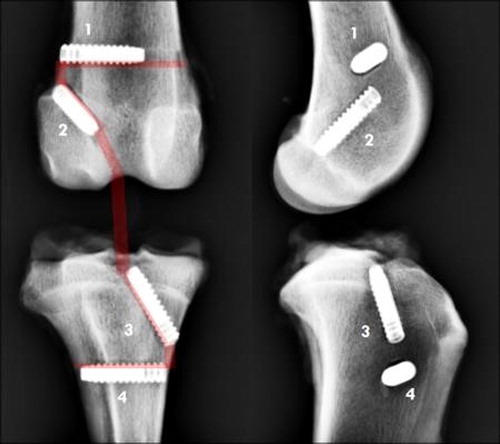
Finally, the last tensile test was performed with a 4 screws fixation of the ligament ().
Figure 1. Cranial and lateral X Rays of implanted knees after traction: The UHMWPE prosthesis is fixed with 4 screws. 1: transverse femoral screw, 2: oblique femoral screw, 3: oblique tibial screw, 4: transverse tibial screw.
Initial failure, failure mode, maximum strength and stiffness were measured and recorded for analysis. Results of each fixation mode were compared to the natural ligament values and between each other.
Results were statistically analyzed using R.
3. Results and discussion
The failure mode was the same for every ligament tested. Natural ligaments were torn on the tibial insertion; ligaments were slipping between the bone and the interference screw during or at the end of the tests.
An ANOVA test revealed a significant difference between 2 screws and 3 screws model compared to natural ligament (p-value < 0.001 in both case) and between 2-screw and 4-screw fixation (p-value = 0.007) 4-screws fixation on the other hand was not statistically different from natural ligament. Values of Initial failure was superior to the maximum estimated value measured during the walk and the trot (for both natural ligament (582 N, SD: 276 N, p-value < 0.001) and 4-screw fixation (347 N, SD: 54 N, p-value = 0.0181) according to a Student univariant t-test, but not for the 4-screw fixation at gallop.
Stiffnesses were similar for all three type of implantation and statistically different from the natural ligament (ANOVA test, p-value < 0.001 for each test). Their values stay quite similar () Moreover, these static tests have shown us that the 2 screws and 3 screws method fail to recreate natural ligament maximum resistance. Because of the slipping of the ligament between bone and the oblique tibial screw in the 2 screws test and the slipping against the oblique femur screw in the 3 screws test. The fixation of the ligament could be the weakest for the oblique screw and the strongest for the transverse one. Oblique and transverse fixations could be stronger in the femur than in the tibia. 4-screw fixation has presented a large standard deviation may be depending the quality of the fixation for the transverse femoral screws.
Table 1. Results, * relates significant difference to sound CCL;° relates significant difference to 4 screws.
Stiffness of this prosthesis is statistically different from the natural ligaments, but the values are of the same magnitude. This could be deemed acceptable as obtaining the exact modulus of a natural ligament with a prosthesis is not the objective.
4. Conclusions
This study shows us that the fixation of the ligament with 4 screws is stronger than with 3 screws, itself stronger than 2 screws. Transverse fixations are stronger than oblique fixations. Fixations in the femur are stronger than those in the tibia but the strength is variable.
UHMWPE intra-articular ligament fixed with 4 interference screws is proved to be able to withstand normal walking and trotting condition in the immediate postoperative period.
This technique could be reconsidered as an option for treatment of ruptured CCL in large dogs.
Further biomechanical fatigue analysis of this prosthetic ligament with these fixations will be needed to validate the efficiency of the bone fixation of the ligament during simulation of locomotion during the healing of conjunctive and bone tissues.
References
- Cabrera SY, Owen TJ, Mueller MG, Kass PH. 2008. Comparison of tibial plateau angles in dogs with unilateral versus bilateral cranial cruciate ligament rupture: 150 cases (2000–2006). J Am Vet Med Assoc. 232(6):889–892.
- Fedorová P, Srnec R, Pěnčík J, Dvořák M, Krbec M, Nečas A. 2015. [Intra-articular reinforcement of a partially torn anterior cruciate ligament (ACL) using newly developed UHMWPE biomaterial in combination with Hexalon ACL/PCL screws: ex-vivo mechanical testing of an animal knee model]. Acta Chir Orthop Traumatol Cech. 82(3):222–228. [in Czech]
- Hospodar SJ, Miller MD. 2009. Controversies in ACL reconstruction: bone-patellar tendon-bone anterior cruciate ligament reconstruction remains the gold standard. Sports Med Arthrosc Rev. 17(4):242–246.
- Kim JY, Hayashi K, Garcia TC, Kim S-Y, Entwistle R, Kapatkin AS, Stover SM. 2012. Biomechanical evaluation of screw-in femoral implant in cementless total hip system. Vet Surg. 41(1):94–102.
- Legnani C, Ventura A, Terzaghi C, Borgo E, Albisetti W. 2010. Anterior cruciate ligament reconstruction with synthetic grafts. A review of literature. Int Orthopaed (Sicot). 34(4):465–471.
- Purchase R, Mason R, Hsu V, Rogers K, Gaughan J, Torg J. 2007. Fourteen-year prospective results of a high-density polyethylene prosthetic anterior cruciate ligament reconstruction. J Long Term Eff Med Implant. 17(1):13–19.