
1. Clinical context and objective
1.1. Diastology in heart failure
Heart failure is the cardiomyopathy that will have the greatest impact on the lives of French in the coming decades. It is a cardiac disease that affects the ability of the heart to meet the oxygen needs of the organs. It involves more than 1 M people in France, and its prevalence will double by 2028. Heart failure can be identified by the reduced ability of the heart to fill with blood (diastolic dysfunction) and/or to empty (systolic dysfunction). Although systolic dysfunction is easily diagnosed, the diagnosis of diastolic dysfunction (diastology) can be challenging. The accessibility of echocardiography, and its ability to provide non-invasive information in real time, make ultrasound the essential technique for evaluating diastolic function of the left ventricle. However, the diagnosis of diastolic dysfunction often remains unclear since the recommended echographic indices may lead to discordant conclusions. As intracardiac blood flow is very sensitive to changes of the myocardium, we hypothesized that a more in-depth examination of the intraventricular blood flow by echocardiographic color Doppler could improve diastology in patients with heart failure.
1.2. Diastolic dysfunction and the intracardiac vortex
During diastole, the mitral valve forces the left intraventricular flow to create a vortex. This vortex naturally redirects blood flow to the left ventricular outflow tract. In healthy subjects, it thus helps the transition from filling to ejection. When the filling is impaired (diastolic dysfunction), a change in blood flow occurs, with a significant impact on the intraventricular vortex. According to the recent literature, it is becoming manifest that the vortex properties are strongly related to the filling function. We present 3-D intraventricular Vector Flow Mapping (3-D iVFM) by echocardiographic color Doppler to decipher the dynamics of the 3-D vortex ring that forms in the left ventricle during diastole.
2. Methods
2.1. Intracardiac velocimetry by iVFM
Intracardiac blood flow can be quantified noninvasively by cardiac magnetic resonance (CMR) or by echo-PIV (“echographic particle image velocimetry”). These techniques, however, cannot be used in a clinical routine. Garcia et al. therefore introduced 2D-iVFM (“intraventricular vector flow mapping”) by conventional echocardiography (Garcia et al. Citation2010). This technique has the advantage of being 100% clinicallycompliant since it uses 2-D color Doppler and nothing more. Due to its commercial availability by Hitachi (Tanaka et al. Citation2015), this innovative modality has been the subject of a number of clinical investigations. We have recently published an updated version of the 2DiVFM (Assi et al. Citation2017), which is much more powerful than the previous form adopted by Hitachi. This new version, based on a robust regularization approach, has the advantage of being transferable to 3-D. Given the clinical success of ultrasound-derived 2D-iVFM, we propose the 3D-iVFM modality.
2.2. Development and validation of 3D-iVFM
To develop a robust and turnkey 3D-iVFM modality, we adopted a methodology similar to the one previously used for 2-D (Assi et al. Citation2017, Faurie et al. Citation2017). Note that color Doppler yields the velocity components projected along the ultrasonic beams, which provides very partial information of the flow. The goal of 3D-iVFM is to generate time-resolved 3-D vector fields from these scalar Doppler fields. Echocardiographic 3D-iVFM was generated from Doppler velocities acquired in the multi-dimensional (tri-plane, ) imaging modes available in the most recent GE Healthcare ultrasound machine and workstation (Vivid e95 and EchoPAC 4D). Doppler data were extracted prior to scan conversion (i.e., in a spherical grid) using EchoPAC and was post-processed offline with Matlab to create 3D-iVFM. We used an optimization strategy and adapted the recently published 2-D algorithm (Assi et al. Citation2017). To obtain a well-posed problem, the constraints in the cost function was related to mass conservation, together with endocardial velocity boundary conditions and smoothness.
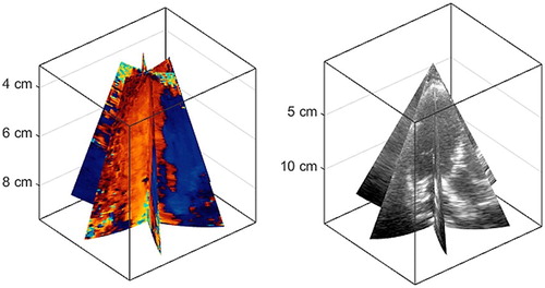
Figure 1. Triplane color Doppler in a left ventricle. Left panel: color Doppler. Right panel: B-mode imaging. The goal of 3D-iVFM is to reconstruct the intraventricular blood flow from these clinical data.
3. Results and discussion
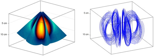
As a first validation step, the 3D-iVFM algorithm was tested in simulations of a divergence-free Hill’s vortex. Three planes of Doppler velocities were simulated (, left) to mimic multi-dimensional color Doppler, as used in clinics (see for a comparison). Different flow and ultrasound configurations (vortex radius, vorticity, Doppler grid and noise) were tested. Volumetric three-component velocity fields were reconstructed (, right) by minimizing the above-mentioned cost function in a least-squares sense. The averaged normalized root-mean-square errors were less than 10%. These in silico results tend to show that it is possible to recover a full 3-D intracardiac velocity field from scalar Doppler fields. Although these findings are promising, they are limited to an axisymmetric vortex under ideal conditions. We will thus optimize and validate 3D-iVFM by using a realistic 3-D dynamic model of intraventricular flow based on CFD (Chnafa et al. Citation2014). Echocardiographic 3D-iVFM will be also compared against CMR-based blood flow velocimetry in volunteers (Faurie et al. Citation2017).
Figure 2. Simulated triplane color Doppler in a Hill’s spherical vortex. Left panel: simulated color Doppler. Right panel: Streamlines of the Hill’s vortex recovered by 3D-iVFM.
4. Conclusions
If successful, we anticipate that 3D-iVFM will be the only fast tool for a thorough clinical analysis of the 3D intraventricular vortex. Fast and 100% clinical, 3DiVFM could offer new echocardiographic insights into left ventricular function, especially for diastology
Acknowledgments
This work was supported by the LABEX CeLyA (ANR-10-LABX-0060) of Université de Lyon, within the program « Investissements d’Avenir » (ANR-16IDEX-0005) operated by the French National Research Agency (ANR).
References
- Assi K.C, Gay E, Chnafa C, Mendez S, Nicoud F, Abascal J.F.P.J, Lantelme P, Tournoux F, Garcia D. 2017. Intraventricular vector flow mapping-a Doppler-based regularized problem with automatic model selection. Phys Med Biol. 62 (17):7131–7147.
- Chnafa C, Mendez S, Nicoud F. 2014. Imagebased large-eddy simulation in a realistic left heart. Comput Fluids. 94:173–187.
- Faurie J, Baudet M, Assi K.C, Auger D, Gilbert G, Tournoux F, Garcia D. 2017. Intracardiac vortex dynamics by high-frame-rate Doppler vortography – in vivo comparison with vector flow mapping and 4-D flow MRI. IEEE Trans Ultrason, Ferroelect, Freq Contr. 64 (2):424–432.
- Garcia D, del Álamo J.C, Tanné D, Yotti R, Cortina C, Bertrand E, Antoranz J.C, Pérez-David E, Rieu R, Fernández-Avilés F, et al. 2010. Two-dimensional intraventricular flow mapping by digital processing conventional color-Doppler echocardiography images. IEEE Trans Med Imaging. 29 (10):1701–1713.
- Tanaka T, Asami R, Kawabata K, Itatani E.K, Uejima T, Nishiyama T, Okada T. 2015. Intracardiac VFM technique using diagnostic ultrasound system. Hitachi Rev. 64(8):488–492.