
1. Introduction
Large animal model such as goat, pigs, sheep are widely used for preclinical research in human medecine to test new spinal implants and surgical procedures. Among those models, the goat is particulary interesting because of her similarity with the anatomy of the human spine and the rapid growth observed in goats between the age of 6 weeks and 8 months which mimics the human’s growth.
Moreover, goats are readily available, inexpensive, easy to handle and well accepted as an ethical animal model. Therefore, precise morphometric data of goat spine are mandatory to improve quality of implant fixation and implantation and thus to avoid loss of animal (Braun et al. Citation2003; Braun and Akyuz Citation2005).
The aim of this study is to provide quantitative reference values and a complete comprehension of the pedicule morphology and angulation of the caprine spine using CT scan.
2. Methods
Eleven adult alpine goats without any history or clinical signs related to spinal diseases were included in this study. All goats came from the same batch. Mean body weight was 42, 5 kg.
The goats were euthanized for other experimental studies unrelated to this study. Goats were positioned in dorsal recumbency in a perpendicular position of the spine relative to the radiographic beam. CT scans were performed on a multi-detector-row helical CT unit (General Electric’s ® BRIGHTSPEED 16 ELITE). The technical settings were 120 kV and 150 mA, and the pitch of 1 slice thickness was 0.625 mm. The data were reconstructed to a transverse, sagittal and frontal image series with slice thickness ranging between 0.2 and 0.8 mm using a high-frequency image reconstruction algorithm (Osirix Imaging Software®).
For each vertebrae, three parameters were obtained from the sagittal images (), while seven parameters were measured from the transverse images (). The multiples parameters were measured as described in human and veterinary literature (McLain et al. Citation2002; Mageed et al. Citation2013).
Figure 1. Parameters measured on each vertebrae on the sagittal CT image: vertebral body height at dorsal border (VBHd), vertebral body height at ventral border (VBHv) and disc thickness (DT).
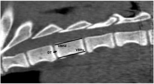
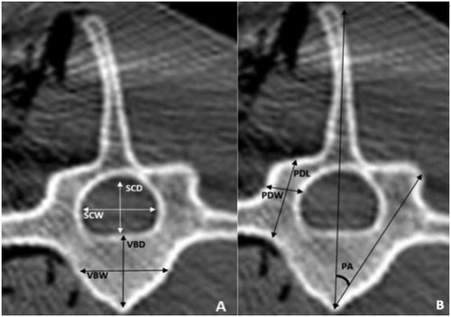
Figure 2. Parameters measured on each vertebrae on the transverse CT image. Measured verterbral body parameters (A) were: vertebral body width (VBW), vertebral body depth (VBD), Spinal canal width (SCW) spinal canal depth (SCD). Measured pedicle parameters (B) were: Pedicle length (PDL), pedicle width (PDW) and pedicle axis angle.
Descriptive data are presented as means and standard deviation (SD). Assessment of intra-observer reliability was calculated by randomly selecting three goats in which measurements were performed four times by the same operator. A coefficient of variation (CV) was used to assess the reliability. MannWhitney/Two-tailed test was used to determine differences between the vertebral levels for each parameter. Statistical analyses were performed with a dedicated software program (Microsoft Excel 2010, XLSTAT ®). The level of significance was set at P < 0.05 and CV< 5% was considered acceptable.
3. Results and discussion
Eleven adults 36 months’ old alpine goats were included in this study and a sample of 198 vertebrae was measured. CT images revealed that all goats had six Lumbar vertebrae and 13 thoracic vertebrae (the first thoracic vertebra was not measured in this study). Assessment of intra-observer reliability revealed a high level of reliability. and represent the mean and standard deviation of the CT measurements in thoracolumbar spines of the eleven goats.
Table 1. Mean and standard deviation of the CT measurement dimensions related to intervertebral disc, vertebral bodies, spinal canal.
Table 2. Mean and standard deviation of CT measurements dimensions of the pedicle.
Vertebral bodies and the spinal canal were wider than they were deep, mostly evident in the lumbar region. The intervertebral discs were as much as 65.7% thicker in the lumbar than in the thoracic spine. The pedicles were longer than wide over the thoracic and lumbar spine. The insertion angles were approximately 30° for T2-T4 segment, 25° for T5T6 segment, 23° for T6 to T11 segment, 20° for T11 to L3, 25° for L4 and 30° for L5 and L6.
4. Conclusions
This study provided a comprehensive quantitative database of the normal goat thoracolumbar spine. This descriptive information can be used to help determine if the goat spine can be a good representative model for preclinical test. The mean length of the adult human lumbar vertebrae is more or less 25 mm and the mean width is 50 mm (Busscher et al. Citation2010). Thus, compare to the human one, the lumbar goat vertebrae is longer but narrower. This difference could be related to the quadruped position of the goat. When selecting a large animal model, for spine study, this difference should be keep in mind. This study provided also important information to select the size of implant used in goat. Based on current recommendations, the ideal pedicle screw size should be 80% or less than the size of the pedicle. Therefore, the current study should be used as a guide to select the correct screw size when using goat as a model for transpedicular fixation research. For example, pedicular screws used in the lumbar vertebra of an adult goat should not be larger than 5 mm. Finally, this study, could be used as a guided to improved implantation of pedicle screw in goat.
References
- Braun JT, Akyuz E. 2005. Prediction of curve progression in a goat scoliosis model. J Spinal Disord Tech. 18(3):272–276.
- Braun JT, Ogilvie JW, Akyuz E, Brodke DS, Bachus KN, Stefko RM. 2003. Experimental scoliosis in an immature goat model: a method that creates idiopathic-type deformity with minimal violation of the spinal elements along the curve. Spine. 28(19):2198–2203.
- Busscher I, Ploegmakers JJW, Verkerke GJ, Veldhuizen AG. 2010. Comparative anatomical dimensions of the complete human and porcine spine. Eur Spine J. 19(7):1104–1114.
- Mageed M, Berner D, Jülke H, Hohaus C, Brehm W, Gerlach K. 2013. Morphometrical dimensions of the sheep thoracolumbar vertebrae as seen on digitised CT images. Lab Anim Res. 29(3):138–147.
- McLain RF, Yerby SA, Moseley TA. 2002. Comparative morphometry of L4 vertebrae: comparison of large animal models for the human lumbar spine. Spine. 27:E200–206.