
1. Introduction
The development of new percutaneous techniques using a balloon inflation for the reduction and cement for the stabilisation for the treatment of tibial plateau fractures (TPF) are promising (Vendeuvre et al. Citation2013; Belaid et al.Citation2018). The biomechanical changes brought by the cement in periarticular fracture are unknown. The measurement of the displacements and strains in the bone and in the cement interface are needed to understand the biomechanical modification that implies the presence of cement in bone at the time of the fracture and after consolidation.
Therefore, the objective of this study was to provide elements of understanding of the bone behaviour on a TPF treated with tuberoplasty and to define the modifications brought by the presence of this cement in the bone. For measurement of the displacement and strain fields’ optical measurement technique was used with the help of which crack initiation and fracture were examined.
2. Methods
2.1. Specimen preparation
An in vitro animal experimentation was conducted in the present study. A metaphyseal region with a good amount of cancellous bone was chosen. The cortico-cancellous fracture protocol reproduced here for preparation and injection of cement. Two samples were prepared and tested, one with a balloon inflation in the hole and the other without any balloon inflation (using a drill bit to enlarge the hole). The first step for the sample preparation was to dig a hole with a square tip. In one group, a balloon was inflated (2 ml and 500PSI of pressure) then the created cavity was filled with PMMA cement at the exact time and texture as it is used in the surgical treatment of a tibial plateau fracture. In the other group (without balloon inflation), the cavity created was further enlarged using a drill bit of 6 mm and the cavity filled with cement. Later, cubic samples were extracted using a mechanical saw with the dimension of (15*15*10mm3).
2.2. Experiments
A uniaxial compression testing was performed using an Instron universal tensile machine (UTM, 5 kN). The compression tests were conducted under a controlled displacement at a strain rate of 1 mm/min until a crack apparition was observed in the samples. The entire experimentation was carried in the presence of a CMOS camera (1280 × 1024 pixels), which was used to record one image every two seconds. For the optical full field measurements, the Heaviside Digital Image Correlation (H-DIC) method (Valle et al. Citation2015) was used to measure the displacement fields. From these displacement fields the Von Mises equivalent strain maps plotted to observe the potential discontinuity (in particular at the interface between the cement and the bone) for the two samples.
3. Results and discussion
The typical load/displacement curve for the sample with balloon inflation is shown in below.
Figure 1. Load displacement curve for the sample with balloon inflation, highlighting the crack initiation and final fracture (marked red symbols) with equivalent strain.
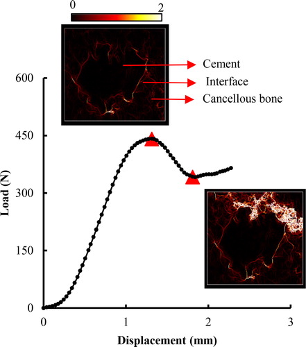
The displacement fields were obtained based upon the degree of similarity between the initial and the deformed state (Valle et al. Citation2015). From the Von Mises equivalent strain maps () the crack evolution at different loading conditions can be observed. It is worth nothing that, just before the fracture (pre-fracture zone) multiple micro cracks were observed. The micro cracks and the discontinuity began very early at the interface between the bone and the cement. Even when the global behaviour was linear, micro cracks were observed and more deformation around the bone and the cement even though there was no strain inside the cement.
The H-DIC method also permits the local analysis of Von Mises equivalent strain in different localisation by selecting a region of interest (ROI) inside the specimen (as shown in ). We compared the strain fields for both the specimens (with balloon inflation and without balloon inflation) as shown in below.
Figure 2. Strain distribution in different zones for the sample with balloon inflation and the sample without balloon inflation.
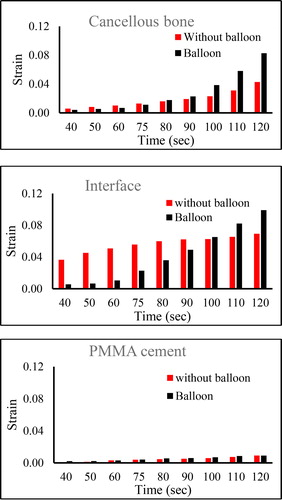
shows Von Mises equivalent strain evolution at three different zones (bone, interface, cement) for the two samples. In the cancellous bone, the strain localisation is similar for both the cases before the fracture, and after the fracture. We can observe that the strain fields in the sample with balloon inflation slightly increased after the fracture.
In the cement, for both specimens, the strain is almost the same with a slight increase after the fracture. At the interface, in the specimen without balloon inflation, the strains were very high from the beginning and were almost constant with a slight increase after the fracture. However, in the specimen with balloon inflation the strain proportionally increased as the load increase even after the fracture.
4. Conclusions
In this work, the compression testing identical to real life loading conditions were performed to study the bone mechanical behaviour when filled with PMMA cement. The strain parameters for the two samples (with balloon inflation and without balloon) were studied for a better understanding of the displacement interaction between the bone and the cement. The use of a novel optical correlation process (Valle et al Citation2015) highlighted the biomechanical role of the cement inside the bone. This work demonstrated that there is a discontinuity in load transfer between the bone and the cement. The cement behaves like a rigid body inside the cancellous bone. If the cement provides a good reduction and a primary stabilisation permitting the patient to undergo rehabilitation with active and passive mobilization. Our work demonstrates that no weight bearing should be authorised while the cortical bone is not consolidated.
References
- Belaid D, Vendeuvre T, Bouchoucha A, Brémand F, Brèque C, Rigoard P, Germaneau A. 2018. Utility of cement injection to stabilize split-depression tibial plateau fracture by minimally invasive methods: a finite element analysis. Clin Biomech (Bristol, Avon). 56:27–35.
- Valle V, Hedan S, Cosenza P, Fauchille AL, Berdjane M. 2015. Digital image correlation development for the study of materials including multiple crossing cracks. Exp Mech. 55(2):379–391.
- Vendeuvre T, Babusiaux D, Brèque C, Khiami F, Steiger V, Merienne J-F, Scepi M, Gayet LE. 2013. Tuberoplasty: minimally invasive osteosynthesis technique for tibial plateau fractures. Orthopaed Traumatol Surg Res. 99(4 Suppl):S267–S272.