
1. Introduction
Scoliosis is defined as tridimensionnal pathological deformation of the spine, and mainly appears at a young age (Boos and Aebi Citation2008). This leads to self-perpetuating aggravations, as the modified biomechanic of the spine worsen the deformations during the growth (Stokes et al. Citation2006).
Early treatment plan is highly recommended. It allows to use spine growth for the deformity correction. Orthopaedic treatment, using external brace, is the first line treatment. Then, surgical treatments, involving vertebral fusion or fusionless techniques, are needed for major deformities. All of them often compromise growth and mobility (Schwab et al. Citation2009).
Fusion-less surgery is considered to be the best treatment to preserve spine growth and, for a few years, spine mobility (Sheng et al. Citation2010).
This work, based on a preliminary study, focuses on a new device which allows growth and mobility of the spine. It is made out of polymer wires (polyetherketon, PEEK) and pedicular screws. Our work hypothesis is (1) the peek wires allow a greater spine mobility than titanium ones and (2) the overall mobility of peek-instrumented spines is close to an intact spine.
2. Methods
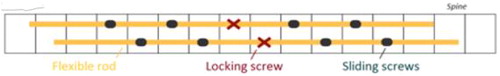
Pig spine is a well-fitted model for human scoliosis (Odent et al. Citation2011; Chun et al. Citation2015). Ten pig spines were used for this study. Three-point flexural tests were done using a vertical traction machine (Shimadzu AGS-X 10 kN) on healthy spines (S group). The spine were then implanted with 10 bicortical pedicle screws, in quincunx using on each side 4 sliding pedicle screws and 1 fixed pedicle screw at the centre of the construct. A 5.5 mm PEEK flexible rod (SP group) or a 5.5 mm titanium rod (ST group) was then inserted into the screws as described in the . A home-made aluminium frame was used to overcome the uneven shape of the pig spines (Korovessis et al. Citation2004) (). Pre-operative sequences were performed for defining the right operative sequence. Then, range of motion (ROM), neutral zone (NZ), overall stiffness (OS), flexion loading stiffness (FLS) and extension loading stiffness (ELS) were measured.
Figure 1. Implantation scheme of the spine. The spine were then implanted with 10 bicortical pedicle screws, in quincunx using on each side 4 sliding pedicle screws and 1 fixed pedicle screw at the centre of the construct. A 5.5 mm PEEK flexible rod (SP group) or a 5.5 mm titanium rod (ST group) was then inserted into the screws.
3. Results and discussion
The S and SP spines have similar results in every parameter. ST spines show a lower ROM, a lower NZ and higher OS, FLS and ELS ().
Table 1. Results, the p value relates to the S group.
Our results are similar to the preliminary work but other studies show that SP group are significantly different from the S group with intermediary results between S and ST group.
For the titanium group, the force limitation is reached for most of the trials. During the flexion phase, the amplitude limitation is reached for every group in all the trials.
4. Conclusions
This study shows the impact of the use of titanium rods devices for fusionless spine surgery. Thus, a new type of rods with intermediary properties is needed in order to lower the impact of surgery. PEEK devices show promising results as it respects the mobility of the spine. Nevertheless, stiffness of the spine implanted with PEEK rod do not differ from normal spine. Thus efficacy of those of construct could be questionable to corrected spinal deformity. This low stiffness could be related to the low force applied to the spine in our study but also to a too small size of the rod. Further studies need to be done, with different protocol limitations of amplitude of motion and vertical force.
Acknowledgements
The authors thank for their supportive work Quentin Blanc and the VetAgro Sup surgery team
References
- Boos N, Aebi M. 2008. Spinal disorders: fundamentals of diagnosis and treatment. Springer-Verlag.
- Stokes IA, Burwell RG, Dangerfield PH. 2006. Biomechanical spinal growth modulation and progressive adolescent scoliosis – a test of the ‘vicious cycle’ pathogenetic hypothesis: Summary of an electronic focus group debate of the IBSE. Scoliosis. 1(1):16.
- Schwab F, Patel A, Lafage V, Farcy JP. 2009. A porcine model for progressive thoracic scoliosis. Spine. 34(11):E397–E404.
- Sheng SR, Wang XY, Xu HZ, Zhu GQ, Zhou YF. 2010. Anatomy of large animal spines and its comparison to the human spine: a systematic review. Eur Spine J. 19(1):46–56.
- Odent T, Cachon T, Peultier B, Gournay J, Jolivet E, Elie C, Abdoul H, Viguier E. 2011. Porcine model of early onset scoliosis based on animal growth created with posterior mini-invasive spinal offset tethering: a preliminary report. Eur Spine J. 20(11):1869–1876.
- Chun K, Yang I, Kim N, Cho D. 2015. Effect of device rigidity and physiological loading on spinal kinematics after dynamic stabilization: an in-vitro biomechanical study. J Korean Neurosurg Soc. 58(5):412–418.
- Korovessis P, Papazisis Z, Koureas G, Lambiris E. 2004. Rigid, semirigid versus dynamic instrumentation for degenerative lumbar spinal stenosis: a correlative radiological and clinical analysis of short-term results. Spine. 29(7):735–742.