
 ?Mathematical formulae have been encoded as MathML and are displayed in this HTML version using MathJax in order to improve their display. Uncheck the box to turn MathJax off. This feature requires Javascript. Click on a formula to zoom.
1. Introduction
The quantitative assessment of cancellous bone tissues mechanical properties became an important task in the biomechanical field. Researchers worked intensively on the evaluation of mechanical properties of cancellous bone using experimental, optical measurement techniques and finite element method. However, as the structural property of bone is its fragility or in other words its resistance to fracture. It is therefore important to evaluate the fracture mechanics (FM) parameters like toughness, fracture energy to understand the fracture mechanics in the cancellous bones. Many researchers have worked on the fracture parameters of cortical bone in the past decade. On the other hand, one can observe that there are very few works done on fracture properties of cancellous bone reported due to their porous nature and lack of tools to extract the parameters (crack length, crack opening displacement COD) for evaluating the fracture properties. Therefore, the present study mainly focusses on evaluating the specific fracture energy (Gf) of the cancellous bone, integrating the wedge-splitting test (WST) and Digital Image Correlation (H-DIC) method. Two approaches were used to evaluate the fracture mechanic properties of the bone. The first method is based on the global approach, which was widely used in the literature and the second method is based on the local approach. In this local approach, the local fracture energy (Gi) during the course of the test was evaluated, which give access to local fracture mechanics and the influence of porosity and pore size on crack propagation.
2. Methods
A bovine shinbone chosen with slaughter age of about six months. The geometry of the WST sample was machined as shown in the (where the length (L) of 17 mm, thickness (B) of 17 mm and width (W) of 15 mm). An initial notch length (ao) of 3 mm grooved carefully. The tests conducted under displacement control at a strain rate of 0.008 mm/sec.
Figure 1. Representation of Wedge Splitting Test.
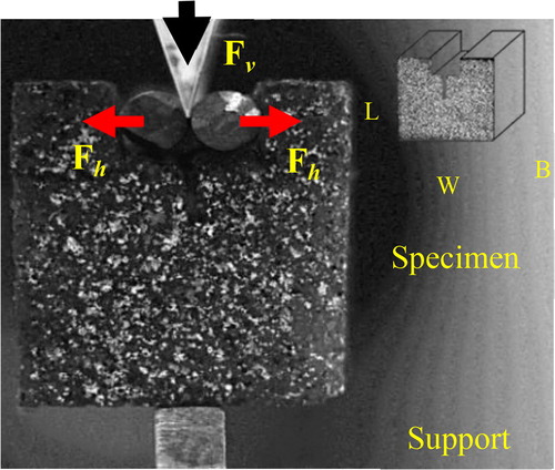
The principle of the wedge splitting method and specimen shapes considered similar as described by Tschegg (Citation1986). The wedge unit transforms the vertical load (Fv) from the compression test machine (under displacement control) into a higher magnitude horizontal force (Fh). This horizontal force Fh (EquationEquation (1)(1)
(1) ) leads to the crack initiation at the notch allowing the crack propagating through the specimen.
(1)
(1)
where, Fv is the vertical load applied by the test machine, 2α is the wedge angle (here α = 15 degrees) and μ is the coefficient of friction for the roller bearing.
The entire experimentation was carried in the presence of a CMOS camera (1280x1024 pixels) which was used to record the images for the optical full field measurements using Digital Image Correlation (HDIC) method (Valle et al. Citation2015) to retrieve the crack opening displacement and corresponding crack length. The image acquisition frequency was one image for every two seconds and 600 images were recorded during the entire test.
3. Results and discussion
3.1. Global fracture analysis
From the principle of WST, we know that load-COD curve contains all information that is necessary to characterize the fracture behaviour. In order to find the fracture energy required to open a crack of unit area, the total dissipated energy calculated from F-COD curve was divided with the fractured area. shows the load to crack opening displacement (COD) curve. Based on the measured load-COD curve, the specific fracture energy can be evaluated using EquationEquation (2)(2)
(2) below.
(2)
(2)
Figure 2. (a) Load COD curves obtained using the correlation process and (b) evolution of local Fracture energy.
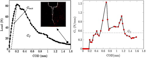
where, A is the ligament area, δ is the COD at the crack initiation i.e., at maximum load, δmax is the value of COD at the end of the test. The fracture energy (Gf) calculated from the above method found to be 0.53 N/mm.
3.2. Evaluation of local fracture energy (Gi)
The local fracture energy Gi can be defined as the instantaneous fracture energy released during each load step (as shown in EquationEquation (3)(3)
(3) ).
(3)
(3)
where n is the number of infinitesimal areas, f is the load value and Δα is the corresponding crack length extension. This evaluation can only be possible if the crack extension is accessible. As it is possible using the H-DIC method to measure the crack extension at each load step, shows the evolution of the local fracture energy in the course of the test. There is a sudden release of energy at the fracture zone followed by spikes (high and low) which was linked to the local porosity in the path of the crack propagation. The fracture energy released Gf can be evaluated from averaging all the local fracture energy (Gi) found to be 0.55 N/mm, which was near compared to the value obtained from EquationEquation (2)(2)
(2) . Evaluating the fracture energy using local approach has advantages as it takes into account the local mechanical aspects of the crack during entire course of propagation when compared to the global approach, which only takes crack initiation and final fracture values (, EquationEquation (2)
(2)
(2) ) for evaluating the fracture energy values. The evaluated values are slightly lower than available literature ((Tschegg et al. Citation2012) i.e., Gf = 3.32 ± 1.77N/mm).
Owing the above, there is a great deal of potentiality to perform more experiments using our method in near future with different variables (density, pore size, shape etc.) governing the fracture mechanics of the cancellous bone and in order to quantify the fracture properties.
4. Conclusions
The study set out to build an integrated methodology (wedge splitting test and digital image correlation) to evaluate the fracture energies of the cancellous bone tissue. In addition evaluating the local fracture energy gave more insight details. The possibility of evaluating the fracture parameters without knowing the actual properties of the material is another feature of the proposed approach. This aspect is interesting in studying the behaviour of the porous cancellous bone.
References
- Tschegg EK. 1986. Equipment and appropriate specimen shapes for tests to measure fracture values. Vienna, Austria: Austrian Patent Office; AT No. 390328.
- Tschegg EK, Celarek A, Fischerauer SF Stanzl-Tschegg S, Weinberg AM. 2012. Fracture properties of growth plate cartilage compared to cortical and trabecular bone in ovine femora. J Mech Behav Biomed Mater. 14:119–129.
- Valle V, Hedan S, Cosenza P, Fauchille A.L, Berdjane M. 2015. Digital image correlation development for the study of materials including multiple crossing cracks. Exp Mech. 55:389391.