
1. Introduction
Tendon rupture is a common injury in animals, which may lead to severe lameness and pain depending on severity and duration. Surgical repair consists in re-apposition of the tendon ends using various suture patterns. Biomechanically, suture techniques using three-loop pulley and modified three-loop pulley patterns offer higher tensile strength than locking-loop sutures (Moores et al. Citation2004; Putterman et al. Citation2019). Synthetic tendon implants have been used to repair chronic ruptures of the insertion of the gastrocnemius tendon in dogs. They seem to offer better mechanical resistance compared to conventional suturing techniques (Morton et al. Citation2015). In the present paper, the authors present the preliminary biomechanical results of a modification of the surgical technique published by Morton and colleagues in 2015.
2. Methods
2.1. Sample preparation protocol
Four hind limbs from 2 adult dogs between 35 and 45 kg were taken. Each anatomic sample was dissected to leave the calcaneum, the gastrocnemius muscle and the femur intact. Each bone extremity was fixed with resin onto two supports. Finally, the calcaneal tendon was cut at the level of the enthesis in order to reproduce a traumatic tendon rupture by avulsion.
2.2. Implantation of UHMWPE implant
For the surgical management of gastrocnemius rupture, the implantation of the UHMWPE (Novaten 8000®, Novetech Surgery, Monaco) synthetic tendon prosthesis requires two fixations. The first one is a proximal fixation in the tendinous part. The gastrocnemius tendon was longitudinally incised on half of its diameter, from the incision made at the level of the enthesis to the musculotendinous junction, over a length of 5 cm. The implant was placed proximally over the whole length of the half-split tendon, then sandwiched inside the tendon incision and secured with 8 simple interrupted sutures of 5 metric polypropylene (Prolene®, Ethicon, Inc., Somerville, N.J.), spaced 5 mm apart, about 4 cm along the implant. The second fixation is a distal one in the calcaneus part. An oblique bone tunnel was drilled from the enthesis of the tendon to the plantar or caudal surface of the bone, using a cannulated drill bit on a 2 mm Kirschner wire. A second perpendicular bone tunnel was drilled a few millimeters distally to the exit of the first one, from the lateral to the medial side. The entry point was defined in order to preserve cranial and caudal bone margins at least equivalent to the diameter of the screw, to avoid the risk of fracture. The tunnel was tapped. The UHMWPE implant was inserted in the tunnels via the puller wire by sliding through grommets. A 1-mm smooth pin was used as a guide to insert the 4.5 × 20-mm interference screw. The screw was inserted with a ratchet screwdriver, respecting the axis of the pin to avoid the risk of fracturing the trans-cortex.
2.3. Biomechanical testing
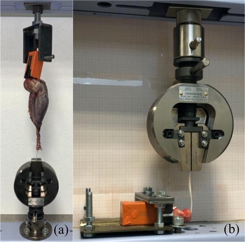
These two types of fixations were tested independently but following the same mechanical test methodology. Eight static tensile tests were performed using a traction system (AGS- X Shimadzu, Japan) with a pre-test of 20 mm/min traction until the load reached 30 N, straightening the system. The tensile test consisted in a 25 mm/min traction until failure and the sampling rate for data acquisition was set at 100 Hz. A total of 8 experimental set-ups were randomly considered: 4 testing the mechanical resistance to tearing of the proximal tendon fixation made using sutures (S1D&G, S2D&G) () and 4 testing the mechanical resistance to tearing of the distal fixation made using an interference screw (C1D&G, C2D&G) ().
Figure 1. Biomechanical setup of (a) proximal fixation along tendon by 8 simple interrupted sutures, (b) the distal fixation in calcaneum by interference screw.
2.4. Data acquisition and processing
During tests, data acquisition was performed using the TrapeziumX software (Shimadzu, Japan). For each implantation technique, two measures were taken: (i) the maximum strength (Ms) and (ii) the failure mode (FM). The data were then processed with Microsoft Excel.
3. Results and discussion
The mode of rupture reported for the proximal and distal fixation proved to be very homogeneous with a single and identical mode of rupture by fixation (). For the proximal fixation, when the most distal suture ruptured, all the others followed a few moments later. For the distal fixation, there was a progressive sliding of the UHMWPE implant at the bone/implant/interference screw interface in the bone tunnel. The maximal strength of the distal fixation was found to be superior to that of the proximal fixation (1025 N ± 107 vs. 692 N ± 102). Considering the preliminary results, using a UHMWPE implant as a mechanical support seems to offer a maximum strength superior to all the conventional suture techniques currently used to treat tendon ruptures in small animals (Putterman et al. Citation2019). Maximum failure strength was increased by 160% with the proximal fixation and by 58% with the distal fixation between our preliminary results and those published by Morton and colleagues for dogs of similar size in similar testing protocol. If our technique is derived from Morton’s work, several modifications have been made. On contrary to our technique, Morton positions the implant along the tendon, not inside it, as described above, and suture it with polydioxanone 3.5 metric. In addition, Morton secured the implant in the calcaneum with an interference screw in a single blind tunnel drill in the long axis of the bone. Our results are encouraging as they approximate the maximal physiological strength of the proximal and distal insertions of the gastrocnemius tendon: proximal fixation (mean ± sd) 692 N ± 102/1031.3 N ± 317.6 and distal fixation 1025 N ± 107/1107.1 N ± 352.7 (Jopp and Reese Citation2009). These preliminary results will have to be confirmed with more mechanical tests in order to conduct a statistical analysis and a more detailed comparison between our results, those of Morton’s study and the physiological findings of Jopp and Reese in 2009.
Table 1. Results, mean and standard deviation of 8 static tensile tests until failure, carried out on two types of proximal and distal fixation.
4. Conclusions
The maximal strength of the proximal fixation achieved by 8 simple interrupted sutures in the gastrocnemius tendon and the UHMWPE implant was (mean ± sd) 692 N ± 102. For the distal fixation, the implant was secured by an interference screw perpendicularly implanted in the calcaneus, with a maximal strength recorded at (mean ± sd) 1025 N ± 107. No implant rupture was observed during any of the mechanical tests.
Disclosure statement
No potential conflict of interest was reported by the authors.
References
- Jopp I, Reese S. 2009. Morphological and biomechanical studies on the common calcaneal tendon in dogs. Vet Comp Orthop Traumatol. 22 (02):119–124.
- Moores AP, Owen MR, Tarlton JF. 2004. The three-loop pulley suture versus two locking-loop sutures for the repair of canine achilles tendons. Vet Surg. 33 (2):131–137.
- Morton MA, Whitelock RG, Innes JF. 2015. Mechanical testing of a synthetic canine gastrocnemius tendon implant: synthetic canine gastrocnemius tendon implant. Vet Surg. 44 (5):596–602.
- Putterman AB, Duffy DJ, Kersh ME, Rahman H, Moore GE. 2019. Effect of a continuous epitendinous suture as adjunct to three‐loop pulley and locking‐loop patterns for flexor tendon repair in a canine model. Vet Surg. 48(7):1229–1236.