
1. Introduction
Osteotomies are surgical procedures used during rhinoplasty, among other interventions in maxillofacial and orthopedic surgery, in order to cut bone tissue (Daniel Citation2010; Dobratz and Hilger Citation2010). They are typically performed using an osteotome (bone chisel) and a surgical mallet. Despite their routine use in clinical practice, Osteotomies still present a number of risks (Rettinger Citation2007) due to the lack of visual control. A preliminary study performed by our group has shown that a hammer instrumented with a force sensor could be used to retrieve information on the mechanical properties (material type and thickness) of plates of various composite materials (Hubert et al. Citation2020). The aim of the present study was to determine whether such approach could be employed in an animal model in order to i) follow the movements of the osteotome in the bone tissue, and ii) detect the arrival of the osteotome at the frontal bone (which corresponds to the end of the osteotomy pathway).
2. Methods
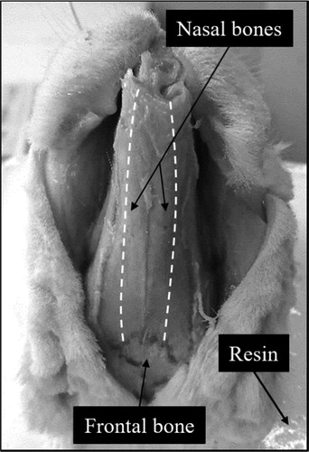
The animal model used in this study was the New Zealand White rabbit since it is widely used to model rhinoplasty (Badran et al. Citation2017) as well as osteotomies (Stein et al. Citation1990; Öğreden et al. Citation2018). Seven rabbit heads were collected from the National Veterinary School of Alfort (Maisons-Alfort, France). For each head, an osteotomy was performed on both the left and right nasal bones using the instrumented hammer (). The osteotomies consisted in series of hammer impacts on the osteotome, alternating between strong impacts (in order to cut bone tissue) and weak impacts (in order to perform localized measurements without modifying the position of the osteotome relatively to bone tissue). For each impact, a signal s(t) corresponding to the evolution of the impact force in time was recorded through the piezoelectric force sensor.
Figure 1. Experimental setup for the realization of the osteotomies. The planned osteotomy paths are represented with white dotted lines.
A first indicator, τ, was derived from the signal, and corresponded to the time between the first two force peaks of s(t). For the strong impacts, a second indicator D, corresponding the displacement of the osteotome with each impact, was measured using the video footage of the experiments.
3. Results and discussion
The results showed that low and repeatable values of τ were obtained for weak impacts. An ANOVA analysis showed that the values of τ in the frontal bone were significantly lower than in the nasal bone (p < 10−10).
Moreover, when considering strong impacts only, a strong correlation was obtained between τ and D, as shown in . , with a spearman correlation coefficient ρ2 = 0,74.
Figure 2. Relationship between τ and D obtained for the strong impacts realized with the instrumented hammer on the osteotome. The dashed line corresponds to the linear regression analysis.
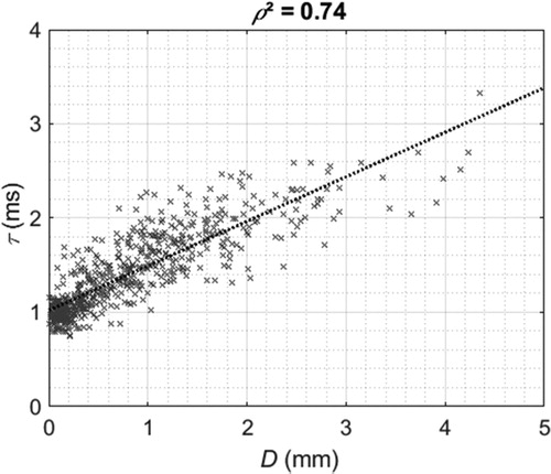
The indicator τ has previously been shown to be related to the biomechanical properties (stiffness and thickness) of the environment surrounding osteotome tip (Hubert et al. Citation2020). When the osteotome cuts through the bone tissue, it first induces a crack opening and then moves into the space created by the crack, which leads to a loss of overall rigidity of the system. Such phenomenon might explain the correlation between τ and D. Similarly, the nasal and frontal bone have different biomechanical properties, the frontal bone being the most rigid and thick. Consequently, the transition between the two different environments is characterized by a change in value of τ.
These results suggest that the values of τ could be used, for each impact, to i) track the movement of the osteotome in the bone tissue and ii) detect the transition between the nasal and frontal bones, which corresponds to the end of the osteotomy pathway.
4. Conclusions
The study of the animal model provides encouraging results which will later need to be confirmed on anatomical subjects. This paves the way for the development of an instrumented hammer that could assist surgeons in their decision-making process during rhinoplasty procedures.
Acknowledgements
The authors would like to acknowledge the support of the “Prématuration programme” of the CNRS through the Osteome project.
Disclosure statement
No potential conflict of interest was reported by the authors.
Additional information
Funding
References
- Badran KW, Chang JC, Kuan EC, Wong BJF. 2017. Anatomy and surgical approaches to the rabbit nasal septum. JAMA Facial Plast Surg. 19(5):386–391.
- Daniel RK. 2010. Mastering rhinoplasty: a comprehensive atlas of surgical techniques with integrated video clips. Springer Sci Bus Media. 127(5):2116–2117.
- Dobratz EJ, Hilger PA. 2010. Osteotomies. Clin Plast Surg. 37(2):301–311.
- Hubert A, Rosi G, Bosc R, Haiat G. (2020). Using an impact hammer to estimate elastic modulus and thickness of a sample during an osteotomy. J Biomech Eng. 142(7):071009.
- Öğreden Ş, Rüzgar S, Tansuker HD, Taşkın Ü, Alimoğlu Y, Aydın S, Oktay MF, Izol U. 2018. Histopathological comparison of bone healing effects of endonasal and percutaneous lateral osteotomy methods in rabbit rhinoplasty model. Braz J Otorhinolaryngol. 84(5):540–544.
- Rettinger G. 2007. Risks and Complications in Rhinoplasty. Head and Neck Surgery. 6:1–14.
- Stein E, Sedlacek T, Fabian RL, Nishioka NS. 1990. Acute and chronic effects of bone ablation with a pulsed holmium laser. Lasers Surg Med. 10(4):384–388.