
1. Introduction
Possible lack of length or over-stiffness of soft tissues may explain some musculo-skeletal disorders. Certain extra length of soft tissues can be recovered by releasing some strain with pie-crusting technique. This technique can be applied to the tendons (Burge et al., Citation2014), the ligaments (He et al., Citation2018), or the fascias (Çatal et al., Citation2019). It is widely used when patients having a too short and too stiff iliotibial band (ITB), suffer from unstable knee, joint line elevation, etc. For example, in valgus knees, ITB is tensed on the lateral side and the main technique used to correct valgus deformity is to perform a pie-crusting technique to release the ITB.
However, soft tissue releasing using the pie-crusting technique is challenging as too extensive releases may lead to complications.
The aim of this preliminary study is to assess the level of strain release of the ITB using pie-crusting technique in view of assessing quantitatively the impact of this surgery on the strain release.
2. Methods
2.1. Specimen preparation
Three fresh post-mortem female subjects numbered 285, 300, and 385 respectively (90–95 yo, 34–58.5 kg, 158–163 cm) from the Department of Anatomy of the University of Rockefeller (DUAR), Lyon, France, were prepared for the strain field measurement on the ITB of the both right and left legs. Because of a preliminary study, no inclusion criteria were considered for subjects’ selection.
Skin and fat tissues were removed carefully so the area of ITB was clean. Then, white clown mask was applied on the ITB surface and speckles pattern was created using a toothbrush with a black ink.
2.2. Test procedure
The 3 D surfacic strain state of the ITB was assessed by digital image correlation (using the VIC3D® software). Digital image acquisition was done using a pair of low noise cameras JAI-GO-5000-USB with objectives KOWA LM12HC with lenses of 12.5 mm and an f-stop range of f/1.4 to f/16. Image size was 2560 × 2048 px. giving a pixel size of 0.2646 mm2. The image correlation was performed using a subset of 31 and a step of 7. Green-Lagrange surfacic strains were computed to obtain principal strains (E1 and E2) and then maximum shear was calculated as 0.5*(E1–E2). During the experiments, each subject was in a sitting posture with a pelvic angle close to 24° and a fully extended knee.
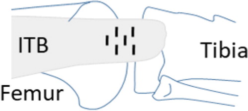
The ITB strain release was performed by pie-crusting cuts done with an 11-blade scalpel and located between the lateral condyle of the tibia and the lateral epicondyle of the femur as shown on .
Figure 1. Schema to perform pie-crusting cuts on the ITB.
The reference strain state was defined as the initial strain state before any strain release. The average strain release on the ITB was assessed after (A) superficial pie-crusting cuts, (B) deep cuts, (C) a full ITB dissection from the tibia insertion (A, B, C states are illustrated on ).
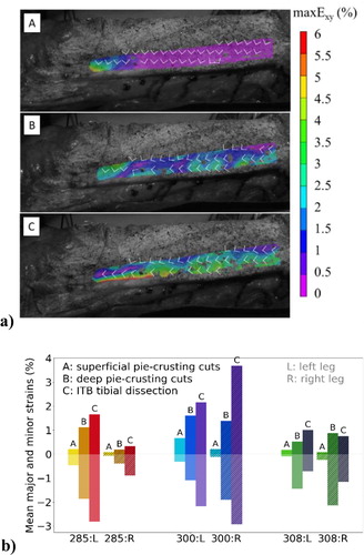
Figure 2. Results representation: a) Maximal shear strain distribution maxExy on the ITB for subject 285, after (A) superficial pie-crusting cuts, (B) deep cuts, and (C) tibial dissection. White arrows show the direction of the major E1 and minor E2 principal strains; b) Average major (positive values) and minor (negative values) principal strain releases measured on the ITB for all subjects.
3. Results and discussion
shows the distribution of the maximal shear strain maxExy on the ITB after each strain release (A, B, C states). With strain release, the principal strains turn relatively to the longitudinal axis of ITB. This may be explained by microstructural mechanisms as two main directions of fibers exists on ITB (Otsuka et al., Citation2018).
presents the average major and minor principal strain release measured for all subjects. It can be observed that average strain release increased along the cutting steps except for the right leg of the subject 308. For subject 285, average strain levels were specifically low for the right leg compared to the left. This was associated to a lack of mobility observed on the right knee. Major and minor strains presented almost opposite values, the minor principal strain was found superior to the major principal strain in absolute values for almost all states. However, it was not the case for subject 300 case C on the left and right legs where the absolute mean value of the ratio major over minor strain release was close to 1, which corresponds to a pure shear strain mechanism. For all subjects, the maximum strain release did not exceed 3% of ITB shortening.
Strain release obtained using the pie-crusting technique was assessed on the ITB using digital image correlation. Even if a general tendency showed the increase of the strain release with deeper pie-crusting cut and full ITB dissection of the tibial attachment, a variability of strain release levels was observed. These differences could be explained by the physiological characteristics of the subjects. The distinction could also be interpreted by the variability of the cutting technique applications. The reproducibility of the location and the size of the cuts could be improved. Moreover, the subjects involved in this ex vivo study were geriatric and had various anthropometries. As demonstrated by Wilke et al. (Citation2019) or Zwirner et al. (Citation2019), age, gender, height and weight are parameters that can affect fascia mechanical properties.
Further tests will be performed on a larger number of subjects, and further analysis will be carried out to associate measured strain release on the ITB and knee stability criteria.
4. Conclusions
Pie-crusting technique is a powerful tool for pain relief and functional improvement of the knee. Although this method is widely used by surgeons, this study is a first step to quantify the ITB strain release. Further work will be done to assess the associated impact on the knee behaviour and stability.
These data could also be used to validate the simulation of the release of the ITB mechanical action on the knee mobility, using FE model of the lower limb. After validation, such models could be used for patient-specific surgery planification.
References
- Burge JR, Sanchez HB, Wagner RA. 2014. Quadriceps and patellar tendon pie-crusting as a treatment for limited flexion in total knee arthroplasty. Am J Orthop. 43(4):E83–E88.
- Çatal B, Keskinbora M, Keskinöz EN, Tümentemur G, Azboy İ, Demiralp B. 2019. Percutaneous plantar fascia release with needle: anatomic evaluation with cadaveric specimens. J Foot Ankle Surg. 58(5):842–846.
- He X, Cai H, Zhang K. 2018. Pie-crusting technique is effective and safe to release superficial medial collateral ligament for total knee arthroplasty. J Orthop Translat. 13:33–40.
- Otsuka S, Yakura T, Ohmichi Y, Ohmichi M, Naito M, Nakano T, Kawakami Y. 2018. Site specificity of mechanical and structural properties of human fascia lata and their gender differences: a cadaveric study. J Biomech. 77(August):69–75.
- Wilke J, Macchi V, De Caro R, Stecco C. 2019. Fascia thickness, aging and flexibility: is there an association? J Anat. 234(1):43–49.
- Zwirner J, Babian C, Ondruschka B, Schleifenbaum S, Scholze M, Waddell NJ, Hammer N. 2019. Tensile properties of the human iliotibial tract depend on height and weight. Med Eng Phys. 69:85–91.