
Abstract
The nematode C. elegans has a contingent of five sod genes, one of the largest among aerobic organisms. Earlier studies revealed each of the five sod genes is capable of making perfectly active SOD proteins in heterologous expression systems therefore none appears to be a pseudogene. Yet deletion of the entire contingent of sod genes fails to impose any effect on the survival of C. elegans except these animals appear more sensitive to extraneously applied oxidative stress conditions. We asked how many of the five sod genes are actually making active SOD enzymes in C. elegans through the usage of in-gel SOD activity analysis and by using KCN as a selective inhibitor against Cu-ZnSOD enzyme(s). Here we provide evidence that out of the five SOD proteins only the mitochondrial SOD is active in the water-soluble fraction of C. elegans extracts albeit at an apparently much lower activity than the multiple active SODs in D. melanogaster and E. coli. We had no opportunity to test the activity of Sod-4a isoform which is possibly a membrane-bound form of SOD. The mutant analysis further confirmed that among the two mitochondrial SOD proteins, SOD-2 is the only naturally active SOD in C. elegans.
Introduction
Aerobic organisms utilize Superoxide Dismutase (SOD) enzymes to scavenge and catabolize partially reduced oxygen molecules (superoxide radicals; O2−) into hydrogen peroxide [Citation1]. Superoxides are generated in the electron transport chain from aerobic respiration. In different aerobes, SOD enzymes entrap superoxide radicals with the help of a variety of redox-active transition metals such as Cu, Mn, Fe, or Ni. Despite all SODs performing the single task of scavenging superoxide radicals, the activity of specific SOD enzymes is noticeably limited to either the mitochondrial matrix, cytoplasm, inter-membrane space, or to the extracellular environment [Citation2–4]. Loss-of-function studies in different species have established the variable requirement of the SOD protection system because the spectrum of defects appearing in SOD mutants is wide-ranging. In general, lack of mitochondrial SOD in vertebrates cause neonatal lethality accompanied by signs of mitochondrial damage, cardiomyopathy, and neuropathology [Citation5,Citation6]. Early lethality is also evident in the Drosophila mitochondrial SOD (SOD2) loss of function condition [Citation7,Citation8] with evidence of neuropathology [Citation9], loss of mitochondrial aconitase activity, and reduced ATP production in the muscle [Citation10].
On the other hand, loss of the Cu-ZnSOD enzyme (SOD1) causes more subtle effects, particularly in higher animals. For example, mice lacking Cu-ZnSOD activity exhibit no effect on longevity although a multitude of other damages are reported [Citation11,Citation12]. Drosophila Cu-ZnSod null mutants survive albeit with a shorter adult life span [Citation13]. Finally, bacterial and yeast mutants of sod1 and sod2 are hypersensitive to hyperbaric Oxygen concentration, and exhibit numerous biochemical defects in these mutants [Citation14].
The nematode C. elegans is a free-living, soil-dwelling aerobe traditionally used as a workhorse in studies related to aging, cell death, and developmental biology [Citation15]. Homology analysis established five sod genes in C. elegans, possibly one of the largest contingents of SOD isoforms among aerobic organisms ([Citation16], Worm Base). C. elegans sod-1, sod-2 and sod-4 represent the traditional cytoplasmic, mitochondrial, and extracellular forms of SODs respectively, while sod-3 and sod-5 are recognized as additional and minor mitochondrial and cytoplasmic SODs [Citation17]. sod-1 and sod-2 mRNAs represent the bulk of the sod RNAs (∼76% and 18% respectively) within the total SOD mRNA populations whereas the remaining three sod mRNAs make up only between 5 and 0.5% of the total [Citation17]. Hence in C. elegans all sod genes are expressed but differentially.
Are the extra copies of sod purposefully acquired to gain additional protection against some pro-oxidative environment or are most of these sod genes evolutionary relics? Mutational analyses have already provided a striking answer as a C. elegans strain containing quintuple deletions of all sod genes survives as long as the wild-type control worms [Citation18]. Additionally, no tissue damage or loss of mitochondrial proteins was evident in this quintuple sod mutant due to the lack of a complete SOD protection system. The only noticeable change seems to be these animals are hypersensitive to different kinds of oxidants and exhibit reduced fertility [Citation27]. However, C. elegans sod genes are not evolutionary relics because sod-2 and sod-3 genes and sod1 and sod-5 genes can make perfectly active SOD proteins in heterologous systems like bacteria and yeast [Citation19,Citation20]. This suggests therefore that no coding sequence deficiencies are expected in these genes like one might observe in a pseudogene. Additionally, SOD enzyme activity assays support that C. elegans does contain active SOD enzyme(s) [Citation17,Citation18] however which one or how many of the five SOD proteins are naturally active remains an open question.
Mutational analyses have begun to provide some intriguing answers to the requirement of SOD enzymes in C. elegans. C. elegans null mutants for either sod-1 or sod-2 exhibit a reduced brood phenotype [Citation21]. For the sod-2 mutant, this was shown to be due to a sperm defect, while the sod-1 mutant fertility defect is hypothesized to be due to defects in oogenesis [Citation21]. The sod-2 null sperm defect can be rescued by treatment with exogenous hydrogen peroxide, suggesting that it is a lack of active SOD enzymatic activity in the sperm upon mutation of sod-2 that causes the defect.
With the help of in-gel SOD activity assays of the soluble protein extract, we demonstrate that only the mitochondrial SOD protein maintains enzymatic activity C. elegans and of the two mitochondrial SODs, SOD-2 remains naturally active in C. elegans.
Materials and methods
C. elegans strains and culture conditions: The following strains were used in this work: wild type N2; CB1370 [daf-2(e1370)III], FX776 [sod-1(tm776)II]; VC433 [sod- 3(gk235)X]; RB1072 [sod-2(ok1030)I]; MQ1766 [sod-2(ok1030)I; sod-5(tm1146) sod- 1(tm783) II; sod-4(gk101)III; sod-3(tm760)X]. All strains were grown and maintained under standard conditions at 20 °C.
Protein extraction
C. elegans protein lysates were prepared by washing the appropriately staged worms off MYOB plates grown at 20 °C with 1X PBS and collected in a 1.5 ml microcentrifuge tube. Worms were pelleted with gentle centrifugation, homogenized in 1 X PBS plus 10ul/ml Halt protease and phosphatase inhibitor (single-use cocktail, Thermo Fisher Scientific Cat. number 78442) on ice for 10 min followed by centrifugation at 16000 G at 4 °C for 15 min. The supernatant was aspirated carefully avoiding the floating lipid layer. Bradford assay was performed to measure the protein concentration. A similar technique is used for extracting Drosophila (Canton-S) and E. coli proteins.
In-gel SOD activity assay
For in-gel SOD activity assay total soluble protein extracts were loaded into a gel (4–20% Criterion Precast Gel, Biorad Cat #3450032) and run at 4 °C. Once completed gels were soaked in 40 ml phosphate buffer containing 80 mg of NBT (2.43 mM), 170 μl of TEMED (28 mM) and 8 μl of stock riboflavin-5′-phosphate (0.14 M [53 mg ml−1] in 50 mM phosphate buffer, pH 7.8) for 20 min under dark. Following incubation, the gels were washed in dH2O three times and put under fluorescent light for 2 h. During this time the gels turned purple and an achromatic band begins to appear. In principle, SOD will compete with NBT for O2−. In the absence of SOD, NBT will react with O2− and form a blue precipitate throughout the gel except in areas where SOD enzyme(s) is active which appears as clear bands (achromatic) because it outcompetes NBT for the O2− in those areas [Citation22]. For KCN treatment 10 mM KCN is added to C. elegans protein extracts and incubated in room temperature for 30 min prior to loading.
Results and discussion
To answer how many and which particular SOD protein is active we performed in situ SOD gel analysis using preparations of native protein extracts from C. elegans and compared this with Drosophila and E. coli extracts. Starting with Drosophila, two active SOD enzymes are identified () with the top band being the mitochondrial SOD (MnSOD or SOD2) and the lower band representing the cytoplasmic SOD (Cu-ZnSOD or SOD1) [Citation7,Citation13]. As expected, the E. coli extract shows three forms of bacterial SOD enzymes MnSOD, Fe-SOD, and the Cu-ZnSOD (). On the other hand, wild type C. elegans (N2) extract shows only a single SOD enzyme band indicating that it carries no more than one active form of a SOD enzyme (). Two things are notable with respect to the single active C. elegans SOD enzyme.
Figure 1. In situ SOD enzyme profiles in three different species. (A) Comparison of SOD activity profiles in D. melanogaster, E. coli and C. elegans. C. elegans has no more than one active SOD enzyme. (B–D) The dauer larval stage, daf-2 mutant, and L4 larvae all carry one active form of SOD.
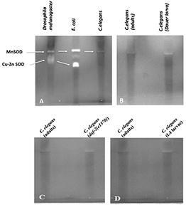
First it lines up perfectly with the MnSOD/SOD2 isoforms of Drosophila and E. coli, suggesting that this SOD activity is from either of the two C. elegans MnSODs- SOD-2 or SOD-3. Second, the C. elegans SOD activity appears as a much less intense band in comparison to the activity observed in Drosophila and E. coli despite loading equal amounts of protein extracts per lane (60 µg/lane). This leads us to hypothesize that either C. elegans makes much less SOD enzyme or that the enzyme itself is less active in this organism compared to the Drosophila and E. coli SODs. Further research should resolve if C. elegans SOD is either expressed at a lesser amount or if it is less active relative to other organisms.
The previous result led us to ask why is there only one naturally active SOD enzyme present in the C. elegans system when there are five sod genes? As none of the five sod genes in C. elegans are pseudogenes since each is capable of making perfectly active SOD protein in a heterologous system we had anticipated multiple active SODs in our in-gel activity assay [Citation19,Citation20]. We tested two possibilities such as some of the SOD enzymes might be made and active during specific developmental stages of the worm [Citation20], or second, that some SODs are active only under high-stress conditions. C. elegans dauer arrested larvae, a stage adopted to survive in high-stress environment show no more than one active SOD enzyme (). Similarly, no additional SOD enzymes appear active in the L4 larval stage protein extracts compared to adult extracts () nullifying the possibility of activation of certain SOD enzymes during the L4 stage of development (). As we have not tested the other worm developmental stages, we cannot comment on each of those. It has previously been reported that daf-2 mutants exhibit overproduction of sod mRNA [Citation23]. In our in-gel SOD activity assay, we observe a slightly more intense SOD band in the daf-2 mutants compared to that observed in the wild-type control supporting the reported increase in sod mRNA (). Taken together our observations raise a strong possibility that only one SOD enzyme is naturally active starting from the L4 stage and throughout adulthood in C. elegans.
The obvious question then is which one of the five SOD proteins remains active in adult C. elegans? We previously mentioned that the single active C. elegans SOD enzyme lines up nicely with the mitochondrial SOD2 band of Drosophila and E. coli (). This suggests that either one of the two mitochondrial SOD proteins (SOD-2 or SOD-3) or maybe both are represented in this single SOD active enzyme band. It has been reported that these two SOD proteins arise from a gene duplication event [Citation19] and the predicted molecular weights of C. elegans SOD-2 and SOD-3 are 24.5 and 24.6kD respectively () which matches very closely with the Drosophila and E. coli SOD2 proteins that are 24.7 and 25.2kD respectively (). The only other C. elegans SOD of similar molecular weight is SOD-4b (a.k.a Sod4-2) isoform that weighs 23.3 kD (). However, SOD-4b with its large trans-membrane domain should remain associated with the cell membrane [Citation26], and therefore it is very likely eliminated with the membrane fraction during protein extraction. On the other hand, the three cytoplasmic SOD proteins in C. elegans, SOD-1, SOD-4a, and SOD-5 are significantly smaller in molecular weight compared to the mitochondrial SODs weighing 18.7, 18.2, and 18.5kD respectively () and thus not likely to contribute to high molecular weight SOD band.
Table 1. Molecular weights of SOD enzymes isomers in C. elegans, D. melanogaster, and E. coli.
Interestingly, the single SOD band in C. elegans was previously designated as the active SOD1 enzyme [Citation20]. To resolve this issue we used various single and multiple sod mutants in our in-gel SOD activity assay. The quintuple sod knockout mutant worms [sod-2(ok1030) I; sod-5(tm1146) sod-1(tm783) II; sod-4(gk101) III; sod-3(tm760) X] [Citation18] displays no active SOD enzyme (). Protein extracts from three single sod null alleles provided a plausible answer. sod-1(tm776) and sod-3(gk235) mutant homozygotes clearly display the single SOD band as in the wild type, whereas the sod-2(ok1030) mutant is completely lacking an active SOD band ().
Figure 2. SOD activity profiles in different C. elegans sod mutants. (A) No active SOD appears in the sod2(ok1030)I null mutant whereas both the sod-1(tm776)II and sod- 3(gk235)X null mutants still possess the single active form of SOD observed in wild type animals. (B) Treatment of wild-type C. elegans with KCN does not inhibit this SOD activity.
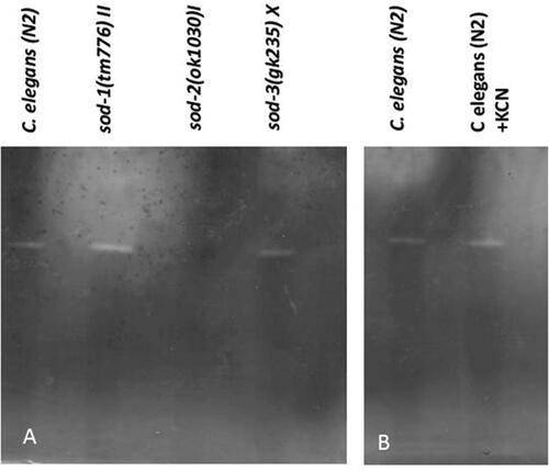
The above observation clearly suggests that the active SOD enzyme in C. elegans is the mitochondrial SOD-2 protein, however as SOD-2 and SOD-4b are quite close in terms of molecular weight there remains the possibility that the single active band may be arising from an active sod-4b allele. We did not think this was likely due to the fact that SOD-4b is a transmembrane protein and all membrane-bound proteins should be eliminated during the extraction procedure. However, to clearly test whether the single active SOD band in C. elegans is due to SOD-4b we treated wild-type protein extract with KCN. SOD-4b isoform is a Cu-ZnSOD [Citation26] and Cu-ZnSOD activities are inhibited by KCN. Incubation of C. elegans extract in the presence of 10 mM KCN solution failed to inhibit the SOD active band proving thereby that the active C. elegans SOD protein it is not a Cu-ZnSOD enzyme and thus not SOD-4b ().
Our attempt to profile the active SOD enzymes in C. elegans led to the finding that the mitochondrial SOD-2 enzyme is the sole active SOD enzyme that is present at least throughout the adult life span of this organism. Since the mitochondria are the main source of superoxide radicals in the cell due to the increased oxygen metabolism in this organelle, keeping the mitochondrial enzyme in an active state makes more sense for any aerobic organism. Why the C. elegans SOD-2 enzyme appears relatively less active compared to other organisms remains unclear (). The nonessential nature of this lone active SOD2 enzyme is demonstrated as the sod-2 deletion mutant positively influences the life span by surviving longer than the wild type and also exhibits other mutant phenotypes such as slow development, a low brood size, and slow defecation [Citation27]. Unlike any other aerobes, total abrogation of all SOD enzymes is tolerated by C. elegans, as well as it is completely resistant to a hypoxic environmen [Citation28]. In fact, hypoxia imposes a positive influence on C. elegans life span. In sum, these observations challenge the necessity of the SOD system as the primary defense mechanism toward ROS detoxification in C. elegans. An important question that remains is what has taken the place of the SOD system requirement in C. elegans?
Supplemental Material
Download PDF (14.4 KB)Acknowledgment
We thankfully acknowledge the National Science Foundation grant 1832026 and NIH grant R25AG047843 awarded to AD. LMF and AKA are supported by a Department of Defense grant W911NF1810465 awarded to AKA. Authors are thankful to Mr. Aon Ali for finishing the KCN sensitivity experiment. Some strains were provided by the CGC, which is funded by NIH Office of Research Infrastructure Programs (PO OD10440).
Disclosure statement
No potential conflict of interest was reported by the author(s).
Additional information
Funding
References
- Fridovich I. Superoxide radical and superoxide dismutases. Annu Rev Biochem. 1995;64:97–112.
- Weisiger RA, Fridovich I. Superoxide dismutase: organelle specificity. J Biol Chem. 1973;248(13):4793–4796.
- Missirlis F, Hu J, Kirby K, et al. Compartment- specific protection of iron–sulfur proteins by superoxide dismutase. J Biol Chem. 2003;278(48):47365–47369.
- Muller FL, Liu Y, Van Remmen H. Complex III releases superoxide to both sides of the inner mitochondrial membrane. J Biol Chem. 2004;279(47):49064–49073.
- Lebovitz RM, Zhang H, Vogel H, et al. Neurodegeneration, myocardial injury, and perinatal death in mitochondrial superoxide dismutase-deficient mice. Proc Natl Acad Sci USA. 1996;93(18):9782–9787.
- Li Y, Huang TT, Carlson EJ, et al. Dilated cardiomyopathy and neonatal lethality in mutant mice lacking manganese superoxide dismutase. Nat Genet. 1995;11(4):376–381.
- Duttaroy A, Paul A, Kundu M, et al. A Sod2 null mutation confers severely reduced adult life span in drosophila. Genetics. 2003;165(4):2295–2299.
- Kirby K, Hu J, Hilliker AJ, Phillips JP. RNA interference-mediated silencing of Sod2 in Drosophila leads to early adult-onset mortality and elevated endogenous oxidative stress. Proc Natl Acad Sci USA. 2002;99(25):16162–16167.
- Paul A, Belton A, Nag S, et al. Reduced mitochondrial SOD displays mortality characteristics reminiscent of natural aging. Mech Ageing Dev. 2007;128(11–12):706–716.
- Godenschwege T, Forde R, Davis CP, et al. Mitochondrial superoxide radicals differentially affect muscle activity and neural function. Genetics. 2009;183(1):175–184.
- Culotta VC, Yang M, O'Halloran TV. Activation of superoxide dismutases: putting the metal to the pedal. Biochim Biophys Acta. 2006;1763(7):747–758.
- Valentine JS, Doucette PA, Potter SZ. Copper-zinc superoxide dismutase and amyotrophic lateral sclerosis. Annu Rev Biochem. 2005;74:563–593.
- Phillips JP, Campbell SD, Michaud D, et al. Null mutation of copper/zinc superoxide dismutase in drosophila confers hypersensitivity to paraquat and reduced longevity. Proc Natl Acad Sci USA. 1989;86(8):2761–2765.
- Culotta VC. Superoxide dismutase, oxidative stress, and cell metabolism. Curr Top Cell Regul. 2000;36:117–132.
- Corsi AK, Wightman B, Chalfie M. A transparent window into biology: a primer on Caenorhabditis elegans. Genetics. 2015;200(2):387–407.
- Landis GN, Tower J. Superoxide dismutase evolution and life span regulation. Mech Ageing Dev. 2005;126(3):365–379.
- Doonan R, McElwee JJ, Matthijssens F, et al. Against the oxidative damage theory of aging: superoxide dismutases protect against oxidative stress but have little or no effect on life span in Caenorhabditis elegans. Genes Dev. 2008;22(23):3236–3241.
- Raamsdonk JM, Hekimi S. Superoxide dismutase is dispensable for normal animal lifespan. Proc Natl Acad Sci USA. 2012;109(15):5785–5790.
- Hunter T, Bannister WH, Hunter GJ. Cloning, expression, and characterization of two manganese superoxide dismutases from Caenorhabditis elegans. J Biol Chem. 1997;272(45):28652–28659.
- Jensen LT, Culotta VC. Activation of CuZn superoxide dismutases from Caenorhabditis elegans does not require the copper chaperone CCS. J Biol Chem. 2005;280(50):41373–41379.
- Sakamoto T, Imai H. Hydrogen peroxide produced by superoxide dismutase SOD-2 activates sperm in Caenorhabditis elegans. J Biol Chem. 2017;292(36):14804–14813.
- Weydert CJ, Cullen JJ. Measurement of superoxide dismutase, catalase and glutathione peroxidase in cultured cells and tissue. Nat Protoc. 2010;5(1):51–66.
- Honda Y, Honda S. The daf-2 gene network for longevity regulates oxidative stress resistance and Mn-superoxide dismutase gene expression in Caenorhabditis elegans. Faseb J. 1999;13(11):1385–1393.
- Hunter GJ, Trinh CH, Bonetta R, et al. The structure of the Caenorhabditis elegans manganese superoxide dismutase MnSOD-3-azide complex. Protein Sci. 2015;24(11):1777–1788.
- Sheng Y, Abreu IA, Cabelli DE, et al. Superoxide dismutases and superoxide reductases. Chem Rev. 2014;114(7):3854–3918.
- Fujii M, Ishi N, Joguchi A, et al. A novel superoxide dismutase gene encoding membrane-bound and extracellular isoforms by alternative splicing in Caenorhabditis elegans. DNA Res. 1998;5(1):25–30.
- Van Raamsdonk JM, Hekimi S. Deletion of the mitochondrial superoxide dismutase sod-2 extends lifespan in Caenorhabditis elegans. PLOS Genet. 2009;5(2):e1000361.
- Mehta R, Steinkraus KA, Sutphin GL, et al. Proteasomal regulation of the hypoxic response modulates aging in C. elegans. Science. 2009;324(5931):1196–1198.