Abstract
A biosensor was developed by immobilizing laccase onto mercury thin film electrode (MTFE) by means of gelatin that is then crosslinked with glutaraldehyde. Mercury thin film (MTF) was deposited onto glassy carbon electrode (GCE) and the obtained biosensor was utilized for the determination of phenolic compounds. The measurement was based on the amperometric detection of oxygen consumption in relation to analyte oxidation. The optimum experimental conditions for the biosensor were investigated and the system was calibrated for both catechol and phenol. A linear relationship between sensor responses and analyte concentrations was obtained in concentration range between 0.5 × 10⁻⁶– 5.0 × 10⁻⁶ M for catechol and 2.5 × 10⁻⁶– 2.0 × 10⁻⁶ M for phenol, respectively. Mercury thin film was also formed onto the surface of screen printed graphite electrodes and applied for the catechol detection. The linearity was observed in concentration range between 2.5 × 10⁻⁶– 3.0 × 10⁻⁵ M.
1. INTRODUCTION
Biosensors are analytical devices combining a biological component with a physical or chemical transducer. Electrochemical biosensor can be explained as a molecular sensing device that combines a biological recognition element to an electrode transducer. For this reason, the success of an electrochemical sensing process relies mainly on a proper choice of the working electrode [Citation[1]]. Solid electrodes like gold, platinum and carbon-based electrodes were utilized as electrode transducers. Apart from these, a mercury thin film electrode (MTFE) based biosensor was developed and used for H2O2 detection [Citation[2]]. Mercury film electrode (MTFE) consists of a very thin film (10–100 µm) layer of mercury that is distributed over the support material. Glassy carbon electrode (GCE) is claimed to be the most favorable supporting material because mercury plated glassy carbon is not contaminated by amalgamation and it does not diffuse into the support material [Citation[1], Citation[3]]. The mercury film formed on a GCE surface is composed of many droplets that lead to a lower hydrogen overvoltage. In addition, MTFE's provide a larger surface to volume ratio and can be utilized in different cell configurations. Furthermore, as small quantities of mercury are used for the preparation of MTFE, the consumption of metallic mercury is minimized. SPGE (screen printed graphite electrode) has also been utilized as the support material for MTFE. These electrodes are based on screen-printing technology and can be employed as low-cost disposable electrochemical sensors [Citation[4]]. The present study includes the use of MTFE as the part of laccase biosensor for the detection of phenolic compounds where GCE and SPGE were used as the support material for MTFE formation. Nowadays, the determination of phenolic compounds is of great importance, since they are widely used in industrial processes, such as the manufacture of plastics, polymers, drugs and dyes [Citation[5]]. These kinds of compounds are also decomposition products from some pesticides and by-products from paper pulp industry, with the types and abundances of phenolic compounds changing with the particular source or mill process [Citation[6]]. Phenolic compounds belong to a class of polluting chemicals that are easily absorbed by animals and humans through the skin and mucous membranes. Their toxicity affects a great variety of organs and tissues, primarily lungs, liver, kidneys and genito-urinary system. In addition, due to their great variety, phenolic compounds show a broad range of toxicity levels, being phenol and its chlorinated or alkylated derivatives, classified as priority pollutants [Citation[7]]. Thus, the development of procedures for detection and simultaneous determination of these compounds in different matrices is highly desired. Analytical methods for the detection and quantification of mixtures of phenols are usually based on analytical separation techniques, which allow the identification and quantification of individual constituents. Many methods have been developed for the determination of phenolic compounds, such as chromatographic, fluorimetric and spectrophotometric techniques [Citation[8-15]]. However, these techniques do not easily allow continuous monitoring, they are expensive, time-consuming, need skilled operators, and sometimes require preconcentration and extraction steps that increase the risk of the sample loss or contamination [Citation[16], Citation[17]]. Thus, the development of new methods that allows the simultaneous determination, without previous separation, of these compounds is a relevant subject of research. The biosensors tend to be more stable and more suitable to environmental monitoring, clinical testing, or food assays compared with the direct electrochemical oxidation of phenols. Biosensors using laccase as detection element have been developed to detect phenols in effluents [Citation[18-20]]. In fact, laccases (benzendiol: oxygen oxidoreductases; EC 1.10.3.2), which are multi-copper enzymes widely distributed in plant and fungal species, oxidize phenols where molecular oxygen is the terminal electron acceptor of the oxidation process [Citation[21]].
2. MATERIAL AND METHODS
2.1. Reagents and Materials
All chemicals were commercially available and of reagent grade. Catechol was obtained from Sigma (Germany) and Phenol was from Merck AG (Darmstadt, Germany). Synthetically concocted wastewater composition; 50 g/L NaCl and 100 g/L phenol in 1.0 M HCl solution [Citation[22]].
Screen Printed Electrodes (SPE): Inks for printing working electrodes were prepared by using a commercially available carbon ink (Du Pont 7101). Printed electrodes were fabricated by depositing several layers of inks on a PVC substrate. The conducting paths and pads were deposited directly on the PVC sheets using Ag/Pd ink (DuPont, 5025). Then, an Ag/AgCl ink was deposited to obtain the reference electrode. Carbon ink was printed to obtain the working electrodes. Finally, an insulator layer was placed over the conducting paths. After each printing step, the paths were treated at 60°C for 60 min [Citation[23]].
2.2. Biological Material
Laccase was isolated from the culture filtrates of the white–rot fungus Trametes versicolor (ATCC 11 235) as described in previous work [Citation[24]]. T. versicolor was maintained at 4°C on 2% malt agar and grown in 100 ml malt extract broth (2%) for three days. The laccase production medium was a nitrogen-limited medium consisting of 10 g glucose, 1 g (NH4)H2PO4, 0.05 g MgSO4 · 7H2O, 0.01 g CaCl2 and 0.025 g yeast extract, per liter. The cultures of T. versicolor were incubated at 26°C on a rotary shaker at 175 rpm. After 72 h cultivation, concentrated solution of phenol was added to the cultures to give 10 mg/l [Citation[24]]. Laccase activity was estimated by oxidation of ABTS [Citation[25]].
2.3. Apparatus
Chronoamperometric measurements were performed using a PALM SENS electrochemical measurement system from PALM Instruments B.V. (Netherlands), while DP-voltammograms were obtained with a Metrohm 693 VA Trace Analyzer and a 694 VA Stand with Ag/AgCl reference and Pt auxiliary electrodes.
2.4. Electrode Preparation
Both glassy carbon (Type I) and screen printed graphite electrode (Type II) surfaces were used as a substrate for the deposition of MTF. The glassy carbon disc electrode (diameter of 2 mm) was cleaned by hand polishing using alumina slurry on a felt pad. MTF electrode was prepared ex-situ by introducing 20 ml of mercury plating solution containing 200 µg/ml Hg (II) ions in 1.0 M HCl solution into the cell. A deposition potential of − 800 mV was applied for 5 min after 5 min deaeration of the solution. Then the electrode was rinsed with double distilled water. In the case of using SPG electrode (diameter of 3 mm), MTF was deposited on the graphite working electrode by using the same procedure as described above.
Laccase and 225 bloom gelatin (10 mg) were mixed at 38°C in potassium phosphate buffer, pH 7.0 (250 µl). 50 µl of mixed solution was spread over the electrode surface and allowed to dry at 4°C for 1 h. Finally; it was immersed in 2.5% glutaraldehyde in 50 mM phosphate buffer (pH 7.0) for 5 min [Citation[2]]. Moreover, the procedure belonging to SPGE was the same as for GCE, except 5 µl of mixed solution was placed on graphite working electrode. Both types of electrode include 7.0 unit of laccase activity.
2.5. Measurements
Dp voltammograms were recorded between − 100 and − 1200 mV with a 50 mV pulse amplitude. Measuring time was 40 ms and the current was sampled in 20 ms.
To determine the concentration of phenolic compounds, oxygen consumption that occurred in the enzymatic reaction was detected. All the measurements were done at 25°C under continuous and constant magnetic stirring and varying substrate concentrations in steady-state conditions in 50 mM oxygen saturated acetate buffer (pH 4.5) without any deaeration. The working electrode was polarized at − 0.3 V vs Ag/AgCl electrode. The duration of each analysis was 200 sec. The residual current was allowed to decay in the presence of working buffer before the addition of substrate solution. The change in the current values was monitored chronoamperometrically. After completion of the measurement, the electrode was rinsed with distilled water and allowed to equilibrate before another measurement. 50 mM of acetate buffer, pH 4.5 was used as working buffer for biosensor.
All the optimization studies were performed with Type I, while both Type I and Type II electrodes were analytically characterized.
3. RESULTS AND DISCUSSION
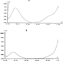
The developed biosensor provides the detection of phenolic compounds by monitoring the consumption of dissolved oxygen. In order to observe the reduction peaks of oxygen, hence explore the optimum operating potential, voltammograms were recorded by using MTFE in the presence of 50 mM acetate buffer (pH 4.5) without any deaeration, as shown in . The first peak at − 81 mV is more significant relative to the second wave and corresponds to the reduction of dissolved oxygen into H2O2. At , the solution was deaerated with N2 for five minutes before the recording of voltammogram. As can easily be seen, the height of the first peak reduces almost 55% while the second peak disappears. As has been known, in amperometric systems the reactions are monitored at the steady state potentials. The choice of the negative potentials accelerates the mass transfer, hence simplify the examination of the mentioned reaction. For these reasons, the operating potential of reduction of oxygen to hydrogen peroxide was chosen as − 300 mV v.s Ag/AgCl and used for further studies.
Figure 1 The dp voltammograms of oxygen reduction obtained with MTFE in the presence of 50 mM acetate buffer (pH 4.5), (a) without any deaeration, (b) after 5 min. N2 deaeration.
3.1. Enzyme Electrode Optimization
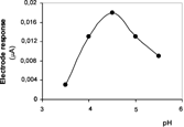
Effect of pH
The effect of pH on the electrode response between pH 3.5–5.5 was investigated. showed that the maximum response was observed in acetate buffer (50 mM) at pH 4.5. This result is in accordance with the previous works of laccase biosensors [Citation[24]].
Figure 2 Optimum pH of laccase biosensor (pH 3.5–5.5; acetate buffer, T; 25°C).
Effect of Temperature
The effect of temperature on the response of the biosensor system was also examined and maximum sensor response was found at 35°C. But, further experiments were performed at 25°C in order to avoid activity loss.
3.2. Analytical Characteristics
Linear Range
A linear relationship between sensor responses and catechol concentration was obtained in concentration range between 0.5 × 10⁻⁶–5.0 × 10⁻⁶ M for Type I biosensor; a linear graph, defined by the equation y = 0.0054x + 0.0035, with a correlation coefficient r2 = 0.990. In the case of using phenol as a substrate, the equation of linear graph becomes y = 0.0272x + 0.003, with a correlation coefficient of r2 = 0.997 in the range of 0.25 × 10⁻⁶ – 2.0 × 10⁻⁶ M. When Type II biosensor was used as support material, linearity was observed in concentration range between 2.5 × 10⁻⁶–3.0 × 10⁻⁵ M of catechol (y = 0.0051x–0.0045, r2 = 0.990). At higher concentrations, standard curves for all systems showed a deviation from linearity. Results of linear regression of the calibration curves are reported in . SPEs sensitivity is higher than GCE sensitivity if the surface area of the electrodes is considered. This is probably due to the rough surface of the SPE.
Table 1. Linear regression coefficients, standard error (S.E), coefficient of variation (cv), Lod and Loq of the biosensors obtained by MTF deposited on GCE and SPE
Sample Application
The next part of the study was the application of developed sensor for determination of phenol in wastewater samples. In these experiments, instead of substrate, wastewater samples with appropriate dilutions were added to the reaction cell. When samples containing 0.25 and 0.5 µM of phenol added the concentrations were calculated as 0.25 ± 0.021 and 0.51 ± 0.005 µM from the calibration curve. Our findings showed that the system could be easily and usefully addressed for the screening of phenolic compounds in industrial waste water samples with acidic nature.
CONCLUSION
The aim of the present study was to develop an alternative procedure for monitoring the consumption of oxygen for the determination of phenolic compounds. As a part of the electrochemical biosensor, the choice of the electrode transducer is important in terms of detection of biologically essential substances. Mercury electrodes are frequently used in electroanalytical techniques because they possess sensitivity and reproducibility and have wide cathodic potential range. As MTFE behaves like a complete mercury electrode and provides a flat surface suitable for immobilization, it allows the usage of mercury electrodes in biosensing systems. On the other hand, the attractive behavior of thin film mercury SPGEs as biosensor compound have proven that these electrodes perform in a manner comparable with conventional electrodes for these systems. The proposed system could be functional as disposable arrays for field use. The immobilization method provides the mild conditions to continue the enzyme activity. Utility of gelatin as an immobilization material provides the use of same electrode for several time without needing any film formation. The advantages of both screen printed disposable electrodes [Citation[26]] and MTFE enable us to get economical and easy–to–use systems for environmental analyses.
REFERENCES
- Wang, J. (2000). Electroanalytical Chemistry, 2nd ed., Wiley: New York.
- Ertaş, N., Timur, S., Akyιlmaz, E., Dinçkaya. (2001). Specific determination of hydrogen peroxide with a catalyse biosensor based on mercury thin film electrode. Turk. J. Chem. 24: 95.
- Frenzel, W. (1993). Mercury films on glassy carbon support: attributes and problems. Analytica Chimica Acta 273: 123. [CSA], [CROSSREF]
- Economou, A., Fielden, P.R. (2003). Mercury film electrodes: developments, trends and potentialities for electroanalysis. Analyst 128: 205. [PUBMED], [INFOTRIEVE], [CSA]
- Jordan, J., Pletschke, B.I., Leukes, W.D. (2004). Purification and partial characterization of a thermostable laccase from an unidentified basidiomycete. Enzyme and Microbial Technology 34: 635. [CSA], [CROSSREF]
- Freire, R.S., Ferreira, M.M.C., Durán, N., Kubota, L.T. (2003). Dual amperometric biosensor device for analysis of binary mixtures of phenols by multivariate calibration using partial least squares. Analytica Chimica Acta 485: 263. [CSA], [CROSSREF]
- Notsu, H., Tatsuma, T. (2004). Simultaneous determination of phenolic compounds by using a dual enzyme electrodes system. Journal of Electroanalytical Chemistry 566: 379. [CSA], [CROSSREF]
- Janda, K.V., Krijt, J. (1984). Recovery of phenols from water by continous steam distillation-extraction. Journal of Chromatography A 283: 309. [CSA], [CROSSREF]
- Alimpiev, S.S., Mlynski, V.V., Belov, M.E., Nikiforov, S.M. (1995). Selective detection of phenol impurities in water. Analytical Chemistry 67: 181. [CSA], [CROSSREF]
- Ortega, F., Dominguez, E., Joensson-Pettersson, G., Gorton, L. (1993). Amperometric biosensor for the determination phenolic compounds using a tyrosinase graphite electrode in a flow injection analysis. J. Biotechnol. 31: 289. [PUBMED], [INFOTRIEVE], [CSA], [CROSSREF]
- Oennerfjord, P., Emneus, J., Marko-Varga, G., Gorton, L., Ortega, F., Dominguez, E. (1995). Tyrosinase graphite-epoxy based composite electrodes for detection of phenols. Biosens. Bioelectron 10: 607. [CSA], [CROSSREF]
- Cosnier, S., Innocent, C. (1993). A new strategy for the construction of a tyrosinase-based amperometric phenol and o-diphenol sensor, Bioelectrochem. Bioenerg. 31: 147. [CSA], [CROSSREF]
- Marko-Varga, G., Emneus, J., Gorton, L., Ruzgas, T. (1995). Development of enzyme based amperometric sensors for the determination of phenolic compounds. Trends Anal. Chem. 14: 319. [CSA]
- Wang, J., Fang, L., Lopez, D. (1994). Amperometric biosensor for phenols based on a tyrosinase-graphite-epoxy biocomposite. Analyst 119: 455. [PUBMED], [INFOTRIEVE], [CSA], [CROSSREF]
- Kim, M.A., Lee, W.-Y. (2003). Amperometric phenol biosensor based on sol-gel silicate/Nafion composite film. Analytica Chimica Acta 479: 143. [CSA], [CROSSREF]
- Bartak, P., Frnkova, P., Cap, L. (2000). Determination of phenols using simultaneous steam distillation-extraction. J. Chromatogr. A 867: 281. [PUBMED], [INFOTRIEVE], [CSA], [CROSSREF]
- Bosch, F., Font, G., Manes, J. (1987). Ultraviolet spectrophotometric determination of phenols in natural and waste waters with iodine monobromide. Analyst 112: 1335. [PUBMED], [INFOTRIEVE], [CSA], [CROSSREF]
- Yaropolov, A.I., Kharybin, A.N., Emnéus, J., Marko-Varga, G., Gorton, L. (1995). Flow injection analysis of phenols at a graphite electrode modified with co-immobilised laccase and tyronisine. Analytica Chimica Acta 308: 137. [CSA], [CROSSREF]
- Freire, R.S., Duran, N., Kubota, L.T. (2002). Development of laccase based flow injection electrochemical biosensor for the determination of phenolic compounds and its application for monitoring remediation of Kraft E1 paper mill effluent. Analytica Chimica Acta 463: 229. [CSA], [CROSSREF]
- Kulys, J., Vidziunaite, R. (2003). Amperometric biosensors based on recombinant laccases for phenol determination, Biosens. Bioelectron 18: 319. [PUBMED], [INFOTRIEVE], [CSA], [CROSSREF]
- Xu, F. (1996). Oxidation of phenols, anilines and benzenethiols by fungal laccases: correlation between activity and redox potential as well as halide inhibitions. Biochemistry 35: 7608. [PUBMED], [INFOTRIEVE], [CSA], [CROSSREF]
- Timur, S., Pazarlιoğlu, N., Pilloton, R., Telefoncu, A. (2003). Detection of phenolic compounds by thick film sensors based on pseudomonas putida. Talanta 61: 87. [CSA], [CROSSREF]
- Timur, S., Della Seta, L., Pazarlιoğlu, N., Pilloton, R., Telefoncu, A. Screen printed graphite biosensors based on bacterial cells. Process Biochem. ( in press). [CSA]
- Timur, S., Pazarlιoğlu, N., Pilloton, R., Telefoncu, A. (2004). Thick film sensors based on laccases from different sources immobilized in polyaniline matrix. Sensors & Actuators B: Chemical 97(1): 132. [INFOTRIEVE], [CSA], [CROSSREF]
- Wolfenden, B.S., Willson, R.L. (1982). Radical cations as reference chromogens in kinetic studies of one electron transfer reactions pulse radiolysis studies of 2,2-azinobis-(3-ethylbenzthiazoline-6-sulphonate). J. Chem. Soc., Perkin Trans. 2: 805. [CSA]
- Meier, P.C., Zund, R.E. (1993). Statistical methods in analytical chemistry, in Chemical Analysis, J.D. Winefordner, Ed., J. Wiley & Sons Inc.: New York, vol. 123, p. 87.