
Article title: Two Cyclin-Dependent Kinases Promote RNA Polymerase II
Transcription and Formation of the Scaffold Complex
Authors: Liu, Y., Kung, C., Fishburn, J., Ansari, A. Z., Shokat, K. M., & Hahn, S.
Journal: Molecular and Cellular Biology
Bibliometrics: Volume 24, Number 4, pages 1721 – 35
DOI: https://doi.org/10.1128/MCB.24.4.1721-1735.2004
In several of the figure panels in this publication (Figs 1A, 2C, 3A), intervening lanes were spliced out to remove data not important for our conclusions and this was not indicated in the published figures. In another figure (Fig 5A), concerns were raised on PUBPEER about whether this image contained unannotated lane splicing and alteration of the image background (https://pubpeer.com/publications/765E62F7376C08BCC55EF73CF232DA). We examined the original data (Western Blot images from the Odyssey imaging system and transcription assay results from phosphorimager data) and made revised figure panels using current accepted standards for figure preparation. There are no changes in any of the conclusions from these corrections and the revised figures look essentially the same as the published figures except that the lane splices are clearly indicated. Examination of the data used to generate Fig 5A showed no lane splicing or alteration of the image background and a high-resolution image of the original data was used to make a revised figure. All original data (uncropped gel images and revised figures) are publicly available at Mendeley data (https://data.mendeley.com/datasets/kkx4tyjyg7/1).
Figure legends associated with revised panels (copied from the original publication with italicized text added for this correction)
FIG. 1. Bur1 and Ctk1 are not PIC components. (A) Nuclear extracts (NE) made from Kin28-Flag, Bur1-Flag, and Ctk1-Flag strains were incubated with the immobilized template for 40 min. PICs were isolated and analyzed by Western blotting. The top panel was probed with the anti-Flag M2 antibody. The asterisk indicates the position of Kin28-Flag, Bur1-Flag, or Ctk1-Flag protein. The lower three panels were probed with antibodies directed against known PIC components. Lanes were spliced out to omit results using the VP16 activator.
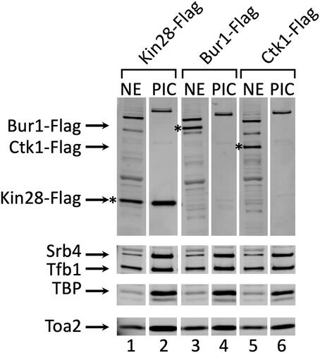
FIG. 2. Inhibition of Kin28-as and Srb10-as activities by NA-PP1. (Only lower panel of 2C shown in revised figure) (C) Immune precipitates from Srb10-as-Flag, Srb10-wt-Flag, and untagged strains were assayed for kinase activities in the presence of increasing amounts of NA-PP1. Reactions and quantitation were performed as described for panel B. Immune precipitate from an untagged strain was used as a control. The lower panel is a Western analysis of equal amounts of the anti-Flag immune precipitates. Lanes were spliced out to omit lanes containing 2-fold lower volume of IP samples so that only the highest volume of IP samples (6 microliters) is shown.
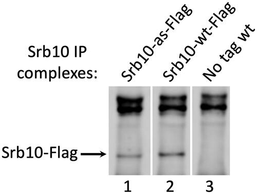
FIG. 3. Both Kin28 and Srb10 can promote transcription. (A) (only upper panel of Fig 3A shown in revised figure) Nuclear extracts (NE) made from wild-type, Kin28-as, Srb10-as, or Kin28-as Srb10-as strains were used as indicated. The transcription reactions were performed as described in Materials and Methods. A lane was spliced out to omit a lower concentration of NA-PP1 used in the experiment.
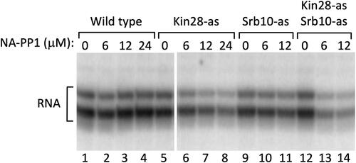
FIG. 5. Inhibition of Kin28 and Srb10 kinases inhibits PIC dissociation. PICs were assembled on the immobilized template using nuclear extracts (NE) made from wild-type, Kin28-as, Srb10-as, and Kin28-as Srb10-as strains. Variable amounts of NA-PP1 along with 600 μM ATP - 10 μCi of [γ-32P]ATP were added for 4 min. (A) (only right panel second row of Fig 5A shown in revised figure) Scaffold complexes (Scaf) were washed, isolated by PstI digestion, and analyzed by Western blotting for the indicated factors. Factors in the PIC without ATP addition (PIC) are shown for comparison.
![FIG. 5. Inhibition of Kin28 and Srb10 kinases inhibits PIC dissociation. PICs were assembled on the immobilized template using nuclear extracts (NE) made from wild-type, Kin28-as, Srb10-as, and Kin28-as Srb10-as strains. Variable amounts of NA-PP1 along with 600 μM ATP - 10 μCi of [γ-32P]ATP were added for 4 min. (A) (only right panel second row of Fig 5A shown in revised figure) Scaffold complexes (Scaf) were washed, isolated by PstI digestion, and analyzed by Western blotting for the indicated factors. Factors in the PIC without ATP addition (PIC) are shown for comparison.](/cms/asset/41425c94-5930-4533-be7d-f35283546250/tmcb_a_2245732_f0004_b.jpg)