Abstract
The sperm morphology of the polychaete Lumbrineris (Scoletoma) impatiens (Claparède, 1868) (Polychaeta, Lumbrineridae) of a sub-tidal population from the Gulf of Naples was studied both by transmission electron microscopy (TEM) and high-resolution scanning electron microscopy (HRSEM). In particular, the ultrastructural characteristics of mature male cells, the shape and structure of the acrosome, the nucleus and the flagellum were investigated by TEM. HRSEM images show the sperm external morphology and allow to classify the male gamete as an ect-aquasperm, by having a short head, four to five rounded mitochondria and a long tail, and generally associated with free-spawning. The structure of the spermatozoon of L. impatiens strictly recalls that of the male cell of the Lumbrineris sp. collected in New South Wales, Australia, the only one of the genus so far examined. On the other hand, by comparing these findings with those obtained on the external morphology of male gametes of the Lumbrineris sp. from Grappler Inlet, Vancouver Island, it emerges once again that significant variations are present. This confirms the extraordinary variability of polychaetes' reproductive characters even at genus level.
Introduction
The identification of Lumbrineris species, as that of other genera of polychaetes, is made difficult by the absence of reliable diagnostic characters and by lack of knowledge of their reproductive features. Environmental constraints also can play an important role in speciation, affecting the structure and shape of sperm cells. Cazaux (Citation1972) and Rouse (Citation1988), respectively, described the larval development of L. impatiens by artificial fertilization and the morphology of sperm of Lumbrineris sp. by TEM. Messina et al. (Citation2005) recently demonstrated the close relationship between free-spawning and the ect-aquasperm morphology in a population of Lumbrineris (Scoletoma) impatiens (Claparède, 1868) (Lumbrineridae) from the Gulf of Naples (Italy), by in vitro experiments on fertilization and by TEM observations. Furthermore, Leander et al. (Citation2003) studied the phylogeny of Lecudina spp., a marine gregarine parasite of Lumbrineris sp. from Grappler Inlet, Vancouver Island, and described by SEM its external morphology: we noticed that on the cortex of a Lecudina trophozoite there were some Lumbrineris sperm cells.
The purpose of this study is to determine the ultrastructural morphology of the male gamete of L. impatiens by TEM and HRSEM, comparing it with the few data available so far for the genus Lumbrineris.
Materials and methods
Individuals of L. impatiens were collected during May 2005 in the Gulf of Naples from professional bait diggers and then transferred to the aquaria of the Zoological Station ‘Anton Dohrn’, Benthic Ecology Laboratory, Ischia, Naples. The mature male gametes were extracted from the coelomatic cavity and diluted in filtered seawater (0.2 μm, salinity 35‰) to activate the cells motility (Pacey et al. Citation1994). The ability of the sperm fertilization was evaluated by means of in vitro fertilization (Eckberg & Hill Citation1996) using eggs obtained from ripe females, as previously described for males. For SEM, the activated sperm cells were treated with paraformaldehyde 1% and 1.25% glutaraldehyde in sodium cacodylate 0.15 M for 1 h. Samples were prepared on round slides (12 mm diameter) covered with poly-l-lysine 0.1% and left in a moist chamber at RT overnight. After washing with buffer (PBS, 0.85% NaCl), the specimens were dehydrated through a graded acetone series, dried with Critical Point Dryer and coated with platinum (3 nm) using an Emitech K575 sputter. The observations of the exterior spermatozoa morphology were made by a Hitachi S4000 FE. For the analysis by TEM, a pellet of motile sperm was fixed for 2 h in paraformaldehyde 1% and 1.5% glutaraldehyde in cacodylate buffer 0.15 M, pre-stained in aqueous uranyl acetate 0.25%, osmicated, dehydrated and embedded in Epon. Ultrathin sections (90nm) were stained with bismuth subnitrate (Riva Citation1974) and observed with a JEOL 100S.
Results
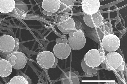
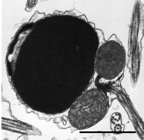
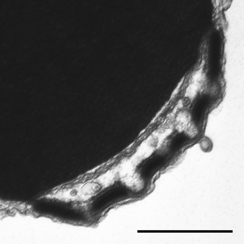
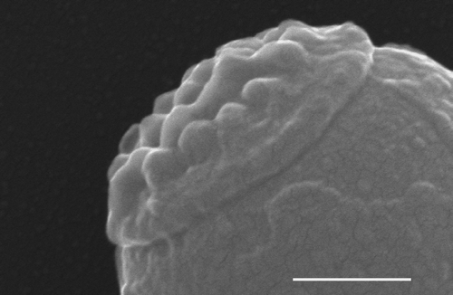
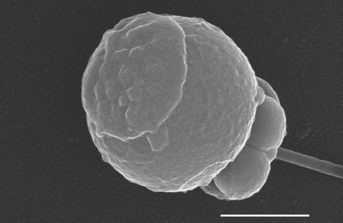
The spermatozoa, observed by light microscopy after dilution in filtered seawater, acquired motility in a few minutes, remaining active for up to an hour (26°C). Fertilization techniques allowed us to obtain the fusion of gametes, and the first development was observed up to the stage of 16 cells, confirming the fertilization capability of sperm. The ultrastructure of the Lumbrineris impatiens spermatozoon is semi-diagramatically illustrated in . By SEM, at low magnification, the mature male gamete of L. impatiens has a subspherical form, with a discoidal acrosome and a long tail ( and ). The plasmalemma, which closely adheres to the whole cell, is smooth to the level of mitochondria and of flagellum, whereas it is granulous in the nuclear and acrosomal regions (). The acrosome, observed by SEM, appears flattened with crenulated edges and occupies a large part of the upper surface ( and ); it is characterized by having the appearance of an apple pie () with hemispherical swellings. These external characteristics correspond to the structures visible by TEM ( and ); they consist of the acrosomal vesicle and of the subacrosomal space, fenestrated by the large protrusions of the subacrosomal material. The longitudinal sections of the acrosomal vesicle () show an inner electron-dense membrane and an outer electron-transparent layer. In transverse sections, the bulges of subacrosomal material, which can be considered sensu Baccetti (Citation1979) as perforatorium, are regularly spaced out across the acrosomal membranes which are in close contact with the plasmalemma. The nucleus is subspherical and covered by the acrosomal complex; below it, there are four to five mitochondria and the centriolar apparatus (). The chromatin, very electron-dense, has a granular appearance with some inclusions of moderate electron density. The large spherical mitochondria show tubular–vesicular cristae. The centriolar complex consists of two centrioles orthogonally arranged and interconnected by very electron-dense material. The distal centriole acts as a basal body for the axoneme which has a 9+2 organization of microtubules. The long tail contains the axoneme.
Figure 1. Schematic drawing of spermatozoon structure in Lumbrineris impatiens. Av, acrosomal vesicle; dc, distal centriole; f, flagellum; m, mitochondrion; n, nucleus; pc, proximal centriole; pl, plasmalemma; sr, satellite ray.
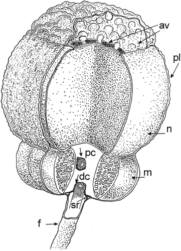
Figure 4. SEM: Mature spermatozoon of L. impatiens. The acrosome occupies a large part of the upper surface. Bar 1 μm.
Discussion
The morphological observations by TEM and by HRSEM, on sperm of L. impatiens, confirm the ect-aquasperm type, closely associated with external fertilization in the aquatic environment. The structure of the spermatozoon of L. impatiens strictly recalls that of the male cell of the Lumbrineris sp. collected in New South Wales, Australia, the only one of the genus so far examined (Rouse Citation1988). On the other hand, the spermatozoon of Lumbrineris sp. from Grappler Inlet, Vancouver Island (Leander et al. Citation2003), compared with those of the species so far studied (Rouse Citation1988; Messina et al. Citation2005), differs greatly in external morphology, in size of the nucleus, which is rather elongated, and in number and shape of mitochondria. These findings highlight the considerable variability of form and structure of sperm in similar polychaetes species which inhabit distant seas and oceans, as noted also by our previous studies (Conti et al. Citation2005).
Acknowledgements
This research was supported by the Campania Regional Administration (funds SFOP, POR 2000–2006, measure 4.23).
References
- Baccetti , B. 1979 . “ The evolution of the acrosomal complex ” . In The Spermatozoonpp , Edited by: Fawcett , DW and Bedford , JM . 305 – 329 . Baltimore, MD : Urban & Schwarzenberg .
- Cazaux , C. 1972 . Développement larvaire d'annélides polychètes (Bassin d'Arcachon) . Archives de Zoologie Expérimentale et générale , 113 : 71 – 108 .
- Conti , G , Loffredo , F and Lantini , MS. 2005 . Fine structure of the spermatozoon of Diopatra neapolitana (Polychaeta, Onuphidae) . Zoomorphology , 124 : 155 – 160 .
- Eckberg WR, Hill SD. 1996. Chaetopterus – Oocyte maturation, early development, and regeneration. Marine Models Electronic Record. http://www.mbl.edu/BiologicalBulletin/MMER/ECK/EckTit.html (http://www.mbl.edu/BiologicalBulletin/MMER/ECK/EckTit.html)
- Leander , BS , Harper , JT and Keeling , PJ. 2003 . Molecular phylogeny and surface morphology of aseptate marine gregarines (Apicomplexa): Selenidium and Lecudina . Journal of Parasitology , 89 : 1191 – 1205 .
- Messina , P , Di Filippo , M , Gambi , MC and Zupo , V. 2005 . In vitro fertilization and larval development of a population of Lumbrineris (Scoletoma) impatiens (Claparède) (Polychaeta, Lumbrineridae) of the Gulf of Naples (Italy) in relation to aquaculture . Invertebrate Reproduction and Development , 48 : 31 – 40 .
- Pacey , AA , Cosson , JC and Bentley , MG. 1994 . The acquisition of forward motility in the spermatozoa of the polychaete Arenicola marina . Journal of Experimental Biology , 195 : 259 – 280 .
- Riva , A. 1974 . A simple and rapid staining method for enhancing the contrast of tissues previously treated with uranyl-acetate . Journal of Microscopy , 19 : 105 – 108 .
- Rouse , GW. 1988 . An ultrastructural study of the spermatozoa of Eulalia sp. (Phyllodocidae), Lepidonotus sp. (Polynoidae), Lumbrineris sp. (Lumbrineridae) and Owenia fusiformis (Oweniidae) . Helgoländer Meeresuntersuchun , 42 : 67 – 78 .
