Abstract
Potato spindle tuber viroid (PSTVd) is an infectious small, circular, non-coding single-stranded RNA that induces disease on many crop species, ornamental plants, weeds and parasitic plants. PSTVd propagate in their host as a population of closely related but non-identical RNA variants referred to as quasispecies. Recently, we have described three de novo arising PSTVd variants in the parasitic plant Phelipanche ramosa after mechanical inoculation with the PSTVd KF440-2 isolate. These P. ramosa derived mutants were designated as G241-C, C208-U and C227-U PSTVd variants. Each of these variants carries a single-nucleotide substitution compared to the PSTVd KF440-2 sequence from which they are considered to have evolved. Here we complement our previous studies on these mutants by exploring their potential to infect the floral organs of tomato plants. We found that the PSTVd G241-C and C208-U variants were able to replicate in systemic leaves and floral organs of tomato plants, while the PSTVd C227-U variant did not develop systemic infection. Furthermore, we analysed the progeny of these PSTVd variants in sepals and petals of tomato plants for retention of the specific mutations.
Introduction
Potato spindle tuber viroid (PSTVd) is a representative member of family Pospiviroidae.[Citation1] Its genome consists of a 359-nt-long non-coding single-stranded RNA, which adopts a rod-like conformation with five distinct domains (from the 5′ terminal end: left (TL), pathogenic (P), central conserved region (CCR), variable (V) and terminal right (TR) domain) built of (paired) stems and (unpaired) loops.[Citation2] The significance and role of more than one domain in a particular function have been reported. However, certain PSTVd structural elements (virulent modulating (VM) region in the P domain; RY motif in the TR domain) or temporally acquired metastable structures (HPII) have been associated with specific functions.[Citation3]
PSTVd replicates in the nucleus via the asymmetric rolling circle pathway of multiplication; it exploits the host RNA polymerase II, host cleavage factors and host ligase for its life cycle.[Citation4,5] PSTVd replicates and moves to sink, but not source, leaves.[Citation6] It was absent from shoot apical meristems. In the flowers of mechanically infected tomato and Nicotiana benthamiana plants, PSTVd was present in the sepals, but was absent in the petals, stamens and ovary.[Citation6] This observation was attributed to restricted traffic of PSTVd into these organs and not to suppression of replication.[Citation7] However, our preliminary analysis showed that the PSTVd KF440-2 isolate could infect both sepals and petals of mechanically infected tomato plants.[Citation8] Successful replication of PSTVd in all floral organs was reported also in transgenic N. benthamiana that expressed cDNA of PSTVd intermediate strain under the 35S promoter.[Citation7]
PSTVd propagates as a population of closely related but not identical RNA variants. The variants appear due to the high rate of replication, disabled error prone activity of RNA polymerase II and selective pressure of the host.[Citation9–13] The effect of sequence variations on disease expression was studied in a pool of single-point mutants,[Citation14–17] ‘thermomutants’ [Citation9] or in libraries of mutants with genomes partially or fully randomized at certain positions (in vivo Selex) [Citation12]. It was found that the mutations affected either replication or movement of the PSTVd.[Citation10–12,Citation18] Thus, many mutations were identified as stably maintained and preserved in the progeny,[Citation17] while others were identified as untolerated in a given host and were substituted by changes in different positions of the genome or by reversion to the wild-type sequence.[Citation10,Citation13,Citation19–21]
Characterization of new PSTVd variants emerging as a result of adaptation to alternative hosts might help for further determination of specific nucleotide changes that contribute to the systemic infectivity of PSTVd.[Citation22] Recently, we reported that the parasitic plant Phelipanche ramosa was able to sustain replication of PSTVd.[Citation23] The analysis of progeny derived from mechanical inoculation of the stem of P. ramosa with the KF440-2 isolate revealed the presence of three variants, with mutations located in the lower strand of the C domain (PSTVd G241-C), in the lower part of the RY motif of the TR domain (PSTVd C208-U) and in the lower part of the V domain (PSTVd C227-U).[Citation23] The secondary structure of these variants was analysed and the possible conformation changes were predicted by an in silico RNA prediction tool (MFold).[Citation24] Bioassays on Rentita tomato plants showed that the PSTVd G241-C and PSTVd C208-U mutants replicated in tomato plants, while PSTVd C227-U did not infect the experimental host.[Citation23] In this study, we complement the current knowledge about these P. ramosa PSTVd variants by exploring their capability to move from an inoculated tomato leaf towards tomato floral organs and to propagate in them.
Materials and methods
Plant material and inoculation of plants
Tomato plants (Solanum lycopersicum) cv. Rentita were grown in a green chamber at 28 °C, at a 14/10 h light/dark cycle. Tomato seedlings were mechanically inoculated with 250 ng of the PSTVd KF440-2 isolate or P. ramosa PSTVd (G208-U, C227-U and G241-C) variants transcribed in vitro. Mock-inoculated plants were treated with 1% potassium phosphate buffer.
Plasmid constructs
P. ramosa PSTVd variants G208-U, C227-U and G241-C were cloned in a pCRII TOPO cloning vector (Invitrogen). BamHI tailed PSTVd oligos (Fw 5′-AGG GAT CCC CGG GGA AAC CTG GAG CGA-3′; Rev 5′-GGG GAT CCC TGA AGC GCT CCT CCG AGC-3′) were used for subcloning of the PSTVd variants in a pHa106 plasmid.
In vitro transcription
The longer-than-unit-length PSTVd(+) RNA of the KF440-2 isolate and each P. ramosa variant (G208-U, C227-U and G241-C) were obtained by in vitro transcription of EcoRI-linearized pHa106 plasmids, using Sp6 RNA polymerase.[Citation25] Approximately 1 μg of HindIII-linearized pHa106 plasmid was used as a template for in vitro transcription of longer-than-unit length PSTVd(−) RNA of the KF440-2 isolate synthesized with T7 RNA polymerase. Digoxigenin (DIG)-labelled PSTVd(−) RNA was prepared with DIG-labelled Uridine triphosphate (UTP), following the instructions of the DIG Northern Starter Kit (Roche).
RNA extraction
Total RNAs were extracted from systemic tomato leaves at 28 days post inoculation (d.p.i) and from floral organs at 65 d.p.i as previously described.[Citation26] The quality of RNA preparations was checked in a 1% agarose gels stained with ethidium bromide and visualized under ultraviolet (UV) light. The RNA quantity was determined spectrophotometrically.
Northern blot analysis
Total RNA isolated from floral organs and systemic leaves (5 μg) was separated in a 1.4% formaldehyde agarose gel. The gel was transferred to a Hybond-XL nylon membrane (Amersham Biosciences) overnight by capillary transfer, using 2 × saline sodium citrate buffer (SSC), and the RNA preparations were immobilized by UV cross-linking. The membrane was stained with methylene blue and then prehybridized with sodium dodecyl sulphate (SDS) hybridization buffer (7% SDS, 50% deionized formamide, 5 × SSC, 2% blocking reagent, 0.1% (w/v) N-laurylsarcosine, 50 mmol/L sodium phosphate, pH 7.0) at 68 °C for 1–2 h. Hybridization was carried out overnight at 68 °C in the same buffer with the addition of DIG-labelled PSTVd(−) RNA probe. Then, the membrane was washed twice in 2 × SSC/0.1% SDS for 5 min at room temperature, followed by two more washes in 0.1 × SSC/0.1% SDS for 15 min at 68 °C. The detection was performed with an anti-DIG-antibody (Ab) conjugated to alkaline phosphatase and visualized with nitroblue tetrazolium (NBT)/5-bromo-4-chloro-3-indolyl phosphate ready-to-use tablets (Roche).
Reverse transcription polymerase chain reaction (RT-PCR)
RT-PCR was used for cDNA synthesis of genomic and anti-genomic PSTVd strands. One microgram of total RNA obtained from sepals and petals of infected and mock-inoculated tomato plants was used for first-strand cDNA synthesis (USB corporation (USB)). To synthesize cDNA specific for PSTVd, the primers described by Weidemann and Buchta [Citation27] were used: cDNA specific for the genomic PSTVd(+) strand was synthesized with PSTVd reverse primer (5′-CCCTGAAGCGCTCCTCCGAG-3′), while the cDNA specific for the antigenomic PSTVd(−) strand was obtained with the forward primer (5′-ATCCCCGGGGAAACCTGGAGCGA-3′). The template RNA was removed by additional ribonuclease (RNase H) treatment at 37 °C for 1 h.
Cloning and sequencing of PSTVd variants from petals and sepals
For sequencing purposes, a PSTVd-specific PCR reaction was set up using Vent DNA polymerase (New England Biolab). Amplification products were cloned into a pCRII TOPO vector system (Invitrogen), according to the manufacturer's instructions. Two independent randomly selected clones derived from sepals and petals were analysed by bidirectional sequencing (T7 and SP6 promotor primers) (AGOWA, Germany).
Trypan blue staining
Cross sections of tomato leaves infected with the PSTVd KF440-2 isolate and P. ramosa variants (G241-C, G208-U, C227-U) were treated with lactophenol–trypan blue solution (10 mL of phenol, 10 mL of 100% glycerol, 10 mL of lactic acid, 0.02 g of trypan blue, 10 mL of water mixed with 96% ethanol (1:2)) and boiled for 2 min. Then, the specimens were incubated for 1 h at room temperature in the same solution. The slices were destained in chloral hydrate solution (pH = 1.2). Total destaining was achieved in approximately 24 h. Observation of leaf necrosis was performed with light microscopy in 50% glycerol.
Results and discussion
Detection of leaf necrosis in tomato plants
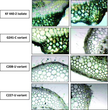
PSTVd symptoms could vary from mild to lethal depending on the host–PSTVd strain combinations and environment conditions. The main symptoms expressed by sensitive cultivars upon PSTVd infection are leaf malformations, chlorotic/necrotic lesions, growth reduction and significant loss in yield. In the present study, successful PSTVd infection on tomato plants was observed by detection of the necrotic lesions in systemic leaves. The trypan blue staining technique assesses the cell viability of leaves. The dead cells are visualized as coloured in blue. In , the leaves infected with PSTVd KF440-2 or PSTVd G241-C showed blue-stained areas, indicating that PSTVd induced necrosis. The PSTVd C208-U variant did not show PSTVd-specific necrosis at 28 d.p.i.; such was observed at a later stage (close to 35 d.p.i). The PSTVd C227-U variant did not induce necrosis and was confirmed as non-infectious on tomato plants.
Northern blot analysis to detect PSTVd(+) RNA in systemic leaves of tomato plants
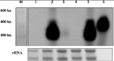
Northern blot analysis showed a size-specific PSTVd signal in tomato plants infected with PSTVd KF440-2 and the PSTVd variant G241-C (). A positive but less intensive signal was observed in tomato plants infected with the PSTVd C208-U variant. No PSTVd specific signal was found in the systemic leaves of tomato plants infected with the PSTVd C227-U variant (). However, this variant was reported to be able to replicate in the inoculated leaves [Citation23] and was further studied.
Figure 2. Northern blot analysis to detect PSTVd infection with P. ramosa PSTVd variants on systemic leaves of tomato plants. Lane M: RNA Ladder 0.1–1 kb (USB); Lane 1: mock-inoculated; Lane 2: infected with PSTVd variant G241-C; Lane 3: infected with PSTVd variant G208U; Lane 4: infected PSTVd variant C227U; Lane 5: infected with PSTVd KF440-2; Lane 6: in vitro synthesized PSTVd(+) RNA. rRNA was used as loading control.

Detection of P. ramosa PSTVd variants in floral organs of tomato plants
RT-PCR analysis
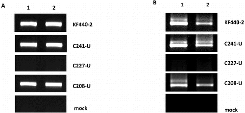
The results from the PSTVd-specific RT-PCR analysis are shown in . PSTVd(+)-specific products were detected both in the sepals and petals of tomato plants infected with the KF440-2 isolate and P. ramosa variants G241-C and C208-U ((A)). PSTVd(−)-specific signals as well as replication intermediates of lower electrophoretic mobility were detected in the sepals and petals of tomato plants infected with the KF440-2 isolate and P. ramosa variants G241-C and C208-U ((B)). No PSTVd signals were observed either in the sepals and petals of tomato plants infected with P. ramosa C227-U or in the mock-inoculated tomato plants ((A) and (B). Our experimental system showed for the first time that the PSTVd KF440-2 isolate and two of the P. ramosa PSTVd variants G241-C and C208-U have the ability to replicate in all floral organs of mechanically infected tomato plants, which had been reported only in transgenic N. benthamiana that expressed cDNA of PSTVd intermediate strain and was not observed in the same mechanically inoculated system [Citation7]. Moreover, our results revealed both replication ability and trafficking competence of the KF440-2 isolate and P. ramosa variants G241-C and C208-U in the floral organs of tomato plants.
Figure 3. RT-PCR to detect PSTVd(+) strand (a) and PSTVd(−) strand (b) in floral organs of tomato plants infected with PSTVd KF440-2 isolate and P. ramosa variants and mock-inoculated tomato plants. Lane 1: sepals; Lane 2: petals. The PSTVd variants used for infection are indicated on the right of each panel.

Northern blot analysis
To confirm the RT-PCR analysis, we applied the Northern blot hybridization technique. PSTVd specific signals were detected both in the sepals and the petals of tomato plants infected with the KF440-2 isolate and the G241-C and C208-U PSTVd variants (). There was no PSTVd signal in the mock-inoculated tomato plants and the PSTVd C227-U variant. Thus, the results from the Northern blot analysis confirmed the RT-PCR data.
Figure 4. Northern blot analysis to detect PSTVd in floral organs of tomato plants. (a) Detection of PSTVd KF440-2. Lane 1: in vitro synthesized PSTVd(+) transcript; Lane 2: PSTVd KF440-2 in sepals; Lane 3 PSTVd KF440-2 in petals. (b) Detection of P. ramosa PSTVd variants. Lane 1: in vitro synthesized PSTVd(+) transcript; Lane 2: mock-inoculated sepals; Lane 3: mock-inoculated petals; lane 4: G241-C variant in sepals; Lane 5: G241-C variant in petals; Lane 6: C208-U variant in sepals; Lane 7: C208-U variant in petals; Lane 8: C227-U variant not detected in sepals; Lane 9: C227-U variant not detected in petals.

Sequence analysis of PSTVd progeny
Analysis of the PSTVd populations in the sepals and petals of tomato plants inoculated with the KF440-2 isolate and the studied P. ramosa variants was performed by bidirectional sequencing of two independently obtained complete cDNA clones from two separate floral organs. We revealed stable maintenance of the KF440-2 isolate in the sepals and petals of tomato plants. The PSTVd G241-C and C208-U sequence variants were also retained in the sepals and petals of tomato plants. However, parallel to parental genomes, we observed a reversion to the wild-type KF440-2 sequence in petals infected with the PSTVd G241-Cvariant and in sepals and petals infected with the PSTVd C208-U variant. These sequencing results are in support of a heterogeneous nature of the PSTVd population in tomato plants infected with P. ramosa variants. The specific nucleotide change characteristic of these variants could be implicated in their replication ability and competence for trafficking towards the floral organs of the infected tomato plant. The PSTVd variants obtained from P. ramosa were previously reported to adopt structures that differ from the wild-type KF440-2 conformation, by using in silico RNA folding package (Mfold).[Citation28] The PSTVd C208-U variant has shown the most distorted structure compared to the wild-type PSTVd KF440-2. Our results indicated that the C208-U substitution most likely affects either the movement or the replication capacity of the mutant variant in systemic leaves () but does not compromise its replication in floral organs ( and ).
According to the Mfold prediction tool results, the G241-C variant showed preservation of the KF440-2 secondary structure. Similarly to KF440-2, this variant showed systemic movement and successful replication in sepals and petals of tomato plants ( and ). The PSTVd variant C227-U did not show PSTVd-specific signals in systemic leaves as well as in sepals and petals of tomato plants, which suggests that it does not move from the inoculated leaf towards the flower organs. The C227-U mutant has been reported to be defective in trafficking in systemic leaves of N. benthamiana by Zhong et al. [Citation29], who showed that this substitution transforms the A135-C227 loop to an elongated stem and this possibly causes disruption in the interaction with cellular factors that assist different aspects of replication and trafficking.
Conclusions
In the present study, PSTVd G241-C and C208-U variants isolated from P. ramosa were identified in the floral organs of mechanically inoculated tomato plants. We showed that these PSTVd variants successfully replicate both in sepals and in petals. The sequence analysis of the population of their progeny in the floral organs of tomato plants showed retention of these mutations in parallel with sequences that are revertant to the wild-type KF440-2 sequence. The PSTVd C227-U variant was not found in sepals and petals, which suggests that it does not move systemically. This work enriches the knowledge on the systemic trafficking of PSTVd in tomato plants by presenting variants that are able to traffic towards the floral organs without being restricted when moving across different cellular boundaries in the tomato plant.
Acknowledgements
We thank Ms Neshe Nazifova-Tasinova for technical assistance.
Additional information
Funding
References
- Diener TO, Smith DR. Potato spindle tuber viroid. VI. Monodisperse distribution after electrophoresis in 20 per cent polyacrylamide gels. Virology. 1971;46:498–499.
- Keese P, Symons RH. Domains in viroids: evidence of intermolecular RNA rearrangements and their contribution to viroid evolution. Proc Natl Acad Sci USA. 1985;82(14):4582–4586. PubMed PMID: 3860809; PubMed Central PMCID: PMC390429.
- Flores R, Serra P, Minoia S, Di Serio F, Navarro B. Viroids: from genotype to phenotype just relying on RNA sequence and structural motifs. Front Microbiol. 2012;3:217. doi: 10.3389/fmicb.2012.00217. PubMed PMID: 22719735; PubMed Central PMCID: PMC3376415.
- Flores R, Gas ME, Molina-Serrano D, Nohales MA, Carbonell A, Gago S, De la Peña M, Daròs JA. Viroid replication: rolling-circles, enzymes and ribozymes. Viruses. 2009;1(2):317–334. doi: 10.3390/v1020317. PubMed PMID: 21994552; PubMed Central PMCID: PMC3185496.
- Flores R, Grubb D, Elleuch A, Nohales MA, Delgado S, Gago S. Rolling-circle replication of viroids, viroid-like satellite RNAs and hepatitis delta virus: variations on a theme. RNA Biol. 2011;8(2):200–206. PubMed PMID: 21358283.
- Zhu Y, Green L, Woo YM, Owens R, Ding B. Cellular basis of potato spindle tuber viroid systemic movement. Virology. 2001;279(1):69–77. doi: 10.1006/viro.2000.0724. PubMed PMID: 11145890.
- Zhu Y, Qi Y, Xun Y, Owens R, Ding B. Movement of potato spindle tuber viroid reveals regulatory points of phloem-mediated RNA traffic. Plant Physiol. 2002;130(1):138–146. doi: 10.1104/pp.006403. PubMed PMID: 12226494; PubMed Central PMCID: PMC166547.
- Vachev T, Ivanova D, Minkov I, Gozmanova M. [Identification of potato spindle tuber viroid KF440-2 isolate in floral organs of tomato (Solanum lycopersicum)]. Trav Sci Univ Plovdiv Plant. 2011;41(6):7–16. Bulgarian.
- Matousek J, Orctova L, Steger G, Skopek J, Moors M, Dedic P, Riesner D. Analysis of thermal stress-mediated PSTVd variation and biolistic inoculation of progeny of viroid “thermomutants” to tomato and Brassica species. Virology. 2004;323(1):9–23. doi: 10.1016/j.virol.2004.02.010. PubMed PMID: 15165815.
- Nie X. Analysis of sequence polymorphism and population structure of tomato chlorotic dwarf viroid and potato spindle tuber viroid in viroid-infected tomato plants. Viruses. 2012;4(6):940–953. doi: 10.3390/v4060940. PubMed PMID: 22816033; PubMed Central PMCID: PMC3397355.
- Podstolski W, Gora-Sochacka A, Zagorski W. Co-inoculation with two non-infectious cDNA copies of potato spindle tuber viroid (PSTVd) leads to the appearance of novel fully infectious variants. Acta Biochim Pol. 2005;52(1):87–98. doi: 055201087. PubMed PMID: 15827608.
- Wiesyk A, Candresse T, Zagorski W, Gora-Sochacka A. Use of randomly mutagenized genomic cDNA banks of potato spindle tuber viroid to screen for viable versions of the viroid genome. J Gen Virol. 2011;92(Pt 2):457–466. doi: 10.1099/vir.0.026286-0. PubMed PMID: 21068216.
- Gora-Sochacka A, Kierzek A, Candresse T, Zagorski W. The genetic stability of potato spindle tuber viroid (PSTVd) molecular variants. RNA. 1997;3(1):68–74. PubMed PMID: 8990400; PubMed Central PMCID: PMC1369463.
- Gozmanova M, Denti MA, Minkov IN, Tsagris M, Tabler M. Characterization of the RNA motif responsible for the specific interaction of potato spindle tuber viroid RNA (PSTVd) and the tomato protein Virp1. Nucleic Acids Res. 2003;31(19):5534–5543. PubMed PMID: 14500815; PubMed Central PMCID: PMC206474.
- Hammond RW. Agrobacterium-mediated inoculation of PSTVd cDNAs onto tomato reveals the biological effect of apparently lethal mutations. Virology. 1994;201(1):36–45. doi: 10.1006/viro.1994.1263. PubMed PMID: 7513926.
- Qi Y, Ding B. Inhibition of cell growth and shoot development by a specific nucleotide sequence in a noncoding viroid RNA. Plant Cell. 2003;15(6):1360–1374. PubMed PMID: 12782729; PubMed Central PMCID: PMC156372.
- Wassenegger M, Spieker RL, Thalmeir S, Gast FU, Riedel L, Sanger HL. A single nucleotide substitution converts potato spindle tuber viroid (PSTVd) from a noninfectious to an infectious RNA for Nicotiana tabacum. Virology. 1996;226(2):191–197. doi: 10.1006/viro.1996.0646. PubMed PMID: 8955038.
- Zhong X, Ding B. Distinct RNA motifs mediate systemic RNA trafficking. Plant Signal Behav. 2008;3(1):58–59. PubMed PMID: 19704772; PubMed Central PMCID: PMC2633962.
- Owens RA, Thompson SM. Mutational analysis does not support the existence of a putative tertiary structural element in the left terminal domain of Potato spindle tuber viroid. J Gen Virol. 2005;86(Pt 6):1835–1839. doi: 10.1099/vir.0.80869-0. PubMed PMID: 15914863.
- Owens RA, Thompson SM, Kramer M. Identification of neutral mutants surrounding two naturally occurring variants of Potato spindle tuber viroid. J Gen Virol. 2003;84(Pt 3):751–756. PubMed PMID: 12604828.
- Owens RA, Steger G, Hu Y, Fels A, Hammond RW, Riesner D. RNA structural features responsible for potato spindle tuber viroid pathogenicity. Virology. 1996;222(1):144–158. doi: 10.1006/viro.1996.0405. PubMed PMID: 8806495.
- Matousek J, Stehlik J, Prochazkova J, Orctova L, Wullenweber J, Fussy Z, Kováčik J, Duraisamy GS, Ziegler A, Schubert J, Steger G. Biological and molecular analysis of the pathogenic variant C3 of Potato spindle tuber viroid (PSTVd) evolved during adaptation to chamomile (Matricaria chamomilla). Biol Chem. 2012;393(7):605–615. doi: 10.1515/hsz-2011-0286. PubMed PMID: 22944665.
- Vachev T, Ivanova D, Minkov I, Tsagris M, Gozmanova M. Trafficking of the Potato spindle tuber viroid between tomato and Orobanche ramosa. Virology. 2010;399(2):187–193. doi: 10.1016/j.virol.2009.12.022. PubMed PMID: 20110096.
- Zuker M. Mfold web server for nucleic acid folding and hybridization prediction. Nucleic Acids Res. 2003;31(13):3406–3415. PubMed PMID: 12824337; PubMed Central PMCID: PMC169194.
- Tsagris M, Tabler M, Sanger HL. Ribonuclease T1 generates circular RNA molecules from viroid-specific RNA transcripts by cleavage and intramolecular ligation. Nucleic Acids Res. 1991;19(7):1605–1612. PubMed PMID: 1709278; PubMed Central PMCID: PMC333922.
- Papaefthimiou I, Hamilton A, Denti M, Baulcombe D, Tsagris M, Tabler M. Replicating potato spindle tuber viroid RNA is accompanied by short RNA fragments that are characteristic of post-transcriptional gene silencing. Nucleic Acids Res. 2001;29(11):2395–2400. PubMed PMID: 11376158; PubMed Central PMCID: PMC55696.
- Weidemann H, Buchta, U. A simple and rapid method for detection of potato spindle tuber viroid by RT-PCR. Potato Res. 1998;41:1–8.
- Zuker, M., 2003. Mfold web server for nucleic acid folding and hybridization prediction. Nucleic Acids Res. 31(13):3406–3415.
- Zhong X, Archual AJ, Amin AA, Ding B. A genomic map of viroid RNA motifs critical for replication and systemic trafficking. Plant Cell. 2008;20(1):35–47. doi: 10.1105/tpc.107.056606. PubMed PMID: 18178767; PubMed Central PMCID: PMC2254921.

