
ABSTRACT
The aim of this comparative study was to evaluate, at one and five years, the implant success rate in the resorbed maxilla after sinus augmentation with platelet-rich plasma (PRP)/deproteinized bovine bone mineral (DBBM) versus DBBM/collagen membrane (CM). Using a split-mouth design, 10 patients with ≤5 mm of residual alveolar bone were treated with PRP/DBBM or DBBM/CM. After eight months, a total of 22 and 21 implants (Osseospeed™, Astra Tech AB, Sweden) were inserted in the PRP/DBBM and DBBM/CM sites, respectively. The implant success and survival rates, modified plaque and bleeding indices, probing depth and changes in bone level were all evaluated one year later, and again five years later. Only one implant was lost before the prosthetic rehabilitation of the PRP/DBBM group. There were no statistically significant differences in the evaluated parameters for the 1- and 5-year follow-up in the two groups (p > 0.05). After five years of loading, no further implants were lost, giving it an overall success rate of 83%. The clinical study showed that a high implant success and survival rate can be achieved at one year and maintained for up to five years, after a sinus-lift procedure utilizing both combinations.
Introduction
Resorption of the residual bone after tooth extraction leads to difficulties in the rehabilitation of the posterior maxilla with dental implants. Although zygomatic and short implants are topics of interest for clinicians, sinus augmentation is considered the standard of care, especially in severely resorbed maxilla [Citation1,Citation2]. Autogenous bone (AB) is regarded as the gold standard for bone regeneration due to its osteoinductive and conductive properties. However, the use of AB has some disadvantages, such as its morbidity, resorption at the recipient site, and prolonged surgical time [Citation3,Citation4]. Deproteinized bovine bone mineral (DBBM) has been shown to be as effective as AB for use in sinus augmentation procedures [Citation5]. It has been also shown that DBBM has high biocompatibility and osteoconductivity when used in sinus augmentation procedures [Citation6–8].
The use of platelet-rich plasma (PRP) as a method has been proposed to introduce concentrated amounts of polypeptide growth factors (PGF) to the wound site. Platelets have a complex membranous structure that allows them to store, and rapidly release, different protein molecules, including adhesion molecules, such as fibronectin and fibrinogen, and PGF [Citation9,Citation10]. Several clinical studies have been performed with the hypothesis that the combination of the graft material with PRP might expedite healing [Citation8,Citation11–14]. Most of the studies have combined PRP with AB in sinus augmentation procedures [Citation12,Citation14,Citation15]. Long-term evaluations (over one, five or ten years) are rare in terms of comparative studies of DBBM and PRP [Citation8,Citation16].
The purpose of the present clinical study was to evaluate and compare the survival and success rates of rough-surface dental implants placed in the resorbed maxilla after sinus augmentation using DBBM/PRP or DBBM/collagen membranes (CMs) at one and at five years.
Subjects and methods
Patients
The study participants included 10 patients (six men and four women) referred to Yeditepe University's Faculty of Dentistry, Department of Periodontology, for insufficient retention in their upper denture. Four of the six patients were partially edentulous (bilateral loss of molars and premolars). Each patient had a corresponding alveolar atrophy class of C or D [Citation17], and less than 5 mm of residual alveolar bone in the vertical direction, as defined by pre-operative panoramic radiographs and computed tomography (CT). After an explanation of all aspects of the study, as well as alternative treatment regimens, an informed consent form was obtained from all patients. The study design and consent forms were reviewed and approved by the University's Institutional Review Board and the Ethics Committee. The study was performed in accordance with the Helsinki declaration.
Study design
All sinus augmentation surgeries were performed between May 2006 and October 2008, and the implant surgeries were performed between February 2007 and August 2009. This study was based on the split-mouth design, where both treatments are randomly assigned to either the right or left halves of the dentition. One side was treated with DBBM/PRP, whereas the other side was treated with DBBM/CM (or vice versa). The randomization procedure was described elsewhere [Citation13].
Preparation of PRP
The PRP preparation is described elsewhere [Citation13]. The thrombin-activated PRP was mixed with DBBM (Bio-Oss®, Geistlich Biomaterials, Wolhusen, Switzerland).
Sinus augmentation and implant installation
The sinus augmentation procedure was explained in a previous study by the same group [Citation13]. Eight months after the sinus augmentation, implants (Osseospeed ™, Astra Tech AB, Mölndal, Sweden) were installed (). A total of 43 implants were placed. After four months, the implants were exposed and healing abutments were placed. Two weeks later, the healing abutments were made permanent and the prosthetic part of the treatment was performed. Then, the patients were summoned for evaluation in six-month intervals. All patients were re-evaluated for the designated parameters in one, three and five years.
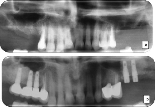
Figure 1. Panoramic radiographs of the representative case: before sinus augmentation (a) and after sinus augmentation and implant placement (b).
Clinical measurements
The day of bridge connection was accepted as baseline day 0. The clinical measurements taken were: modified plaque index (mPLI) [Citation18]; modified sulcus bleeding index (mSBI) [Citation18]; peri-implant suppuration and a probing depth (PD) with a 15-mm UNC periodontal probe (PCP 15 UNC; Hu Friedy®, Limen, Germany). Measurements were taken at four sites on each implant (mesial, mid-buccal, distal and mid-lingual/palatinal); the data were averaged and the resulting median and mean values with standard deviation (±SD) were calculated.
Radiographic measurements
Periapical radiographs were obtained to evaluate the marginal bone level changes at baseline, and at one and five years of loading with a long cone technique [Citation19,Citation20]. The linear distance between the first visible bone--implant contact point and the reference point on the neck of the implant (DIB) was measured at the mesial and distal sites of each implant (in millimetres) and was averaged for each implant to evaluate the crestal bone level changes [Citation19,Citation21]. Then, the median and the mean values were calculated.
Survival and success rates
Implant survival and success rates were assessed according to Buser's criteria [Citation19]. Implants not removed but not meeting the criteria for success were considered to have survived [Citation3]. Implant stability at one and five years of function was evaluated by means of a mobility test with forceps to determine it as mobile or immobile [Citation3].
Statistical analysis
All analyses were conducted using IBM SPSS Statistics 22 (IBM SPSS, Turkey) software. In the analysis, the experimental unit was accepted as an individual implant. The normal distribution of the parameters was evaluated using the Kolmogorov–Smirnov test. Quantitative data were recorded as mean values ±SD for the mPLI, mSBI, PD and DIB. The median (min–max) values were also calculated. The Wilcoxon signed-rank test was used to evaluate inter-group comparisons of the clinical and radiographic parameters, whereas the Friedman test was used for an intragroup comparison of parameters according to years. Then, to detect the significance related to the time point, a Bonferroni-corrected Wilcoxon signed-rank test was used. Survival and success rates were evaluated using the Kaplan–Meier analysis and the log-rank test. The statistical significance was set at p < 0.017 for the Bonferroni-corrected Wilcoxon signed-rank test. The general significance was set at p < 0.05.
Results and discussion
Twenty-five patients were screened for study eligibility. Five patients did not meet the inclusion criteria and five patients refused to participate in the study. Five patients dropped out of the study; one patient left after one year and four patients following the three-year follow-up. The remaining ten patients were included in the study and were, thus, evaluated for five years. The patients’ mean age was 65 years with a range of 53–72.
Twenty-two implants were placed at sites grafted with PRP and 21 implants were placed at non-PRP sites (). One implant was lost at the PRP site before prosthetic rehabilitation. A local peri-implant infection of two implants at the two-year mark and two implants at the three-year mark was observed in the DBBM/CM group. Clinically, all 38 inserted implants showed no signs of infection during the study period. In general, the level of the oral hygiene of the patients was satisfactory throughout the study period. The mean mPLI and mSBI values are presented in . These values were noted to have increased slightly at the 1- and 5-year examinations with no difference between the time periods and groups (, p > 0.05). The measurements related to the PD and DIB at the baseline, at one and at five years are presented in . The difference was significant compared to the baseline for the 1-year (p:0.005, p:0.005) and 5-year (p:0.005, p:0.005) results in PRP/DBBM and DBBM/CM sites, respectively. When PD at 1 year was compared to PD at 5 years, the increase was significant in PRP/DBBM and DBBM/CM sites, respectively (p:0.014, p:0.007). The radiographs revealed no signs of continuous peri-implant radiolucency throughout the 5-year observation period even if the implants were defined as surviving ( and ). The mean DIB increased at the 1- and 5-year marks in both groups with no difference between the groups (, p > 0.05). The difference was significant compared to the baseline for the 1-year (p:0.005, p:0.007) and 5-year (p:0.005, p:0.007) results at the PRP/DBBM and DBBM/CM, respectively (). When a comparison was performed between the results scored at one and five years, the increase was significant at PRP/DBBM and DBBM/CM sites, respectively (p:0.007, p:0.012). The study started with a total of 42 implants. In total, seven implants were classified as surviving. The data and corresponding details related to the success rate and cumulative success rate are presented in . At the PRP/DBBM sites, four implants were defined as surviving (9.1%). There was no difference between the groups in terms of success rate.
Table 1. Length and width of inserted implants (n = 43).
Table 2. Clinical and radiographic parameters of dental implants evaluated at follow-up visits.
Figure 2. Radiographical results of the right side of the patient (PRP/DBBM side) at baseline (B), 1 and 5 years.
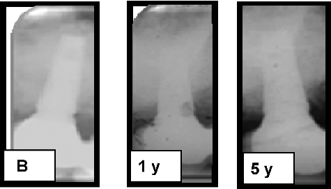
Figure 3. Periapical results of the left side of the patient (DBBM/CM side) at baseline (B), 1 and 5 years.
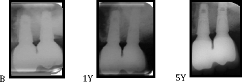
Table 3. Success and survival rates of implants after MSFA.
Various studies have demonstrated that DBBM is slowly resorbed in the normal bone-remodelling process [Citation22,Citation23]. Therefore, it appears to serve as a long-lasting matrix for new bone and helps to maintain the graft volume [Citation23]. Since primary stability is crucial for the long-term survival rates of dental implants, the quality and quantity of post-regenerative existing bone is important [Citation22]. PRP, as a storage vehicle for GF with angiogenic and mitogenic effects, has a possible advantage when it is used in conjunction with osteoconductive scaffolds, especially during the early healing stages [Citation8,Citation16]. Different autologous platelet-based formulations with therapeutic potential have been used in MSFA with mainly autogenous and DBBA bone grafts [Citation8,Citation11,Citation14–16,Citation24]. A very limited number of controlled studies have directly evaluated the long-term results [Citation8,Citation14,Citation16]. There is great heterogeneity among different studies regarding the study design, surgical technique, graft material, healing time for implant placement and the method of preparation of the platelet concentrate [Citation25,Citation26].
In a very recent systematic review, the authors concluded that there is no consensus about the additive effect of PRP on implant survival in sinus augmentation [Citation26]. In this study, although we have presented [Citation13] histological data, more prominent and mature bone formation was observed at sites treated with PRP/DBBA rather than with DBBA/CM, and similar implant survival and success rates were found in both groups without any superiority in five years concomitant with the other reported data [Citation8,Citation16].
The additional effects of absorbable CMs over a lateral window as a physical barrier are still a source of debate. However, in a recent review, the positive effects of membranes were mentioned [Citation27]. In this study, to cover the lateral window of the sinuses on one side, CM in the other PRP membrane was used. In the literature, several studies pointed out the advantages of using these platelet-based membranes because their adhesive properties improve the handling and administration of the bone substitute, while avoiding the uncontrolled displacement of particular graft materials into the sinus cavity, especially in the case of Schneiderian membrane perforation [Citation16]. In addition, platelet-derived bioactive molecules might limit the inflammation [Citation28].
Long-term studies are essential for evaluating the clinical variables influencing implant success and survival rate after MSFA. At least five years of follow-up is necessary to give reliable information about the survival and success rate of the implants [Citation29]. Our study represents one of the few reports of long-term results of implant performance after MSFA using PRP/DBBA with a 5-year follow-up. On the other hand, more data are available after MSFA with DBBA with long-term results [Citation11,Citation14,Citation15]. The present study revealed no statistically significant differences in either the survival or the success rates of the implants after the 5-year follow-up between the groups. The survival rate of implants placed in both PRP/DBBA and DBBA/CM groups was 100%. There is only one similar study to compare our results within the PRP group: Torres et al. [Citation8] studied bilateral MSFA using PRP/DBBA and DBBA alone in a delayed approach in five edentulous patients. After a 2-year follow-up, the authors obtained a survival rate of 98.2% and 90.7% for the PRP/DBBA and DBBA groups, respectively. The study reported implant survival rates varying between 91.3% and 100% for delayed-placed implants after 14–60 months when DBBA was used as grafting material [Citation3,Citation8,Citation30,Citation31]. Our results concur with the these. No additive effect of PRP was observed in the long-term, as expected. The recovery time after surgery and the healing of the wound started almost immediately. Although tissue maturation is a long process, the early phase of wound healing is important, and PRP used in this study might contribute to only the early phase of healing. Furthermore, it may be speculated that the limited bone contact area might camouflage the true effect of PRP. It has been stated that PRP was not beneficial in MSFA in the absence of osteoblasts and osteocytes [Citation32].
Since the analysis of the implant success involves more complex parameters and criteria, assessment of implant success is often ignored as a subject of long-term studies. In this study, the success rate for implants in PRP/DBBA and DBBA/CM was found to be 82% and 85%, respectively. The overall cumulative success rate was 83% for all implants. To the best of our knowledge, there is no study that compares to our results obtained in the PRP/DBBA group. On the other hand, there is only one report on the five-year success rate of DBBA in sinus augmentation. In that study, Mordenfeld et al. [Citation3] reported the cumulative success rate to be 91.3% for DBBA at the five-year follow-up. Although our results are slightly lower, this difference should be regarded with caution, and clinicians should also consider the lack of standardized and internationally accepted success criteria [Citation33]. The peri-implant bone was stable during these follow-up periods, with mean PDs and the mean DIB levels demonstrating moderate changes over time in both groups (, p > 0.05). The changes observed in the DBBA and CM groups are in agreement with previously published limited data [Citation1,Citation3]. In the related literature, a range of 0.08–2.79 and 1.0–2.87 have been reported for the baseline to 1-year and 1- to 5-year comparisons, respectively. Based on our data, the satisfactory success and survival rate with minimum crestal bone level change can be achieved in either PRP/DBBM or DBBM/CM in MSFA in 5 years with implant-supported bridges. Although both combinations exhibited similar outcomes, during MSFA, PRP usage may offer advantage in case of Schneiderian membrane perforation. That will limit excess membrane usage for covering the perforation. On the other hand, PRP preparation, rather than CM, prolonged the surgery time.
Conclusions
Our results indicate that successful clinical outcomes can be achieved after one and five years with implant-supported bridges following grafting of either PRP/DBBM or DBBM/CM in MSFA. Although PRP is not a decisive factor for implant survival in sinus augmentation, its combined effects on angiogenesis and tissue remodelling, along with improved handling, can ease the graft application process done by clinicians. This study demonstrated successful clinical outcomes after one and five years with implant-supported bridges following sinus augmentation of severely resorbed edentulous maxilla with either a PRP/DBBM or DBBM/CM. Considering both treatment modalities, only one implant was lost before prosthetic rehabilitation at the PRP/DBBM site, which resulted in a survival rate of 100% after five years at both sites.
Disclosure statement
No potential conflict of interest was reported by the authors.
References
- Urban IA, Lozada JL. A prospective study of implants placed in augmented sinuses with minimal and moderate residual crestal bone: results after 1 to 5 years. Int J Oral Maxillofac Implants. 2010;25:1203–1212.
- Lindgren C, Mordenfeld A, Johansson CB et al., A 3-year clinical follow-up of implants placed in two different biomaterials used for sinus augmentation. Int J Oral Maxillofac Implants. 2012;27:1151–1262.
- Mordenfeld A, Albrektsson T, Hallman M. A 10-year clinical and radiographic study of implants placed after maxillary sinus floor augmentation with an 80:20 mixture of deproteinized bovine bone and autogenous bone. Clin Implant Dent Relat Res. 2014;16:435–446.
- Nkenke E, Schultze-Mosgau S, Radespiel-Tröger M et al., Morbidity of harvesting of chin grafts: a prospective study. Clin Oral Implants Res. 2001;12:495–502.
- Esposito M, Grusovin MG, Rees J et al., Effectiveness of sinus lift procedures for dental implant rehabilitation: a Cochrane systematic review. Eur J Oral Implantol. 2010;3:7–26.
- Hallman M, Sennerby L, Lundgren S. A clinical and histologic evaluation of implant integration in the posterior maxilla after sinus floor augmentation with autogenous bone, bovine hydroxyapatite, or a 20:80 mixture. Int J Oral Maxillofac Implants. 2002;17:635–643.
- Yildirim M, Spiekermann H, Biesterfeld S et al., Maxillary sinus augmentation using xenogenic bone substitute material Bio-Oss in combination with venous blood. A histologic and histomorphometric study in humans. Clin Oral Implants Res. 2000;11:217–229.
- Torres J, Tamimi F, Martinez PP et al., Effect of platelet-rich plasma on sinus lifting: a randomized-controlled clinical trial. J Clin Periodontol. 2009;36:677–687.
- Arora NS, Ramanayake T, Ren YF et al., Platelet-rich plasma: a literature review. Implant Dent. 2009;18:303–310.
- Wrotniak M, Bielecki T, Gazdik TS. Current opinion about using the platelet-rich gel in orthopaedics and trauma surgery. Ortop Traumatol Rehabil. 2007;9:227–238.
- Thor A, Wannfors K, Sennerby L et al., Reconstruction of the severely resorbed maxilla with autogenous bone, platelet-rich plasma, and implants: 1-year results of a controlled prospective 5-year study. Clin Implant Dent Relat Res. 2005;7:209–220.
- Kassolis JD, Reynolds MA. Evaluation of the adjunctive benefits of platelet-rich plasma in subantral sinus augmentation. J Craniofac Surg. 2005;16:280–287.
- Yilmaz S, Karaca EO, Ipci SD et al., Radiographic and histologic evaluation of platelet-rich plasma and bovine-derived xenograft combination in bilateral sinus augmentation procedure. Platelets. 2013;24:308–315.
- Dasmah A, Thor A, Ekestubbe A et al., Marginal bone-level alterations at implants installed in block versus particulate onlay bone grafts mixed with platelet-rich plasma in atrophic maxilla. A prospective 5-year follow-up study of 15 patients. Clin Implant Dent Relat Res. 2013;15:7–14.
- Schaaf H, Streckbein P, Lendeckel S et al., Sinus lift augmentation using autogenous bone grafts and platelet-rich plasma: radiographic results. Oral Surg Oral Med Oral Pathol Oral Radiol Endod. 2008;106:673–678.
- Anitua E, Prado R, Orive G. Bilateral sinus elevation evaluating plasma rich in growth factors technology: a report of five cases. Clin Implant Dent Relat Res. 2012;14:51–60.
- Misch CE. Division of available bone in implant dentistry. Int J Oral Implantol. 1990;7:9–17.
- Mombelli A, van Oosten MA, Schurch E Jr et al., The microbiota associated with successful or failing osseointegrated titanium implants. Oral Microbiol Immunol. 1987;2:145–151.
- Buser D, Weber HP, Lang NP. Tissue integration of non-submerged implants: 1-year results of a prospective study with 100 ITI hollow-cylinder and hollow-screw implants. Clin Oral Implants Res. 1990;1:33–40.
- Brägger U, Häfeli U, Huber B et al., Evaluation of postsurgical crestal bone levels adjacent to non-submerged dental implants. Clin Oral Implants Res. 1998;9:218–224.
- Weber HP, Buser D, Fiorellini JP et al., Radiographic evaluation of crestal bone levels adjacent to nonsubmerged titanium implants. Clin Oral Implants Res. 1992;3:181–188.
- Esposito M, Grusovin MG, Coulthard P et al., The efficacy of various bone augmentation procedures for dental implants: a Cochrane systematic review of randomized controlled clinical trials. Int J Oral Maxillofac Implants. 2006;21:696–710.
- Traini T, Valentini P, Lezzi G et al., A histologic and histomorphometric evaluation of anorganic bovine bone retrieved 9 years after a sinus augmentation procedure. J Periodontol. 2007;78:955–961.
- Consolo U, Zaffe D, Bertoldi C et al., Platelet-rich plasma activity on maxillary sinus floor augmentation by autologous bone. Clin Oral Implants Res. 2007;18:252–262.
- Beretta M, Poli PP, Grossi GB et al., Long-term survival rate of implants placed in conjunction with 246 sinus floor elevation procedures: results of a 15-year retrospective study. J Dent. 2015;43:78–86.
- Del Fabbro M, Bortolin M, Taschieri S et al., Effect of autologous growth factors in maxillary sinus augmentation: a systematic review. Clin Implant Dent Relat Res. 2013;15:205–216.
- Wallace SS, Tarnow DP, Froum SJ et al., Maxillary sinus elevation by lateral window approach: evolution of technology and technique. J Evid Based Dent Pract. 2012;12(Suppl 3):S161–S171.
- Woodall J Jr, Tucci M, Mishra A et al., Cellular effects of platelet rich plasmainterleukin1 release from prp treated macrophages. Biomed Sci Instrum. 2008;44:489–494.
- Needleman I, Chin S, O'Brien T et al., Systematic review of outcome measurements and reference group(s) to evaluate and compare implant success and failure. J Clin Periodontol. 2012;39(Suppl 12):122–132.
- Oliveira R, El Hage M, Carrel JP et al., Rehabilitation of the edentulous posterior maxilla after sinus floor elevation using deproteinized bovine bone: a 9-year clinical study. Implant Dent. 2012;21:422–426.
- Ferreira CE, Novaes AB, Haraszthy VI et al., A clinical study of 406 sinus augmentations with 100% anorganic bovine bone. J Periodontol. 2009;80:1920–1927.
- Wiltfang J, Schlegel KA, Schultze-Mosgau S et al., Sinus floor augmentation with beta-tricalciumphosphate (beta-TCP): does platelet-rich plasma promote its osseous integration and degradation? Clin Oral Implants Res. 2003;14:213–218.
- Moraschini V, Poubel LA, Ferreira VF et al., Evaluation of survival and success rates of dental implants reported in longitudinal studies with a follow-up period of at least 10 years: a systematic review. Int J Oral Maxillofac Surg. 2015;44:377–388.