
Abstract
Objectives: Elevated oxidative stress and reduced heart rate variability (HRV) is prevalent in patients with chronic kidney disease (CKD) and is associated with increased morbidity and mortality. Previous studies have identified a positive association between elevated oxidative stress and autonomic dysfunction, however this relationship has not yet been investigated in the CKD population.
Methods: Plasma was collected from 78 patients with stage 3–4 CKD (estimated glomerular filtration rate 25–60 ml/min/1.73 m2) for the assessment of oxidative stress, including plasma total F2-isoprostanes, glutathione peroxidase activity and total antioxidant capacity. Time and frequency HRV parameters were measured from a three lead electrocardiogram.
Results: Participants with elevated F2-isoprostanes had reduced HRV compared to patients with normal levels of F2-isoprostanes. A number of HRV parameters were found to be inversely correlated with F2-isoprostanes in all CKD patients, including SDNN (r = −0.337; P < 0.01), VLF (r = −0.281, P = 0.01), LF (r = −0.315, P < 0.01) and total power (r = −0.288, P = 0.01). Multiple linear regression found F2-isoprostanes to be an independent predictor of SDNN (r2 = 0.287, β = −0.272, P = 0.01).
Discussion: Oxidative stress is significantly and independently associated with HRV in patients with CKD. Identifying oxidative stress in the pathogenesis of autonomic dysfunction may help target therapeutic strategies.
Introduction
Elevated oxidative stress is common in patients with chronic kidney disease (CKD)Citation1 and occurs when there is a disruption in redox signalling and control pathways.Citation2 Elevated oxidative stress may contribute to a multitude of disease pathways including cardiovascular conditions,Citation3 the mortality rates of which are increased in the CKD cohort.Citation4,Citation5 The gold standard for the measurement of oxidative stress injury is plasma total F2-isoprostanes, a by-product of lipid peroxidation.Citation6
Patients with CKD often have a reduction in heart rate variability (HRV).Citation7 HRV is a measure of the constantly deviating normal beat to beat interval and, thus the balance between the dual sympathetic/parasympathetic stimulation of the pacemaking sino-atrial node in the heart.Citation8 A reduction in HRV is indicative of cardiac autonomic dysfunction and is associated with increased morbidity and mortality.Citation9,Citation10 Indeed, sudden cardiac death is a significant problem reportedly contributing to up to 60% of cardiovascular mortality in dialysis patients, increasing with each stage of CKD.Citation11 It has been suggested that impaired baroreflex effectiveness through autonomic dysfunction can initiate fatal arrhythmias and sudden cardiac death.
A number of time and frequency parameters can be attained from a single HRV measure. However, the standard deviation of normal beat to beat intervals (SDNN) is used as a global measure of HRV.Citation12 Generally, time domain parameters are thought to provide a global measure of autonomic activity, whereas frequency parameters provide an indication of autonomic balance. A reduced SDNN and/or total power indicates reduced HRV and therefore autonomic dysfunction.Citation13
Previous studies have investigated the relationship between HRV and elevated oxidative stress in diseases such as hypertensionCitation14,Citation15 and diabetes.Citation16,Citation17 Indeed, Ziegler et al.Citation17 found that oxidative stress biomarkers were increased and antioxidant levels decreased, in diabetic patients with peripheral neuropathy. The authors also found oxidative stress to be greater in patients who had dual peripheral and cardiac neuropathy. However, the relationship between CKD and oxidative stress in the pathogenesis of cardiac autonomic neuropathy has not previously been studied. Investigating the relationship between oxidative stress and HRV is of particular importance as it may assist in further developing appropriate treatments in CKD patients. Therefore, the purpose of this study was to determine the association between oxidative stress and HRV in the CKD population. It was hypothesized that elevated plasma F2-isoprostanes would be associated with reduced HRV in patients with stage 3–4 CKD.
Subjects and methods
The data from this study is a cross-sectional analysis of the ‘LANDMARK III’ randomized control trial (Longitudinal Assessment of Numerous Discrete Modifications of Atherosclerotic Risk in Kidney Disease), which compares the effects of a lifestyle intervention to standard nephrological care in CKD patients. Seventy-eight subjects with stage 3 or 4 CKD (MDRD estimated glomerular filtration rate (eGFR) 25–60 ml/min/1.73 m2) were included in this cross-sectional analysis. Patients were aged between 18 and 75 years and had at least one of the following risk factors – blood pressure or lipids not at target; overweight (BMI >25 kg/m2); or poor diabetic control. Exclusion criteria were life expectancy less than 6 months, pregnancy, enrolment in another research study or organ transplant recipient.
The study protocol was approved by the Princess Alexandra Human Research Ethics Committee (HREC 2007/190), and was registered at www.anzctr.org.au (Registration Number ANZCTR12608000337370). Patients provided written and informed consent.
Routine blood biochemistry
Blood measures of lipids, haemoglobin, phosphate, creatinine, C-reactive protein (CRP), magnesium, albumin, glucose, and insulin were conducted using standard laboratory techniques, through venipuncture, after an overnight fast. Patients were also asked to abstain from any stimulants, such as caffeine and smoking, on the morning of the test. Insulin resistance was computed using the homeostatic model assessment of insulin resistance method (HOMA-IR).Citation18 eGFR was determined using the Modification of Diet in Renal Disease-175 formula.Citation19 eGFR rate of change was calculated as the change in eGFR at 12 months from baseline after participation in the LANDMARK III randomized control trial (both control and intervention groups),Citation20 divided by 12 (months in a year).
Heart rate variability
After 5 minutes of supine rest HRV was recorded from a supine 5 minute three lead ECG recording that was conducted in a quiet room with no disturbances (such as talking or coughing). The R–R interval tachogram was visually inspected and any ectopic beats were removed by using commercially available software (Kubios version 2.1, Kuopio, Finland). The software was used to de-trend the data using a smoothness priors regularization procedure before calculating the time, frequency and non-linear domain HRV parameters. A number of time and frequency parameters of HRV were assessed. Time parameters included the standard deviation of normal beat to beat (N–N) intervals (SDNN), the root mean square of differences in successive N–N intervals (RMSSD), the number of successive N–N intervals greater than 50 ms (NN50), the proportion of NN50 intervals in the entire recording (pNN50) and the standard deviation of heart rate (STD HR). Frequency parameters included a spectral power analysis incorporating very low frequency (VLF; ≤0.04 Hz), low frequency (LF; 0.04–0.15 Hz), high frequency (HF; 0.15–0.4 Hz) and total power (TP, the addition of all frequencies). All HRV measures are described in Table . The LF/HF ratio was computed using these values. SDNN was used in regression analyses as it is recognized as a robust global marker of HRV.Citation12 Patients with a pacemaker were excluded from analysis. Patients were asked to abstain from taking their beta-blocker medication at least 12 hours prior to HRV testing. However, to ensure residual beta-blocker medication was not influencing the findings, a sub-analysis on patients not on beta-blockers was also performed (normal F2-isoprostanes n = 39, elevated F2-isoprostanes n = 14).
Table 1 Time and frequency HRV parameter descriptionsCitation42
Plasma F2-isoprostanes
Total F2-isoprostanes were analysed using a gas chromatography mass spectrometry protocol developed in our laboratory.Citation21 Samples were analysed in duplicate and the coefficient of variation for this assay is 7.6%. Patients were divided into two groups based on their plasma F2-isoprostane levels: normal (<250 pg/ml) and elevated (≥250 pg/ml). This was based on using a value 1.5 standard deviations from mean values from a previous study on apparently healthy 18–30 year old males and females from our laboratory.Citation22
Plasma glutathione peroxidase activity
Samples were measured in duplicate using an automated method adapted from Wheeler et al.Citation23 as the rate of oxidation of NADPH at 340 nm in a coupled reaction, cycling oxidized glutathione to reduced glutathione using glutathione reductase. These measures were performed on an autoanalyser (Cobas Mira, Roche Diagnostica, Switzerland). The laboratory coefficient of variation for this assay is 2.4%.
Plasma total antioxidant capacity
Total antioxidant capacity was measured using a modified version of an assay previously describedCitation24 and adapted for a Cobas Mira autoanalyser (Cobas Mira, Roche Diagnostica, Switzerland). Briefly, plasma was incubated with met-myoglobin and 2,2′-azino-bis(3-ethylbenzothiazoline-6-sulphonic acid (ABTS). After incubation, hydrogen peroxide was added and the sample incubated again. Absorbance was measured spectrophotometrically to determine total antioxidant capacity. The laboratory coefficient of variation for this assay is 1.9%.
Statistics
Mean ± standard deviation (SD) was used for the description of normally distributed baseline characteristics. Log transformed and not normally distributed variables were reported as median (interquartile range [IQR]). Frequencies were used to describe categorical variables. Study variables that did not exhibit normal distribution were transformed using the natural logarithm. These included: F2-isoprostanes, VLF, LF, HF, LF: HF, TP, triglycerides (TG), and CRP. A series of Independent Sample T tests were used to compare variables between the normal and elevated F2-isoprostanes group. Non-parametric tests were performed on not normally distributed variables that did not normalize after log transformation. As such, the Mann–Whitney-U tests were used for the remaining variables that were not normally distributed, which included: age, NN50, pNN50, high density lipoprotein (HDL), total cholesterol (TC), HOMA-IR, magnesium, albumin, and HbA1c. A Pearson's Chi square test was used for categorical variables. Bivariate correlations between plasma F2-isoprostanes and HRV variables were assessed using Pearson's correlations for normally distributed data and Spearman's Rho for data that was not normally distributed or categorical. A multiple linear regression using the enter method was undertaken with SDNN as the outcome measure against all variables that exhibited a significant univariate association with SDNN. All variables were assessed for multi-collinearity by variance inflation factor. Significance for all tests was assumed at P < 0.05. The statistical program used for the analysis was SPSS 20 (IBM, New York, USA).
Results
The characteristics, medications, HRV parameters and biochemical measures of the study participants are presented in Table . Sixteen patients were identified as having elevated levels of F2-isoprostanes. Total cholesterol (P = 0.01) and HDL (P < 0.01) was significantly higher in patients with elevated oxidative stress.
Table 2 Characteristics, medications and biochemical measures of CKD patients
Table compares the HRV measures for all patients based on plasma isoprostane values (normal vs elevated). In addition, because of concerns that patients who ceased their beta-blocker medication the in the 12 hours prior to testing may have had an amplified reactive increase in HRV. Patients with elevated F2-isoprostanes had significantly (p < 0.05) reduced HRV measures of SDNN, VLF, and LF (Table ). In addition to SDNN and LF, the sub-analysis of patients not on beta-blockers also identified STD HR to be lower in the elevated F2-isoprostanes group. In the sub-analysis VLF was trending towards significance (p = 0.08) (Table ).
Table 3 Heart rate variability parameters of all and patients not on beta-blockers
Table shows F2-isoprostanes of all patients to be inversely correlated with SDNN, STD HR, VLF, LF, and TP. Mean RR, RMSSD, and HR also had an inverse correlation which was approaching significance. The sub group analysis on patients not on beta-blockers also identified the same significant correlates, as found with all patients with F2-isoprostanes (STD HR, SDNN, VLF, LF, and TP).
Table 4 The association between plasma F2-isoprostanes and HRV parameters in all patients and those not on beta-blockers
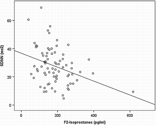
A multiple linear regression model (Table ) found increased F2-isoprostanes to be an independent predictor of reduced SDNN (β = −0.272, P = 0.01), along with female sex (β = −0.263, P = 0.01) (Fig. ). The multiple linear regression model including other significant bivariate associates with SDNN (HbA1C, diabetes status, F2-isoprostanes, and sex), found F2-isoprostanes and sex to explain 28.7% of the variance in SDNN (P < 0.01).
Table 5 Bivariate and multivariate associations with SDNN
Discussion
This is the first study to show that CKD patients with elevated oxidative stress have evidence of autonomic dysfunction. The main findings from the study are that, (1) time and frequency parameters of HRV were significantly reduced in patients with elevated plasma F2-isoprostanes compared to patients with normal F2-isoprostanes, (2) there were significant inverse relationships between F2-isoprostanes and a number of HRV parameters and (3) plasma F2-isoprostanes independently predicted SDNN, a global measure of HRV. It was also identified that the use of beta-blockers in a sub-set of patients did not affect these findings.
Owing to the complexity of measuring HRV, there are a number of time and frequency parameters that are considered with these analyses. Generally, time domain parameters are thought to give a global measure of autonomic activity, whereas frequency parameters provide an indication of autonomic balance.Citation25 Our study found significant reductions in a number of both time and frequency parameters in patients with elevated plasma F2-isoprostanes. The reduction in SDNN indicates an overall decrease in autonomic function. Furthermore, the frequency data showed that reductions in VLF and LF were associated with increased plasma F2-isoprostanes. It is commonly accepted that HF relates to parasympathetic function and that VLF and LF are more representative of sympathetic contribution.Citation12 Although not statistically significant, HF was also considerably lower in the elevated F2-isoprostanes group, suggesting global autonomic dysfunction of both parasympathetic and sympathetic nervous systems. This is supported by the considerably lower global measure of frequency, TP, in the elevated F2-isoprostanes group, which was approaching significance. This study also identified no significant differences between GPX and TAC in the normal and elevated F2-isoprostanes groups. The disconnect between F2-isoprostanes and GPX and TAC in our findings may be suggesting that an increase in oxidants is occurring without a compensatory increase in antioxidants.Citation26 This imbalance may be important in identifying the pathogenesis of oxidative injury and therefore autonomic dysfunction in CKD patients.Citation27
It is understood that elevated oxidative stress is present relatively early during kidney disease progression.Citation28 Kidney disease is associated with a decrease in antioxidant scavenging enzymes and increase in the production of reactive oxygen species (ROS), thereby leading to elevated oxidative stress.Citation29 Furthermore, previous studies have found that the level of oxidative stress continues to increase as CKD progresses.Citation26,Citation30 A study by Dounousi et al.Citation30 found a significant correlation between increasing oxidative stress levels and declining kidney function, as measured by eGFR. A number of studies have suggested a role of oxidative stress in the development of autonomic neuropathy in other populations, such as patients with diabetes.Citation16 Oxidative stress has been associated with a decrease in cardiovagal modulation in pre-hypertensive subjects.Citation15 Additionally, reduced HRV has been associated with air pollutant-induced oxidative stress.Citation31 A study similar to the current investigation, in patients with diabetes, found that oxidative stress levels are enhanced prior to the development of polyneuropathy and remain high within this cohort.Citation17 The interplay of kidney disease, diabetes and reduced heart rate variability is rather complex. As mentioned previously, multiple studies have demonstrated that uraemic neuropathy may be implicated in the pathogenesis of autonomic dysfunction. However, a recent study demonstrated a significant relationship between diabetic status and autonomic neuropathy in patients with CKD stages 4 and 5.Citation32 Both diabetic nephropathy and diabetic neuropathy (including cardiac autonomic neuropathy) are well known complications of diabetes. Various hypotheses have been postulated to explain the pathogenesis of diabetic neuropathy, including several studies that have examined the role of oxidative stress.Citation33,Citation34 In these studies, oxidative stress was found to play a role amongst other factors. Indeed, the contribution of uraemic and glycaemic neuropathy in patients with CKD and diabetes warrants further investigation and is currently being investigated by our group.
The precise mechanistic pathways through which ROS cause autonomic damage remains unclear and it has been hypothesized that oxidative stress leads to neuronal injury in a variety of different ways. Indeed, a suggested mechanism of autonomic dysfunction is through damage to proteins and lipids by oxidative stress, which influences axonal transport.Citation35 It has also been well documented that cellular stressors, such as ROS, can lead to mitochondrial damage, causing damage to the electron transport system and upregulating the release of pro-apoptotic proteins, such as cytochrome C. This, in turn, activates the caspase cascade system, which subsequently leads to apoptosis of neurons.Citation36,Citation37 Therefore, large-scale neuronal apoptosis could be considered a potential mechanism for the observed decrease in autonomic stimulation.Citation16 It should also be noted that a build-up of uraemic toxins, as seen in CKD, may be a source of ROS, although the role that uric acid plays in the genesis of ROS remains unclear.Citation28,Citation38
Clinically, a greater understanding of the pathological processes leading to decreased HRV can provide a potential therapeutic target. Some studies have suggested possible antioxidant therapies to specifically target ROS (e.g. α-tocopherol, vitamin C, N-acetylcysteine). However, the lack of a clinically valid global marker for oxidative stress means that the current evidence of their efficacy is lacking.Citation39 Other studies have established that regular exercise can protect against oxidative stress by promoting up-regulation of antioxidant enzymes in response to the acute increases in ROS which occur with exercise training.Citation40 The effects of exercise training in CKD patients considered to have elevated oxidative stress is currently being investigated by our group.
Owing to the cross-sectional design of this study, a direct causative link between oxidative stress and reduced HRV cannot be established. The use of a 5 minute HRV recording, rather than longer, 24 hour monitoring, may be a limitation to the study. Although, it has been suggested that 5 minute recordings made under physiologically stable conditions are appropriate for frequency analysis.Citation41 Indeed, the strong association between F2-isoprostanes and both time and frequency parameters suggest that the use of only a 5 minute recording may not be a limitation to the findings. The use of negative chronotropic drugs such as beta-blockers may influence HRV. Participants were asked to refrain from taking heart rate lowering medications at least 12 hours prior to testing, however some residual medication effect may still occur. Nonetheless, the normal F2-isoprostanes group actually had greater beta-blocker use (which would lower HRV), so it does not appear that the difference in beta-blocker medication between the groups would be influencing the current findings. Furthermore, the difference in heart rate variability measures in patients with elevated oxidative stress was similar between patients taking beta-blocker medication and not taking beta-blocker medication.
Conclusion
In summary, CKD patients with elevated oxidative stress display evidence of autonomic dysfunction. It was also identified by a multiple linear regression model that F2-isoprostanes independently predicted SDNN measures, a global measure of HRV. Although due to the cross-sectional design of this study, causation cannot be deduced. These findings are clinically relevant, as identifying oxidative stress in the pathogenesis of autonomic dysfunction may help target therapeutic strategies for decreasing the high cardiovascular mortality seen in the CKD population (Fig. ).
Figure 1 Relationship between SDNN and plasma F2-isoprostanes in patients with CKD (r = 0.34, P < 0.01).
Disclaimer statements
Contributors Shannon Fadaee analysed and interpreted the data, and drafted the manuscript. Kassia Beetham was involved in data collection, analysed and interpreted the data, and revised the manuscript. Erin Howden was involved in data collection and revision of the manuscript. Nicole Isbel was involved in conception of the study, data analysis, and revision of the manuscript. Tony Stanton was involved in data analysis and revision of the manuscript. Jeff Coombes was involved in conception of the study, data analysis, revision and final approval of the manuscript.
Funding This research was supported by the National Health and Medical Research Council (NHMRC)-funded Centre for Clinical Research Excellence – Vascular and Metabolic Health (CCRE), University of Queensland (UQ) (#000759) and the Department of Nephrology, Princess Alexandra Hospital. The work was also supported by NHMRC Australia, through Australia Fellowship award #511081, CKD.QLD (Chronic Kidney Disease in Queensland) and Centre for Chronic Disease at the University of Queensland.
Conflict of interest The results in this paper have not previously been published in whole or part. There are no conflicts of interests for all authors.
Ethics approval The study protocol was approved by the Princess Alexandra Human Research Ethics Committee (HREC 2007/190).
References
- Oberg BP, McMenamin E, Lucas FL, McMonagle E, Morrow J, Ikizler TA, et al. Increased prevalence of oxidant stress and inflammation in patients with moderate to severe chronic kidney disease. Kidney Int 2004;65(3):1009–16. doi: 10.1111/j.1523-1755.2004.00465.x
- Jones DP. Redefining oxidative stress. Antioxid Redox Signal 2006;8(9–10):1865–79. doi: 10.1089/ars.2006.8.1865
- Finkel T, Holbrook NJ. Oxidants, oxidative stress and the biology of ageing. Nature 2000;408(6809):239–47. doi: 10.1038/35041687
- Shlipak MG, Fried LF, Cushman M, Manolio TA, Peterson D, Stehman-Breen C, et al. Cardiovascular mortality risk in chronic kidney disease: Comparison of traditional and novel risk factors. JAMA 2005;293(14):1737–45. doi: 10.1001/jama.293.14.1737
- Go AS, Chertow GM, Fan D, McCulloch CE, Hsu C-Y. Chronic kidney disease and the risks of death, cardiovascular events, and hospitalization. New Engl J Med 2004;351(13):1296–305. doi: 10.1056/NEJMoa041031
- Roberts LJ, Morrow JD. Measurement of F(2)-isoprostanes as an index of oxidative stress in vivo. Free Radic Biol Med 2000;28(4):505. doi: 10.1016/S0891-5849(99)00264-6
- Brotman DJ. Heart rate variability predicts ESRD and CKD-related hospitalization. J Am Soc Nephrol 2010;21(9):1560–70. doi: 10.1681/ASN.2009111112
- Lewis MJ. Heart rate variability analysis: a tool to assess cardiac autonomic function. Comput Inform Nurs 2005;23(6):335–41. doi: 10.1097/00024665-200511000-00011
- Burger AJ, D'Elia JA, Weinrauch LA, Lerman I, Gaur A. Marked abnormalities in heart rate variability are associated with progressive deterioration of renal function in type I diabetic patients with overt nephropathy. Int J Cardiol 2002;86(2):281–7. doi: 10.1016/S0167-5273(02)00346-7
- Ranpuria R, Hall M, Chan CT, Unruh M. Heart rate variability (HRV) in kidney failure: measurement and consequences of reduced HRV. Nephrol Dial Transplant 2008;23(2):444–9. doi: 10.1093/ndt/gfm634
- Shamseddin MK, Parfrey PS. Sudden cardiac death in chronic kidney disease: epidemiology and prevention. Nat Rev Nephrol 2011;7(3):145–54. doi: 10.1038/nrneph.2010.191
- Stauss HM. Heart rate variability. Am J Physiol Regul Integr Comp Physiol 2003;285(5):R927. doi: 10.1152/ajpregu.00452.2003
- Buchman TG, Stein PK, Goldstein B. Heart rate variability in critical illness and critical care. Curr Opin Crit Care 2002;8(4):311–5. doi: 10.1097/00075198-200208000-00007
- Pavithran P, Nandeesha H, Sathiy Appriya V, Bobby Z, Madanmohan T. Short-term heart variability and oxidative stress in newly diagnosed essential hypertension. Clin Exp Hypertens 2008;30(7):486–96. doi: 10.1080/10641960802251875
- Thiyagarajan R, Pal P, Pal GK, Subramanian SK, Bobby Z, Das AK, et al. Cardiovagal modulation, oxidative stress, and cardiovascular risk factors in prehypertensive subjects: cross-sectional study. Am J Hypertens 2013;26(7):850–7. doi: 10.1093/ajh/hpt025
- Vincent AM, Russell JW, Low P, Feldman EL. Oxidative stress in the pathogenesis of diabetic neuropathy. Endocr Rev 2004;25(4):612–28. doi: 10.1210/er.2003-0019
- Ziegler D, Sohr CG, Nourooz-Zadeh J. Oxidative stress and antioxidant defense in relation to the severity of diabetic polyneuropathy and cardiovascular autonomic neuropathy. Diabetes Care 2004;27(9):2178–83. doi: 10.2337/diacare.27.9.2178
- Matthews DR, Hosker JP, Rudenski AS, Naylor BA, Treacher DF, Turner RC. Homeostasis model assessment: insulin resistance and beta-cell function from fasting plasma glucose and insulin concentrations in man. Diabetologia 1985;28(7):412–9. doi: 10.1007/BF00280883
- Levey AS, Coresh J, Greene T, Stevens LA, Zhang Y, Hendriksen S, et al. Using standardized serum creatinine values in the modification of diet in renal disease study equation for estimating glomerular filtration rate. Ann Intern Med 2006;145(4):247–54. doi: 10.7326/0003-4819-145-4-200608150-00004
- Howden EJ, Leano R, Petchey W, Coombes JS, Isbel NM, Marwick TH. Effects of exercise and lifestyle intervention on cardiovascular function in CKD. Clin J Am Soc Nephrol 2013;8(9):1494–501. doi: 10.2215/CJN.10141012
- Briskey DR, Wilson GR, Fassett RG, Coombes JS. Optimized method for quantification of F2-isoprostanes using gas chromatography–tandem mass spectrometry. J Pharm Biomed Anal 2013; In Press.
- Mullins AL, van Rosendal SP, Briskey DR, Fassett RG, Wilson GR, Coombes JS. Variability in oxidative stress biomarkers following a maximal exercise test. Biomarkers 2013;18(5):446–54. doi: 10.3109/1354750X.2013.810668
- Wheeler CR, Salzman JA, Elsayed NM, Omaye ST, Korte DW. Automated assays for superoxide dismutase, catalase, glutathione peroxidase, and glutathione reductase activity. Anal Biochem 1990;184(2):193–9. doi: 10.1016/0003-2697(90)90668-Y
- Miller NJ, Rice-Evans C, Davies MJ, Gopinathan V, Milner A. A novel method for measuring antioxidant capacity and its application to monitoring the antioxidant status in premature neonates. Clin Sci 1993;84(4):407–12. doi: 10.1042/cs0840407
- Wang H-M, Huang S-C. SDNN/RMSSD as a surrogate for LF/HF: a revised investigation. Model Simul Eng 2012;2012(2):1. doi: 10.1155/2012/931943
- Karamouzis I, Sarafidis PA, Karamouzis M, Iliadis S, Haidich AB, Sioulis A, et al. Increase in oxidative stress but not in antioxidant capacity with advancing stages of chronic kidney disease. Am J Nephrol 2008;28(3):397–404. doi: 10.1159/000112413
- Beetham KS, Howden EJ, Small DM, Briskey DR, Rossi M, Isbel N, et al. Oxidative stress contributes to muscle atrophy in chronic kidney disease patients. Redox Rep 2014; 20(3):126–32.
- Small DM, Coombes JS, Bennett N, Johnson DW, Gobe GC. Oxidative stress, anti-oxidant therapies and chronic kidney disease. Nephrology 2012;17(4):311–21. doi: 10.1111/j.1440-1797.2012.01572.x
- Zalba G, Fortuño A, Díez J. Oxidative stress and atherosclerosis in early chronic kidney disease. Nephrol Dial Transplant 2006;21(10):2686–90. doi: 10.1093/ndt/gfl398
- Dounousi E, Papavasiliou E, Makedou A, Ioannou K, Katopodis KP, Tselepis A, et al. Oxidative Stress Is Progressively Enhanced With Advancing Stages of CKD. Am J Kidney Dis 2006;48(5):752–60. doi: 10.1053/j.ajkd.2006.08.015
- Chuang K-J, Chan C-C, Su T-C, Lee C-T, Tang C-S. The Effect of Urban Air Pollution on Inflammation, Oxidative Stress, Coagulation, and Autonomic Dysfunction in Young Adults. Am J Respir Crit Care Med 2007;176(4):370–6. doi: 10.1164/rccm.200611-1627OC
- Clyne N, Hellberg M, Kouidi E, Deligiannis A, Höglund P. Relationship between declining GFR and measures of cardiac and vascular autonomic neuropathy. Nephrology 2015; in press.
- Naruse R, Suetsugu M, Terasawa T, Ito K, Hara K, Takebayashi K, et al. Oxidative stress and antioxidative potency are closely associated with diabetic retinopathy and nephropathy in patients with type 2 diabetes. Saudi Med J 2013;34(2):135.
- Figueroa-Romero C, Sadidi M, Feldman EL. Mechanisms of disease: The oxidative stress theory of diabetic neuropathy. Rev Endocr Metab Disord 2008;9(4):301–14. doi: 10.1007/s11154-008-9104-2
- Metodiewa D, Kośka C. Reactive oxygen species and reactive nitrogen species: relevance to cyto(neuro)toxic events and neurologic disorders. An overview. Neurotoxicity Res 2000;1(3):197–233. doi: 10.1007/BF03033290
- Simon H-U, Haj-Yehia A, Levi-Schaffer F. Role of reactive oxygen species (ROS) in apoptosis induction. Apoptosis 2000;5(5):415–8. doi: 10.1023/A:1009616228304
- Green D, Reed J. Mitochondria and apoptosis. Science 1998;281(5381):1309–12. doi: 10.1126/science.281.5381.1309
- Himmelfarb J, Stenvinkel P, Ikizler TA, Hakim RM. The elephant in uremia: oxidant stress as a unifying concept of cardiovascular disease in uremia. Kidney Int 2002;62(5):1524–38. doi: 10.1046/j.1523-1755.2002.00600.x
- Coombes JS, Fassett RG. Antioxidant therapy in hemodialysis patients: a systematic review. Kidney Int 2012;81(3):233–46. doi: 10.1038/ki.2011.341
- Powers SK, Nelson WB, Hudson MB. Exercise-induced oxidative stress in humans: Cause and consequences. Free Radic Biol Med 2011;51(5):942–50. doi: 10.1016/j.freeradbiomed.2010.12.009
- Malik M, Bigger JT, Camm AJ, Kleiger RE, Malliani A, Moss AJ, et al. Heart rate variability standards of measurement, physiological interpretation, and clinical use. Eur Heart J 1996;17(3):354–81. doi: 10.1093/oxfordjournals.eurheartj.a014868
- Heart rate variability. Standards of measurement, physiological interpretation, and clinical use. Task Force of the European Society of Cardiology and the North American Society of Pacing and Electrophysiology. Eur Heart J 1996;17(3):354–81. doi: 10.1093/oxfordjournals.eurheartj.a014868