Abstract
Objectives. Knowledge of normal aortic diameters is important in the assessment of aortic disease. The aim of this study was to determine normal thoracic aortic diameters. Design. 77 patients undergoing computed tomography of the thorax were studied. The diameter of the thoracic aorta was measured at three levels in the ascending aorta and at three levels in the descending aorta. The diameter was studied in relation to age, sex, weight and height. Results. We found that aortic diameter is increasing with increasing age. Even sex and BMI influence the aortic diameter but to a lesser extent than age. The upper normal limit for ascending aorta can be calculated with the formula D(mm) = 31 + 0.16*age and for descending aorta with the formula D(mm) = 21 + 0.16*age. Thus a 20-year-old person has an upper normal limit for ascending aorta of 34 mm and an 80-year-old person has a limit of 44 m. Conclusions. The thoracic aortic diameter varies with age, sex and body weight and height. The strongest correlation can be seen with age. Age should therefore be taken into consideration when determining whether the thoracic aorta is dilated or not.
Computed tomography (CT) is frequently used for the evaluation of different aortic diseases, in particular aortic dissection and aneurysm Citation1–3. However, very little has been published regarding normal thoracic aortic diameters especially in relation to variables such as age, sex, height and weight Citation4–7. Aortic dissection is generally preceded by a dilatation of the aorta Citation8–12. In patients with increased risk of aortic dissection, e.g. first grade relatives to patients with hereditary aortic dissection, measurement of aortic diameter with CT is used for diagnosis of aortic disease and for decision making regarding operation. Thus, knowledge of normal aortic diameters is very important in the assessment of aortic disease, especially if there is a hereditary form of aortic dissection.
Material and methods
We studied prospectively 77 consecutive individuals above 18 years undergoing CT examination of the thorax. Of the patients 41 (53%) were men and 36 (47%) were women. The age range was 18 – 82 the mean being 54 years. The patients were further divided into three age groups: 18 – 40 years, (n = 16), 41 – 60 years, (n = 25) and those over 60 years, (n = 36). Indications for the CT were follow-up of lymphoma (29%), search for metastasis to different primary tumours (32%), trauma (5%), tamponade (4%), suspected aortic dissection (4%, those with dissection were excluded), pulmonary embolus (3%) and other (10%, e.g. emphysema, infection, hemoptysis). The only exclusion criteria were ongoing aortic dissection and age below 18 years.
The patients were examined by a spiral CT scanner (Somatom Plus4, Siemens-Elema, Erlagen, Germany). Slice collimation, reconstruction interval and table speed per gantry rotation were all 10 mm. The gantry rotation time was 0.75 seconds. A soft tissue algorithm (AB 50) was used for the image reconstruction.
A standard dose of 90 ml of a low osmolar non-ionic contrast medium (Ultravist 300mg I/ml, Schering, Germany) was used in all patients. The flow rate was 1.5 ml/second and the scan delay was 60 seconds.
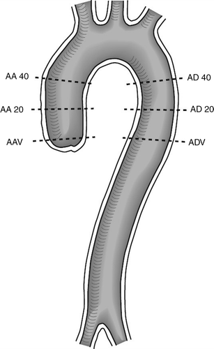
Each examination was evaluated on the same workstation (Magic View, Siemens-Elema, Erlagen, Germany). The diameter of the ascending aorta (AA) was measured at the level of the aortic valve (V), 20 mm and 40 mm above the aortic valve and at the corresponding levels in the descending aorta (AD) ().
Figure 1. Sagittal view over the thoracic aorta. The figure shows were the measurement were made. AV = the level of the aortic valve, AV20 = 20 mm over the aortic valve, AV40 = 40 mm over the aortic valve, AD40 – AD20 – ADV = corresponding levels in the descending aorta.

In order to find a statistical model that gives both a good prediction of the diameter and is useful in practice, several linear regression models were applied. Initially, a univariate regression model including the predictor age was used. In addition, more advanced multiple models including also sex, body mass index (BMI) and significant interactions were investigated. A combination of p-values, coefficient of determination and practical aspects was used to decide which model should be used. Body mass index (BMI) and body surface area (BSA) was highly correlated hence BMI was used in the models since it is easier to measure.
The upper limit of the normal diameter was set to the upper limit of a 95% prediction interval, approximated by the predicted value plus 1.96 times the estimated standard deviation of the model residuals.
Results
The aortic diameter ranged in different subjects from a maximum of 41 mm in the ascending aorta to a minimum of 16 mm in the descending aorta and the diameter diminished progressively. The ascending aorta was larger than the descending aorta at every corresponding level.
In the univariate model, age was significantly associated to the aortic diameter at all levels. The R-square values were at acceptable levels (0.20 – 0.50, the mean 0.40). In the multiple model, sex and BMI also correlated significantly with the aortic diameter at most of the levels but not all (). The R-square values increased somewhat compared to the univariate model (0.44–0.65, the mean 0.55).
Table I. Results of the multivariate analyses. In the age-sex-BMI model, sex did not add significantly to the model. In the age-sex model, sex did not add significantly to the model. SE: standard error. CV: coefficient of variation.
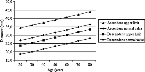
Thus, the results showed that the aortic diameter increases with increasing age and this could be seen at every level investigated (). The diameter increased by 0.12 – 0.20 mm (mean 0.17 mm) per year and was dependent on level. Thus, by thirty years of age the aortic diameter increased by 5.1 mm. The descending aorta shows the same age dependent dilation as the ascending aorta but the ascending aortic diameter increases slightly faster than the descending aorta.
The sex-related difference in diameter was 1.28 – 2.60 mm, mean 1.99 mm, men having larger aortic diameter than women. It seems that this difference decreases with increasing age.
Inclusion of BMI in the model showed statistical correlation with aortic diameter in the descending aorta and even in some levels in the ascending aorta. The mean difference per unit BMI was 0.27mm (0.14 – 0.44 mm). In other words a difference of 5 units in BMI between two persons of same age and sex gives a difference of 1.35mm.
In order to get a single measurement for the ascending and descending aorta, respectively, we measured the mean of AA20 and AA40 to represent the ascending aorta and the mean of ADV, AD20 and AD40 to represent the descending aorta. We excluded the level of the valve in the ascending aorta because the variability in the measurements at this level was quite large. The variations in the measurements can be explained by the configuration of the aortic root (annulus, sinus and sinotubular junction). In these measurements age was the only variable to correlate to aortic diameter at both levels. The age dependent growth was 0.16 mm/year for both the ascending and descending aorta.
shows the formulas for calculating normal aortic dimension at the different levels we measured.
Table II. Formulae for calculating the upper limit for thoracic aorta
The formula for calculating the upper normal limit for ascending aorta (AA20/40) is D = 23.64 + 0.16*age + 1.96*3.79 in which 0.16*age is the factor for age and 1.96*3.79 is 2 standard deviations over the normal value. This formula can be rewritten as D = 31 + 0.16*age. If the body size is extreme this can be taken into consideration by using formula D = 21 + 0.14*age + 0.41*BMI. It is possible using one of these formulae to calculate an upper limit for the diameter, beyond which the ascending aorta should be considered dilated. The formula for the descending aorta is D = 21 + 0.16*age and D = 15 + 0.15*age + 0.22*BMI, respectively.
Discussion
The present study was designed to examine the influence of age, sex and body size on aortic diameter and to define normal thoracic aortic dimensions. We have found only two previous papers that study the correlation between these variables and the thoracic aortic diameter Citation4, Citation5. Even these studies showed that age and sex had influence on aortic diameter and that age was highly significant. The authors also presented average aortic diameters at different levels but did not give any exact limits for normal thoracic aorta at different ages.
Computed tomography is a good method of measuring the aortic diameter because it is widely available, relatively fast and easy to use and offer good reproducibility Citation1, Citation14. The fact that the study was prospective and consecutive resulted in twice as many subjects in the oldest group (60 + ) compared with the youngest group (18 – 40) and that a majority (61%) of patients had a malignant disease. In order to have a more correct body size for patients with malignant disease they were asked for their normal weight before onset of their disease. Previous studies have showed that hypertension only has a slight influence on aortic diameter and therefore was not studied Citation15.
Our data show that the aortic diameter varies with age, sex and body weight and height. The strongest correlation can be seen with age. The diameter increases with increasing age at every level measured and age should thus be taken into consideration when determining normal diameters for the thoracic aorta.
Sex only showed statistical correlation with the descending aortic diameter. This can probably be explained by a smaller variation in aortic diameter in the descending aorta compared with ascending aorta. The mean sex-related difference in millimetres was 1.99. Our opinion is that this can be held clinically irrelevant.
BMI was significant at most of the levels measured. The mean difference per unit BMI was 0.27 mm. An adjustment for body size might be appropriate. From a practical point of view, only very large variations in body size would significantly alter the normal ranges or be of clinical interest.
By using the formulas presented in , the upper limit for the normal thoracic aortic diameter can be calculated. The possibility of relating the diameter of a measured aorta to age and body size can make the decision whether the aorta is pathologically dilated or not somewhat easier.
References
- Nienaber CA, von Kodolitsch Y, Nicolas V, Siglow V, Piepho A, Brockhoff C, et al. The diagnosis of thoracic aortic dissection by non-invasive imaging procedures. N Engl J Med. 1993; 328: 1–9
- Cigarroa JE, Isselbacher EM, DeSantis RW, Eagle KA. Diagnostic imaging in the evaluation of suspected aortic dissection. N Engl J Med. 1993; 328: 35–43
- Willoteaux S, Lions C, Gaxotte V, Negaiwi Z, Beregi JP. Imaging of aortic dissection by helical computed tomography (CT). Eur Radiol. 2004; 14: 1999–2008
- Aronberg DJ, Glazer HS, Madsen K, Sagel SS. Normal thoracic aortic diameters by computed tomography. J Comput Assist Tomogr. 1984; 8: 247–50
- Hager A, Kaemmerer H, Rapp-Bernhardt U, Blucher S, Rapp K, Bernhardt TM, et al. Diameters of the thoracic aorta throughout life as measured with helical computed tomography. J Thorac Cardiovasc Surg. 2002; 123: 1060–6
- Pearce WH, Slaughter MS, Le Maire S, Salyapongse AN, Feinglass J, McCarthy WJ, et al. Aortic diameter as a function of age, gender and body surface area. Surgery. 1993; 114: 691–7
- Tzourio C, Cohen A, Lamisse N, Biousse V, Bousser MG. Aortic root dilatation in patients with spontaneous cervical artery dissection. Circulation. 1997; 95: 2351–3
- Pitt MPI, Bonser RS. The natural history of thoracic aortic aneurysm disease: An overview. J Card Surg 1997; 12(Suppl)270–8
- Westaby S. Management of aortic dissection. Curr Opin Cardiol. 1995; 10: 505–10
- Kouchoukos NT, Dougenis D. Surgery of the thoracic aorta. N Engl J Med. 1997; 336: 1876–88
- Shimada I, Rooney JR, Pagano D, Farneti PA, Davies P, Guest PJ, et al. Prediction of thoracic aortic aneurysm expansion: Validation of formulae describing growth. Ann Thorac Surg. 1999; 67: 1968–70
- Hirose Y, Takamiya M. Growth curve of ruptured aortic aneurysm. J Cardiovasc Surg. 1998; 39: 9–13
- Boyer JK, Gutierrez F, Braverman AC. Approach to the dilated aortic root. Curr Opin Cardiol. 2004; 19: 563–9
- Shimada I, Stephen JR, Farneti PA, Riley P, Guest P, Davies RS. Reproducibility of thoracic aortic diameter measurement using computed tomografic scans. Euro J Cardiothorac Surg. 1999; 16: 59–62
- Kim M, Roman MJ, Cavallini MC, Schwartz JE, Pickering TG, Devereux RB. Effect of hypertension on aortic root size and prevalence of aortic regurgitation. Hypertension. 1996; 28: 47–52
