Abstract
Objective. The aim of this study was to determine the prevalence of moderate ischemic mitral regurgitation (IMR) in the contemporary CABG population. We also aimed to correlate the effective regurgitant orifice area (ERO) of any regurgitant mitral valve in patients with coronary artery disease with the semiquantitative integrated scale of IMR. Design. From March 15 through June 15, 2006, 510 consecutive CABG patients in three tertiary centres were included in the study. All patients showing any sign of mitral regurgitation (MR) at the referring hospital underwent a preoperative transthoracic echocardiographic estimation of the degree of MR using the integrated scale (1–4) and ERO. Results. IMR was found in 141 patients (28%). The prevalence of moderate 2+ or worse IMR was 4% (95% CI; 2.5–6.1%) and the ERO corresponding to 2+ IMR or more ranged from 5 to 30 mm2. Fourteen patients had an ERO between 15–30 mm2. Conclusions. According to our study, patients with moderate IMR, defined as an ERO between 15–30 mm2, account for only 2.7% (95% CI; 1.5–4.7%) of a non-emergency CABG population.
Ischemic mitral regurgitation (IMR) has been shown to have prognostic implications Citation1 where an “effective regurgitant orifice area” (ERO) of more than 20 mm2 results in a significantly reduced long-term survival Citation2, Citation3. The presence of mitral regurgitation (MR) in patients undergoing isolated coronary artery by-pass surgery (CABG) is also associated with an increased mortality Citation4–8.
The benefit of adding mitral valve surgery to CABG is well documented in the combination of coronary artery disease and severe IMR Citation9–12. In many centres clinical practice is to refrain from repairing the mitral valve in patients with mild to moderate IMR undergoing CABG. However, there are no available conclusive data to support this practice. Existing studies are small, retrospective, and the results contradictive Citation5, Citation13–17. Properly powered prospective randomized trials are missing. One of the reasons for this might be that the prevalence of mild to moderate IMR may have decreased in the current era of primary percutaneous coronary intervention (PCI) for ST-elevation myocardial infarction (STEMI). Contemporary aggressive medical treatment of heart failure might also contribute to a reduction of the prevalence of IMR. Moreover, the change of echocardiographic criteria to quantitate mitral regurgitation from the integrated semiquantitative scale to quantitative calculation of the ERO might have caused some confusion concerning definition. However, in our opinion, the controversy of whether or not a moderate IMR in patients undergoing CABG should be repaired, needs to be studied in a prospective randomized design. In order to include an adequate number of patients i.e., secure an adequate power of the study, the prevalence of moderate MR needs to be assessed. Hence, the aims of this study were to correlate ERO with the integrated scale in a contemporary CABG population with IMR and to determine the present prevalence and surgical handling of moderate IMR in three Scandinavian tertiary centers.
Methods
This study was approved by the Ethics Committee at The University of Aarhus (ref nr 20040224). All patients referred for CABG between March 15 through June 15, 2006 at the Aarhus University Hospital, Skejby, in Denmark, Lund University Hospital, and Sahlgrenska University Hospital in Sweden were screened for mitral regurgitation. All patients had a diagnostic echocardiography at their referring centre and those who had any degree of mitral regurgitation were referred for a transthoracic echocardiography at the tertiary centre on the day before surgery. The degree of IMR was first characterized according to the integrated scale 1–4+ Citation18 and independently calculated as ERO using the Proximal Isovelocity Surface Area (PISA) method Citation2, Citation3, Citation19, Citation20. The patients underwent CABG alone or a concomitant mitral valve repair at the discretion of the responsible surgeon. Patients undergoing emergency surgery were excluded as well as patients with structural mitral valve disease.
Results
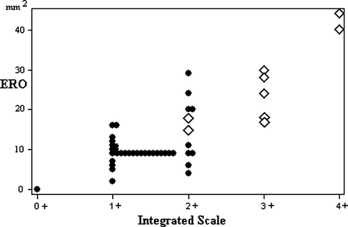
During the inclusion period, a total of 510 patients underwent either isolated CABG or CABG with concomitant mitral valve surgery for IMR. Of these 510 patients, 141 (28%) had signs of mitral insufficiency at the referring hospital. At the preoperative echocardiography the day before CABG the prevalence according to the integrated semiquantitative scale was 1+ IMR in 23% (19–27%, 95% confidence limits), 2+ in 4% (2.5–6.1%), 3+ in 0.6% (0.1–0.7%), and 4+ in 0.4% (0.05–1.4%), see . The table also includes the corresponding ERO intervals. The relation between the IMR of each patient determined by the integrated semi-quantitative scale and ERO is depicted in . All patients with IMR and ERO above 30 mm2 underwent mitral valve repair performed together with the CABG, whereas none of the patients with an ERO below 15 mm2 underwent mitral valve repair. Fourteen of the 510 patients [2.7%, 95% CI 1.5–4.7%] had an ERO between 15–30 mm2 and seven (50%) of these had a concomitant mitral valve repair at the discretion of the responsible surgeon ().
Figure 1. The distribution of patients with IMR according to the semiquantitative scale, ERO, and their treatment. Filled dots denotes CABG alone and open rhombi denotes CABG with mitral repair.

Table I. Distribution of mitral regurgitation the day before CABG, according to integrated semi-quantitative echocardiographic scale and the corresponding ERO interval. (All patients with any sign of mitral regurgitation at referral were included).
Discussion
In this study, 2+ IMR in the integrated scale corresponded to ERO of 5–30 mm2, and the actual prevalence of 2+ IMR was 4% (2.5–6.1% with 95% confidence intervals). This is a little lower than reported earlier Citation7, Citation8, Citation17, Citation21. However, the prevalence of moderate IMR seems to be dependent upon the year of study, the type of diagnostic method, as well as on the definition of moderate MR, and on the population studied (). The significance of the study year may reflect the study method and the current treatment of coronary artery disease, myocardial infarction, and heart failure at each époque. Previously, ventriculography was used as the gold standard and in a large series published by Hickey et al. in 1988, MR was seen in 19% of 11 748 patients during cardiac catheterisation due to symptomatic coronary disease. In the majority of cases the regurgitation was mild, but 5.7% had grade 2+ MR Citation22. In more recent publications IMR has predominantly been diagnosed by echocardiography with a prevalence of IMR varying between 1.9 and 11.8%. In these studies, IMR was classified into a semiquantitative scale, 1–4+, both by ventriculography and echocardiography using an integrated visual approach. With the introduction of an assessment of MR by means of calculation of ERO Citation19, a more precise quantification of the MR could be made. Several studies have confirmed the usefulness of ERO as an outcome predictor for different types of MR Citation2, Citation3, Citation20. However, the correlation between ERO and estimates of MR by the semiquantitative echocardiographic scale has to the best of our knowledge not been undertaken in a CABG population with IMR. According to the American Association of Echocardiography, the definition of a moderate MR is an ERO between 20–40 mm2 Citation18. This refers to a 3-graded scale (mild-moderate-severe), while the integrated semiquantitative scale is a 4-graded scale (1–4 + ). Moreover, the integrated scale can also be divided into “mild, mild-moderate, moderate, moderate-severe, and severe”. In our investigation, the corresponding ERO interval for each regurgitation grade on the semi-quantitative scale had a wide overlap, see . However, the treatment dilemma seemed to be in the ERO interval between 15–30 mm2.
Table II. Studies reporting on the prevalence of moderate IMR in CABG patients (TTE and TEE: transthoracic and transesophageal echocardiography, respectively).
The impact of having a moderate IMR has been well studied Citation4–8, Citation23. In one of the more recent publications Shroeder et al. demonstrated that a moderate or even a mild MR with the integrated scale, determined by intraoperative transesophageal echocardiography, meant an increased risk for heart failure and mortality Citation7. Paparella et al. showed that patients undergoing CABG with mild to moderate MR had significantly lower freedom from heart failure needing hospitalization and death Citation8. On the other hand there are no conclusive data to support that correction of a moderate IMR improves the outcome in a CABG population. It can be hypothesized that concomitant correction of the moderate IMR will improve the outcome, and this needs to be determined in a large prospective randomized study.
Limitations
Our study was not designed to answer the question; if moderate IMR still constitutes a risk in the current CABG population. Hypothetically, primary PCI in acute myocardial infarction and improved medical treatment of heart failure might in general have improved postinfarction myocardial function, and hence reduced the prevalence of IMR. However, the results of this study did not show a definite reduction of IMR in patients scheduled for CABG. A comparison of mortality in patients with and without moderate IMR would necessitate a multivariate analysis considering at least age, sex, and overall left ventricular function. However, the prevalence of only 4% in patients with moderate MR in our population of 510 patients renders such a comparison meaningless.
We did not repeat the echocardiography on those patients who did not show signs of MR on referral, except for those who experienced a new ischemic event awaiting surgery. There are variations in the degree of MR over time, but we do not believe that patients who are free from any sign of MR will develop significant IMR in the interval between referral and surgery.
We did not register the prevalence and location of previous myocardial infarctions but this study represents a consecutive series of all patients who underwent CABG at our three tertiary referral centers, each with a well defined geographical referring area. Moreover, the aims of this study were to correlate the ERO of the insufficient valve with the semiquantitative integrated scale of IMR in a contemporary CABG population and to determine the present prevalence of moderate IMR in this entity. This has been undertaken as an initial and necessary step in designing the planned prospective multi-center trial for patients with moderate mitral regurgitation undergoing CABG.
In conclusion, moderate IMR, here defined as grade 2+ or ERO 5–30 mm2 does still occur in CABG patients and the present prevalence is about 4% (2.5–6.1%, 95% confidence intervals). The optimal treatment remains uncertain and the treatment dilemma seemed to be the ERO interval between 15–30 mm2. This forms the basis to proceed with a multicenter trial, with randomisation of patients with IMR and ERO 15–30 mm2 to receive either CABG alone or CABG with mitral repair. It should be stressed though, that such patients only seem to constitute 2.7(1.5–4.7)% of a contemporary non-emergency CABG population and hence, the controversy on whether a moderate mitral regurgitation should be corrected concomitantly with the CABG can only be solved in a large scale prospective multicenter investigation.
Acknowledgements
Declaration of interest: The authors report no conflict of interest. The authors are responsible for the content and writing of the paper.
References
- Lamas GA, Mitchell GF, Flaker GC, Smith SC, Jr, Gersh BJ, Basta L, et al. Clinical significance of mitral regurgitation after acute myocardial infarction. Survival and Ventricular Enlargement Investigators. Circulation. 1997; 96: 827–33
- Grigioni F, Enriquez-Sarano M, Zehr KJ, Bailey KR, Tajik AJ. Ischemic mitral regurgitation: Long-term outcome and prognostic implications with quantitative Doppler assessment. Circulation. 2001; 103: 1759–64
- Lancellotti P, Gerard PL, Pierard LA. Long-term outcome of patients with heart failure and dynamic functional mitral regurgitation. Eur Heart J. 2005; 26: 1528–32
- Adler DS, Goldman L, O'Neil A, Cook EF, Mudge GH, Jr, Shemin RJ, et al. Long-term survival of more than 2,000 patients after coronary artery bypass grafting. Am J Cardiol. 1986; 58: 195–202
- Duarte IG, Shen Y, MacDonald MJ, Jones EL, Craver JM, Guyton RA. Treatment of moderate mitral regurgitation and coronary disease by coronary bypass alone: Late results. Ann Thorac Surg. 1999; 68: 426–30
- Sergeant P, Blackstone E, Meyns B. Validation and interdependence with patient-variables of the influence of procedural variables on early and late survival after CABG. K.U. Leuven Coronary Surgery Program. Eur J Cardiothorac Surg. 1997; 12: 1–19
- Schroder JN, Williams ML, Hata JA, Muhlbaier LH, Swaminathan M, Mathew JP, et al. Impact of mitral valve regurgitation evaluated by intraoperative transesophageal echocardiography on long-term outcomes after coronary artery bypass grafting. Circulation. 2005;112(9 Suppl):I293–I298.
- Paparella D, Mickleborough LL, Carson S, Ivanov J. Mild to moderate mitral regurgitation in patients undergoing coronary bypass grafting: Effects on operative mortality and long-term significance. Ann Thorac Surg. 2003; 76: 1094–100
- Balu V, Hershowitz S, Zaki Masud AR, Bhayana JN, Dean DC. Mitral regurgitation in coronary artery disease. Chest. 1982; 81: 550–5
- Chaffin JS, Daggett WM. Mitral valve replacement: A nine-year follow-up of risks and survivals. Ann Thorac Surg. 1979; 27: 312–9
- Cohn LH, Couper GS, Kinchla NM, Collins JJ, Jr. Decreased operative risk of surgical treatment of mitral regurgitation with or without coronary artery disease. J Am Coll Cardiol. 1990; 16: 1575–8
- Radford MJ, Johnson RA, Buckley MJ, Daggett WM, Leinbach RC, Gold HK. Survival following mitral valve replacement for mitral regurgitation due to coronary artery disease. Circulation. 1979;60(2 Pt 2):39–47.
- Christenson JT, Simonet F, Bloch A, Maurice J, Velebit V, Schmuziger M. Should a mild to moderate ischemic mitral valve regurgitation in patients with poor left ventricular function be repaired or not?. J Heart Valve Dis. 1995; 4: 484–8
- Czer LS, Maurer G, Bolger AF, DeRobertis M, Chaux A, Matloff JM. Revascularization alone or combined with suture annuloplasty for ischemic mitral regurgitation. Evaluation by color Doppler echocardiography. Tex Heart Inst J. 1996; 23: 270–8
- Mihaljevic T, Lam BK, Rajeswaran J, Takagaki M, Lauer MS, Gillinov AM, et al. Impact of mitral valve annuloplasty combined with revascularization in patients with functional ischemic mitral regurgitation. J Am Coll Cardiol. 2007; 49: 2191–201
- Vaskelyte J, Stoskute N, Ereminiene E, Zaliunas R, Benetis R, Sirvinskas E. The impact of unrepaired versus repaired mitral regurgitation on functional status of patients with ischemic cardiomyopathy at one year after coronary artery bypass grafting. J Heart Valve Dis. 2006; 15: 747–54
- Grossi EA, Crooke GA, DiGiorgi PL, Schwartz CF, Jorde U, Applebaum RM, et al. Impact of moderate functional mitral insufficiency in patients undergoing surgical revascularization. Circulation. 2006;114(1 Suppl):I573–I576.
- Zoghbi WA, Enriquez-Sarano M, Foster E, Grayburn PA, Kraft CD, Levine RA, et al. Recommendations for evaluation of the severity of native valvular regurgitation with two-dimensional and Doppler echocardiography. J Am Soc Echocardiogr. 2003; 16: 777–802
- Bargiggia GS, Tronconi L, Sahn DJ, Recusani F, Raisaro A, De SS, et al. A new method for quantitation of mitral regurgitation based on color flow Doppler imaging of flow convergence proximal to regurgitant orifice. Circulation. 1991; 84: 1481–9
- Enriquez-Sarano M, Avierinos JF, Messika-Zeitoun D, Detaint D, Capps M, Nkomo V, et al. Quantitative determinants of the outcome of asymptomatic mitral regurgitation. N Engl J Med. 2005; 352: 875–83
- Ryden T, Bech-Hanssen O, Brandrup-Wognsen G, Nilsson F, Svensson S, Jeppsson A. The importance of grade 2 ischemic mitral regurgitation in coronary artery bypass grafting. Eur J Cardiothorac Surg. 2001; 20: 276–81
- Hickey MS, Smith LR, Muhlbaier LH, Harrell FE, Jr, Reves JG, Hinohara T, et al. Current prognosis of ischemic mitral regurgitation. Implications for future management. Circulation. 1988;78(3 Pt 2):I51–I59.
- Lam BK, Gillinov AM, Blackstone EH, Rajeswaran J, Yuh B, Bhudia SK, et al. Importance of moderate ischemic mitral regurgitation. Ann Thorac Surg. 2005; 79: 462–70