ABSTRACT
Reports of sequential occurrence of two or more types of lymphoma are rare, especially when they involve different cell lineages. Herein, we report a rare case of sequential development of peripheral t-cell lymphoma following treatment of diffuse large B cell lymphoma. In a 73-year-old Chinese male patient, diffuse large B-cell lymphoma (DLBCL) was diagnosed in September 2011 based on the result of a tongue biopsy. Afterwards, he received rituximab combined with chemotherapy and local radiotherapy. Though he achieved completed remission, he had a new symptom of one enlarged left inguinal lymph node in November of 2015. A new biopsy was then performed. Immunohistochemistry and polymerase chain reaction (PCR) for gene rearrangements proved monoclonal T-cell lymphoma. We didn't detect EBV infection in either of two biopsies, nor any evidence of immune dysfunction complications. Sequential development of B-cell and T-cell malignancy in this patient maybe an example of treatment-related secondary lymphoma.
Dear editor
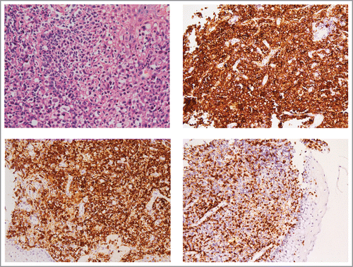
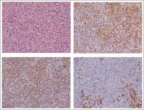
A 73-year-old Chinese male was initially diagnosed as “lymphoma” in September 2011. He had a sore throat and enlargement of bilateral cervical lymph nodes, while fever, night sweat or weight loss were not observed.Positron emission tomography (PET) scan showed that there were several enlarged lymph nodes and soft tissue masses with FDG uptake above the diaphragm. Tongue biopsy was performed subsequently. Microscopically, there were diffuse infiltrations of copious lymphocyte. The nuclei were large, heteromorphic, round in shape and cytoplasm was abundant. By immunohistochemistry, lymphoma cells were reactive for CD20, CD79α,MUM1, CD10 (focal positive) and negative for CD3,CD2 (). Only IGH gene rearrangement was observed by using PCR. He was eventually diagnosed as diffuse large B-cell lymphoma (DLBCL). Afterwards, he received rituximab combined with chemotherapy for 6 cycles and local radiotherapy. Though he achieved completed remission, he had a new symptom of one enlarged left inguinal lymph node in November of 2015. A new biopsy was then performed. Immunohistochemical detection showed that lymphocytes were positive for CD2, CD3,CD4 and negative for CD20, CD8, CD10, BCL−2, BCL−6, PAX5 and MUM-1(). TCR gene rearrangements were detected by PCR, meanwhile, rearrangements of IGH, IGK, IGL were not found. Diagnose of peripheral T-cell lymphoma (PTCL) was made. A full- body computed tomography (CT) scan was done and displayed multiple enlarged lymph nodes only under the diaphragm. We didn't detect EBV infection in either of 2 biopsies by using in situ hybridization, nor any evidence of immune dysfunction complications. Results of bone marrow were normal. So we consider this case as a sequential discordant lymphoma which maybe caused by the immunochemotherapy of DLBCL. By May of 2016, the patient had received 6 cycles of reduced ESHAP regimen and achieved a stable disease so far.
Figure 1. DLBCL in the root of tongue. (A) Histopathology of tongue showed diffuse infiltration of large B-cell lymphocytes (H&E,×400). (B) Immunohistochemical stain with anti-CD20 antibody (×200). (C) Anti-CD79α antibody (×200). (D) Anti-CD3 antibody (×200).

Figure 2. PTCL in inguinal lymph node. (A) Lymph node biopsy showed small-to-medium-sized T-cell lymphocytes in the setting of arborizing endothelial venules (H&E,×400). (B) Immunohistochemical stain with anti-CD2 antibody (×200). (C) Anti-CD3 antibody (×200). (D) Anti-CD20 antibody (×200).

Unlike composite lymphoma which has 2 or more diverse lymphoma types in a single anatomic site, the situation that different types of lymphomas occurring in different anatomic sites concurrently or sequentially is defined as discordant lymphoma.Citation1 Reports of sequential occurrence of DLBCL and PTCL are rare.Citation2,3 Some researchers hypothesized that immunodeficiency and hypogammaglobulinemia caused by R-CHOP therapy led to the development of such a phenomenon.Citation3 In study by Tarella et al, they estimated 1,347 patients with lymphoma. Multivariate analysis demonstrated that 3 factors had an independent association with secondary solid tumor occurrence: advanced age, rituximab addition to high-dose sequential (HDS) program, and radiotherapy after HDS.Citation4 Due to the negative effect on immune system, rituximab combined with chemotherapy has potential possibility to induce a secondary lymphoma.
Disclosure of potential conflicts of interest
No potential conflicts of interest were disclosed.
Funding
Shanghai Key Laboratory of Clinical Geriatric Medicine, 13dz2260700.
References
- Kim H. Composite lymphoma and related disorders. Am J Clin Pathol 1993; 99(4):445-51; PMID:8475911; http://dx.doi.org/10.1093/ajcp/99.4.445
- Micallef IN, Kirk A, Norton A, Foran JM, Rohatiner AZ, Lister TA. Peripheral t-cell lymphoma following rituximab therapy for b-cell lymphoma. Blood 1999; 93(7):2427-8; PMID:10215354
- Kilner MF, Merante S, Svec A. The development of peripheral T-cell lymphoma after successful treatment for diffuse large B-cell lymphoma in a patient with suspected adult onset immunodeficiency: more questions than answers? BMJ Case Rep 2013; 2013:pii: bcr2013200079; PMID:24343800; http://dx.doi.org/10.1136/bcr-2013-200079
- Tarella C, Passera R, Magni M, Benedetti F, Rossi A, Gueli A, Patti C, Parvis G, Ciceri F, Gallamini A, et al. Risk factors for the development of secondary malignancy after high-dose chemotherapy and autograft, with or without rituximab: a 20-year retrospective follow-up study in patients with lymphoma. J Clin Oncol 2011; 29(7):814-24; PMID:21189387; http://dx.doi.org/10.1200/JCO.2010.28.9777
