
ABSTRACT
Primary small cell carcinoma (SCC) of the esophagus is characterized by high malignancy with a tendency to metastasize early through lymph and blood circulation. Metastasis of esophageal SCC frequently occurs to distant organs such as liver and lung. However, few cases of appendiceal metastasis have been reported. This paper first presents a pathologically confirmed case with metastasis of esophageal SCC to the appendix. This particular case highlights the importance of pathological diagnosis and provides new evidence of appendiceal metastasis from esophageal SCC.
Introduction
In 1952, Mckeown first reported 2 cases of esophageal small cell carcinoma (SCC).Citation1 According to recent literature, SCC accounts for 2.4%-2.8% of esophageal tumors,Citation2,Citation3 which usually occurs in the lower and the middle thirds of the esophagus.Citation4,Citation5 Distant metastasis has mostly already occurred when the patients are diagnosed, and the 5-year survival rate of esophageal SCC is merely about 4.8%.Citation6 Therefore, earlier discovery and accurate assessment are extremely important for clinical diagnosis.
Here, we report a rare case of esophageal SCC with metastasis to the appendix to provide new reference for diagnosis and treatment of this kind of tumor. To the best of our knowledge, this is the first report of metastasis of esophageal SCC to the appendix. Immunohistochemical staining of lymph node metastases from esophageal SCC demonstrated strong positivity of synaptophysin (Syn) and CD56, while cells of metastatic SCC in appendix expressed Syn and panCK.
Case report
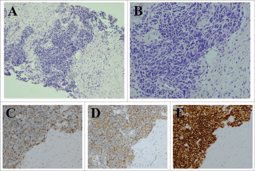
A 57-year-old gentleman was admitted to hospital in April 2016 because of increasing dysphagia and retrosternal pain of one month. He was a former alcohol drinker with a history of smoking cigarettes and severe heart dysfunction. On physical examination an enlarged supraclavicular lymph node was palpable. Computed tomography (CT) scan confirmed a tumor of the lower third of esophagus with enlarged mediastinal and supraclavicular lymph nodes. There was no abnormality in the chest, abdominal space, bone and brain. Gastroscopy demonstrated a submucous protruded lesion of esophagus with a redness of mucosa, arising at 30 cm from the incisors and extending to 35 cm. No biopsy had been taken through gastroscope in consideration of the risk of narcosis for the patient with cardiac insufficiency and fast heart rate. The pathological examination of biopsy of the supraclavicular lymph node demonstrated SCC ( and ). Immunohistochemical staining was analyzed as follows: the staining index for Ki-67: 40% (), the cells expressing Syn () and CD56 ().
Figure 1. Pathological images from esophageal SCC. A. Hematoxylin-eosin stain (× 100). B. Hematoxylin-eosin stain (× 200). C.D.E. Immunohistochemical staining in esophageal SCC (× 200). Ki-67: 40%, Syn (+), CD56 (+).
The patient was treated with a total of 5 cycles of chemotherapy consisting of cisplatin and etoposide and sequential radiotherapy (56Gy/28fractions). The planning target volume included gross tumor volume and draining lymph nodes. After treatment the clinical symptoms were improved significantly and the supraclavicular lymph node could not be palpated. A repeat esophageal barium meal examination in September 2016 indicated that the esophageal lesions disappeared. However, an additional CT scan revealed some residual tumor of esophagus complicated with pneumonia. There was no evidence for extrathoracic extension of the tumor.
In December 2016, the patient was readmitted to hospital reporting right lower quadrant pain 3 months after treatment. Physical examination showed tenderness in the right lower abdominal region and no rebound pain. The abdominal pain had resolved completely after injection of bucinnazine in the emergency department. No fever and vomiting were observed. The laboratory values revealed a leukocyte count of 8.66 × 10⁹/L with 84.4% neutrophils, 8.9% lymphocytes, and 5.4% monocytes. C reactive protein level was 141.3mg/L.
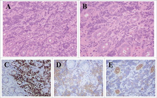
An abdominal ultrasound indicated a swollen appendix with a large well-defined hypoechoic mass lesion. The CT scan of the chest, abdomen and pelvis showed pulmonary inflammation, hepatic and mesentery lymph node metastases. In particular, the appendix was significantly swollen with the size of 7.2 cm × 2.7 cm (). To find out the nature of the swelling appendix, the electronic colonoscopy was performed and found that the appendix hyperaemia erosion. The pathological report of the biopsy described the presence of metastatic SCC in appendix ( and ). Using immunohistochemical analyses, the staining index for Ki-67 was 80% (). The cells expressed Syn () and panCK ().
Figure 2. CT scan of abdomen demonstrating a swollen appendix with an unclear border, uneven density, and heterogeneous enhancement is visible (arrow).
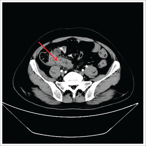
Figure 3. Pathological results from SCC of appendix. A. Hematoxylin-eosin stain (× 100). B. Hematoxylin-eosin stain (× 200). C.D.E. Immunohistochemical staining in SCC of appendix. Ki-67: 80% (× 100), Syn (+) (× 200), panCK (+) (× 200).
The patient was given symptomatic relief and supportive treatment. Unfortunately, he refused to accept further work-up and anti-tumor therapy and left hospital. The patient died of heart failure and liver failure caused of extensive liver metastases in March 2017. The overall survival time was 11 months from initial diagnosis.
Discussion
The SCC is generally found in the bronchial tubes. Primary SCC of the esophagus is thought to be the most common type among extrapulmonary SCC.Citation4 Like small cell lung cancer, esophageal SCC is characterized by high malignancy with a tendency to metastasize early through lymph and blood circulation associated with a poor prognosis.Citation7 Metastases of esophageal SCC have mostly already occurred when the patients are diagnosed, and the 5-year survival rate is about 4.8%.Citation6 The clinical and imaging features of esophageal SCC are lack of specificity. As a result, the correct diagnosis of esophageal SCC mainly depends on histological examination. The esophageal cancer may be incurable when the patient has symptom of dysphagia.Citation8
The causes of death in patients are closely associated with local recurrence and distant metastasis.Citation9 The clinical feature of metastatic carcinoma is various, and it is important for clinicians to detect the metastatic tumor earlier. There is evidence indicating esophageal SCC may metastasize to the liver, lung and draining lymph nodes frequently.Citation2 Mandard et al reported that the lymph node metastases were present in 91% of the esophageal SCC cases by autopsy. 61% of these cases revealed pulmonary metastases and 43% had hepatic metastases.Citation10 Additionally, metastases in the kidneys,Citation9 pancreas,Citation11 adrenal gland,Citation11 bone,Citation12 spleen,Citation13 pleuraCitation14 and multiple metastatic skin nodulesCitation11 have been reported in published literature. Patients usually had extensive dissemination at death.Citation9 However, it is seldom reported the appendiceal tumor caused by esophageal SCC.
The chemotherapy associated with local treatment (radiotherapy or surgery) could be used as standard therapy for SCC of the esophagus. Casas et al concluded that multimodality therapy such as chemotherapy combined with radiotherapy might improve resectability and overall outcome.Citation15
The appendix is attached to the large intestine. According to reported cases, primary carcinoma of the appendix is a rare malignancy of the gastrointestinal tract, which mostly consists of carcinoid tumor, adenocarcinoma, and mucinous adenocarcinoma.Citation16,Citation17 O'Kane et al first presented a case of SCC arising from appendix. CT showed an appendiceal lesion and multiple liver metastases. Histopathological analysis confirmed a primary SCC of the appendix.Citation18 For the appendiceal tumor, the most common presentation is acute appendicitis.Citation19 Involvement of the appendix by secondary tumors is few.Citation20 What's more, it is rare for SCC to metastasis to the appendix.
The patient in our case appeared lower quadrant abdominal pain 3 months after sequential chemoradiotherapy. On physical examination he had tenderness in the right lower abdomen without rebound pain and there were no other typical symptoms of appendicitis. Laboratory data were normal except for elevated neutrophil cell count and C reactive protein level, which the attending doctor attributed to the pneumonia. The ultrasound and CT could not confirm the nature of enlarged appendix (inflammation or tumor). Generally speaking, the metastatic appendiceal carcinoma was rare, but it could not be ruled out. The pathological examination of biopsy by colonoscopy led to the diagnosis of appendiceal SCC eventually. In consideration of the poor heart function of the patient, local radiotherapy for appendix was recommended. Chemotherapy could be given if the heart function improved well. The risks of radiation therapy included appendicular perforation and intestinal obstruction. Unfortunately, the patient rejected further anti-tumor therapy and left hospital because of the risks and costs of treatments. Therefore, no therapeutic effects were reported yet.
When patients with SCC show symptoms of right lower abdominal pain or acute appendicitis, the clinicians may take the appendiceal tumor into consideration though it is rare clinically. This paper first presents a case with a diagnosis of metastasis of esophageal SCC to the appendix confirmed by pathology of appendix biopsy. Through consulting literature, there are 2 case reports presenting the metastasis from small cell lung cancer to appendix, producing acute appendicitis.Citation21,Citation22 Both of the patients had surgery and found gangrenous appendicitis with perforation induced by metastatic SCC of the appendix. The 2 patients were given a new course of chemotherapy. One of them died of acute pneumonia with respiratory function failure. Tumor reduction appeared in the other patient, but there was still an active disease in the abdomen.
Conclusion
To the best of our knowledge, this is the first report of metastasis of esophageal SCC to the appendix in current publications. Primary SCC of the esophagus with high malignancy is likely to metastasize early. It should be noted that whether cancer spreads to uncommon organs. Further examination should be performed in time to avoid diagnostic errors and delayed treatment. This particular case has added to the knowledge of esophageal SCC and highlights the importance of the pathological examination for comprehensive assessment of patient's condition.
Disclosure of potential conflicts of interest
No potential conflicts of interest were disclosed.
Funding
This work was supported by National Nature Science Foundation of China under Grant No. 81530060.
References
- Mckeown F. Oat-cell carcinoma of the oesophagus. J Pathol Bacteriol. 1952;64(4):889-91. doi:10.1002/path.1700640420. PMID:13000600
- Briggs JC, Ibrahim NB. Oat cell carcinomas of the oesophagus: a clinico-pathological study of 23 cases. Histopathology. 1983;7(2):261-77. doi:10.1111/j.1365-2559.1983.tb02240.x. PMID:6133829
- Bennouna J, Bardet E, Deguiral P, Douillard JY. Small cell carcinoma of the esophagus: analysis of 10 cases and review of the published data. Am J Clin Oncol. 2000;23(5):455-9. doi:10.1097/00000421-200010000-00005. PMID:11039503
- Ibrahim NB, Briggs JC, Corbishley CM. Extrapulmonary oat cell carcinoma. Cancer. 1984;54(8):1645-61. doi:10.1002/1097-0142(19841015)54:8<1645::AID-CNCR2820540828>3.0.CO;2-Q. PMID:6089995
- McFadden DW, Rudnicki M, Talamini MA. Primary small cell carcinoma of the esophagus. Ann Thorac Surg. 1989;47(3):477-80. doi:10.1016/0003-4975(89)90404-9. PMID:2539065
- Yun JP, Zhang MF, Hou JH, Tian QH, Fu J, Liang XM, Wu QL, Rong TH. Primary small cell carcinoma of the esophagus: clinicopathological and immunohistochemical features of 21 cases. BMC Cancer. 2007;7:38. doi:10.1186/1471-2407-7-38. PMID:17335582
- Sabanathan S, Graham GP, Salama FD. Primary oat cell carcinoma of the oesophagus. Thorax. 1986;41(4):318-21. doi:10.1136/thx.41.4.318. PMID:3016939
- Belsey RH. Palliative management of esophageal carcinoma. Am J Surg. 1980;139(6):789-94. doi:10.1016/0002-9610(80)90384-0. PMID:6155789
- Sarma DP. Oat cell carcinoma of the esophagus. J Surg Oncol. 1982;19(3):145-50. doi:10.1002/jso.2930190307. PMID:6279973
- Mandard AM, Chasle J, Marnay J, Villedieu B, Bianco C, Roussel A, Elie H, Vernhes JC. Autopsy findings in 111 cases of esophageal cancer. Cancer. 1981;48(2):329-35. doi:10.1002/1097-0142(19810715)48:2<329::AID-CNCR2820480219>3.0.CO;2-V. PMID:6453643
- Doherty MA, McIntyre M, Arnott SJ. Oat cell carcinoma of esophagus: a report of six British patients with a review of the literature. Int J Radiat Oncol Biol Phys. 1984;10(1):147-52. doi:10.1016/0360-3016(84)90421-8. PMID:6321409
- McCullen M, Vyas SK, Winwood PJ, Loehry CA, Parham DM, Hamblin T. Long-term survival associated with metastatic small cell carcinoma of the esophagus treated by chemotherapy, autologous bone marrow transplantation, and adjuvant radiation therapy. Cancer. 1994;73(1):1-4. doi:10.1002/1097-0142(19940101)73:1<1::AID-CNCR2820730102>3.0.CO;2-E. PMID:7506114
- Imai T, Sannohe Y, Okano H. Oat cell carcinoma (apudoma) of the esophagus: a case report. Cancer. 1978;41(1):358-64. doi:10.1002/1097-0142(197801)41:1<358::AID-CNCR2820410148>3.0.CO;2-F. PMID:203379
- Walker SJ, Steel A, Cullen MH, Matthews HR. Treatment of oesophageal small cell carcinoma by combined chemotherapy and surgical resection: report of two cases and review of published cases. Thorax. 1989;44(9):751-2. doi:10.1136/thx.44.9.751. PMID:2555933
- Casas F, Ferrer F, Farrús B, Casals J, Biete A. Primary small cell carcinoma of the esophagus: a review of the literature with emphasis on therapy and prognosis. Cancer. 1997;80(8):1366-72. doi:10.1002/(SICI)1097-0142(19971015)80:8<1366::AID-CNCR2>3.0.CO;2-D. PMID:9338459
- Jetmore AB, Ray JE, Gathright JB, Jr, McMullen KM, Hicks TC, Timmcke AE. Rectal carcinoids: the most frequent carcinoid tumor. Dis Colon Rectum. 1992;35(8):717-25. doi:10.1007/BF02050318. PMID:1643994
- Sandor A, Modlin IM. A retrospective analysis of 1570 appendiceal carcinoids. Am J Gastroenterol. 1998;93(3):422-8. doi:10.1111/j.1572-0241.1998.00422.x. PMID:9517651
- O'Kane AM, O'Donnell ME, Shah R, Carey DP, Lee J. Small cell carcinoma of the appendix. World Journal of Surgical Oncology. 2008;6:4. doi:10.1186/1477-7819-6-4. PMID:18197972
- Benedix F, Reimer A, Gastinger I, Mroczkowski P, Lippert H, Kube R, Study Group Colon/Rectum Carcinoma Primary T. Primary appendiceal carcinoma–epidemiology, surgery and survival: results of a German multi-center study. European Journal Of Surgical Oncology: The Journal of the European Society of Surgical Oncology and the British Association of Surgical Oncology. 2010;36(8):763-71. doi:10.1016/j.ejso.2010.05.025. PMID:20561765
- Deans GT, Spence RA. Neoplastic lesions of the appendix. Br J Surg. 1995;82(3):299-306. doi:10.1002/bjs.1800820306. PMID:7795991
- González-Vela MC, García-Valtuille AI, Fernández FA, Val-Bernal JF. Metastasis from small cell carcinoma of the lung producing acute appendicitis. Pathol Int. 1996;46(3):216-20. doi:10.1111/j.1440-1827.1996.tb03601.x. PMID:10846573
- Pang LC. Metastasis-induced acute appendicitis in small cell bronchogenic carcinoma. South Med J. 1988;81(11):1461-2. doi:10.1097/00007611-198811000-00033. PMID:2847329