ABSTRACT
Leptomeningeal carcinomatosis (LMC) from gastrointestinal cancer is rare. A 56-year-old man with complaint of upper abdominal pain exhibited adenocarcinoma upon histopathologic examination of biopsy specimens. At the end of adjuvant chemotherapy, the patient was affected by hearing loss. Malignant cells were observed by cerebrospinal fluid (CSF) cytology. Therefore, the patient received intrathecal methotrexate and oral temozolomide chemotherapy and radiotherapy. The progress-free survival was approximately 11 months. To our knowledge, such cases of LMC from gastric cancer are very rare. Here, we describe the case of a patient with LMC from gastric cancer and review the literature associated with treatment. We hope that the present report may be helpful when considering how to improve treatment of LMC in gastric cancer patients and offer some tips for the adjuvant treatment modality.
Introduction
Leptomeningeal carcinomatosis (LMC), also known as carcinomatous leptomeningitis, is characterized by diffuse infiltration of metastatic carcinoma into the meninges. LMC is most commonly observed in patients with leukaemia, breast cancer, and lung cancer.Citation1 The prevalence of LMC in these malignancies is estimated at approximately 5–8%.Citation2 LMC does not commonly occur along with gastric cancer, and it is a rare initial manifestation in gastric cancer.Citation3 The prevalence of LMC in gastric cancer patients is as low as 0.16–0.69%.Citation4
The diagnosis of LMC is often difficult to confirm, even when strongly suspected clinically. Definitive diagnosis in most patients is established by the presence of malignant cells in the cerebrospinal fluid (CSF). The prognosis for LMC is very poor because most treatments are palliative. The median survival of LMC patients with adenocarcinoma of the gastrointestinal tract is 3–4 weeks.Citation5
No golden standard treatment for LMC exists. Modalities used in patients with LMC to date include radiation therapy (RT), systemic therapy, and intrathecal therapy.Citation6 Such treatment may improve the neurologic status of the patient and prolong survival.
We herein report an unusual case of sudden bilateral sensorineural hearing loss caused by involvement of the meninges with metastatic gastric cancer, which led us to identify the disease in a timely manner. Subsequently, this patient received intrathecal (methotrexate) and systematic chemotherapy (temozolomide) and radiotherapy. The patient showed a prolonged progress-free survival of 11 months.
Case presentation
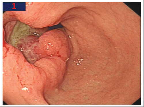
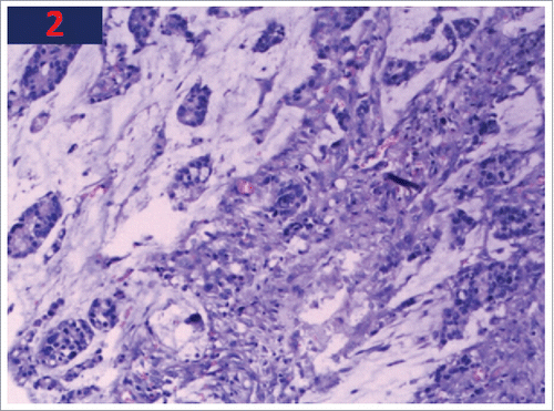
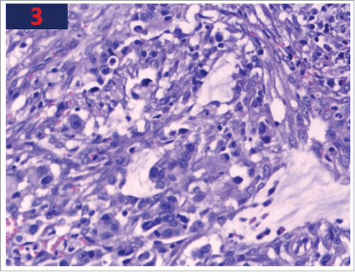
A 50-year-old man presented with upper abdominal pain that worsened after meals in May 2013. The pathological examination by endoscopic biopsy showed gastric pyloric adenocarcinoma (). The patient received radical surgery (R0) with D2 dissection at a local hospital. Tissue samples from this patient were sent for laboratory analysis after surgery. The cytological examination revealed poorly differentiated adenocarcinoma, partly with mucinous adenocarcinoma (, ). The tumour size was approximately 5 × 4 cm. The post-surgery pathological examination also showed invasion of the serosa layer by tumour tissues; 11 lymph nodes in the greater curvature of the stomach were all positive (11/11) for infiltration along with 7 lymph nodes in the lesser curvature (7/7). In addition, the 6 lymph nodes belonging to the “8,9,12 group” were also positive. Both the upper and lower margins were negative. Therefore, the pathological stage of this patient was classified as T3N3M0 (stage III). After surgery, the patient received 6 cycles of combination chemotherapy with oxaliplatin and fluorouracil. The last chemotherapy cycle was completed in December 2013, and no recurrence was observed during regular follow-up.
Figure 1. Endoscopy shows an ulcero-infiltrative lesion with irregular base and abnormalities of surrounding folds.

Figure 2. The biopsy of stomach shows cellular infiltration with poorly differentiated adenocarcinoma.

Figure 3. The biopsy of stomach shows cellular infiltration with poorly differentiated adenocarcinoma.

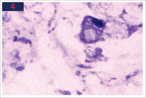
In April 2015, the patient was diagnosed with neurological deafness due to hearing loss. Treatment was unfortunately ineffective. The patient complained of gradually severe headaches and vomiting. Brain MRI found no positive signs. Subsequently, a lumbar puncture was performed. The initial CSF pressure was up to 230 mm/H2O (normal range: 80–180 mm/H2O), and adenocarcinoma cells were observed in the CSF (), which suggested the occurrence of leptomeningeal carcinomatosis from gastric cancer.
Subsequently, the whole brain was irradiated with 6MV X-ray 30 Gy in 10 fractions, with concurrent oral temozolomide 75 mg/m2 daily until the end of radiotherapy. Meanwhile, 5 mg of methotrexate was administered intrathecally twice a week until the malignant cells could not be found in the CSF on three consecutive examinations. Mannitol and dexamethasone were used to reduce the intracranial pressure during treatment. At the conclusion of radiotherapy, the CSF pressure was reduced to 80 mm/H2O. Headache and hearing loss were relieved, and nausea and vomiting disappeared. After the end of radiotherapy, the patient received six cycles of modified FolFox6 chemotherapy. The disease was stable at regular follow-ups every three months. In March 2016, the patient died in a traffic accident.
Discussion
Leptomeningeal carcinomatosis (LMC) occurs in approximately 5–8% of all patients with cancer and is the third most common metastatic complication of the central nervous system.Citation2 Leukaemia, lymphoma, melanoma, breast, and lung cancer are the common offenders, but LMC can complicate any tumour including brain tumours. However, LMC from a gastric cancer is extremely rare. The prevalence of LMC in gastric cancer patients is as low as 0.16–0.69%.Citation4 Of note, the prevalence of LMC in Asian countries, particularly Korea and Japan, has been reported as high compared with other countries.Citation7 LMC diagnosis has increased with improved neuroimaging techniques; however, a true increase in incidence probably results from more effective therapy for systemic cancer resulting in longer survival. The histopathologic type of gastric cancer is signet ring cell carcinoma in most cases.Citation8 In our case, the histopathologic type was adenocarcinoma.
On average, the time lapse between the first recognition of cancer and establishing the diagnosis of LMC presenting with heavy neurological symptoms is approximately 12 months. However, in our case, the interval was much longer at 24 months. This could be explained by the lower tumour burden present at the time of the diagnosis and proper adjuvant treatment. However, since imaging modalities do not aid in diagnosis in many cases, the diagnosis for many patients may be delayed until after the onset of neurological symptoms.Citation9
LMC may lead to multifocal neurologic deficits, which may be associated with infiltration of the cranial and spinal nerve roots, direct invasion of the brain or spinal cord, obstructive hydrocephalus, or some combination of these factors. As a result, the presenting manifestations are usually headache, nausea, vomiting, visual disturbances and seizures. A variety of other neurologic deficits may be present. In the case described in this report, however, the main symptom was hearing difficulty. This symptom has been rarely described in the literature.
Patient prognosis for LMC arising from gastric cancer is poor. Even in patients who underwent chemotherapy and RT following the diagnosis of LC, the mean survival has been reported to be only approximately 4–6 weeks.Citation5 The reason for this poor survival may include not only the late diagnosis but also the lack of established treatments.
Currently, there is no established single diagnostic test for LMC, although CSF cytology with spinal tapping and MRI are useful in diagnosing LC. In the reported literature, the sensitivity of cranial MRI in the diagnosis of LMC had been reported as between 65% and 75%.Citation7 Park et al.Citation10 reported that with the additional consideration of positive CSF cytology results, the diagnostic sensitivity of MRI increased up to 91%, although the sensitivity of CSF cytology alone for LC was at most 54%. CSF samples collected from patients with LMC usually exhibit increased pressure, pleocytosis, elevated levels of protein and lactate dehydrogenase (LDH), and hypoglycaemia.Citation11 The definitive diagnosis of LMC can only be documented by the presence of malignant cells in the CSF. In our case, the first CSF sampling documented the presence of malignant cells. In cases in which the cytological examination of the CSF is negative for malignant cells, one should not hesitate to repeat the procedure, because the likelihood of detecting malignant cells increases in repeated samplings.Citation12
Currently, there are no golden standard treatment guidelines for LMC due to the lack of standardization.Citation13,14 Chemotherapy or RT is the important treatment option for patients with LMC, although these are performed as palliative treatments, and the resultant patient outcomes are suboptimal.Citation15 Although systemic chemotherapy is a crucial treatment for LMC, most anti-cancer agents have difficulty in penetrating the blood-brain barrier. For this reason, whole brain irradiation and intrathecal chemotherapy, each alone or in combination with systemic chemotherapy, have been attempted to treat patients with LMC.
Drugs that could be used for IT chemotherapy include methotrexate, thiotepa, and cytarabine in combination with steroids.Citation16 There is no evidence to support the superiority of combined therapy over single agent therapy.Citation17 Previous reports suggested that patients receiving IT chemotherapy live longer than those receiving best supportive care.Citation18 Methotrexate is one of most useful agents for IT chemotherapy. High dose intravenous methotrexate (3.5 g/m2) injection showed a 28% partial response with stable disease and 44% progressive disease.Citation19
Radiation therapy (RT) is the most important treatment modality for symptomatic or bulky LMC. RT can rapidly relieve pain, particularly radicular pain, and other neurologic symptoms and improve quality of life.Citation20 RT should be employed to treat symptomatic areas and sites of bulky disease even if asymptomatic. It can preserve neurologic function and improve CSF flow, making intrathecal chemotherapy more effective.Citation21
Numerous reports have suggested that systemic therapy improves survival for patients with LMC. Temozolomide is an orally bioavailable drug that reaches CSF levels at approximately 20% of those in the serum. After oral administration, bioavailability is nearly 100%, with excellent penetration into all body tissues, including the brain.Citation22 This agent is the gold standard treatment for glioblastoma and its efficacy in the treatment of brain metastases has been proven.Citation23 Temozolomide used for LMC has been discussed in phase II trials.Citation24 These studies showed that the agent is well tolerated and did not adversely affect the quality of life of patients with LMC. In addition, published case report studies have proven the efficiency of Temozolomide.Citation25,26 Therefore, oral temozolomide was well-tolerated in this patient.
Although systemic and intrathecal chemotherapy as well as radiotherapy are both palliative, they improved the symptoms of meningeal irritation lead to a better quality of life. In addition, as the patient's LMC was diagnosed early and adjuvant treatment was administered in a timely manner, all his neurological symptoms regressed after the initiation of treatment. Moreover, he showed a longer duration of progress-free survival (11 months) than did previously described patients. Accordingly, we hope that the present report may be helpful when considering how to improve the treatment of LMC in gastric cancer patients and offer some tips for the adjuvant treatment modality.
References
- Grossman SA, Krabak MJ. Leptomeningeal carcinomatosis. Cancer treatment reviews. 1999;25(2):103-19. doi:10.1053/ctrv.1999.0119.
- Pentheroudakis G, Pavlidis N. Management of leptomeningeal malignancy. Expert Opin Pharmacother. 2005;6(7):1115-25. doi:10.1517/14656566.6.7.1115.
- Chamberlain MC. Leptomeningeal metastasis. Paper presented at: Seminars in neurology 2010.
- Kim M. Intracranial involvement by metastatic advanced gastric carcinoma. J Neurooncol. 1999;43(1):59-62. doi:10.1023/A:1006156204385.
- Groves MD. New strategies in the management of leptomeningeal metastases. Arch Neurol. 2010;67(3):305-12. doi:10.1001/archneurol.2010.18.
- Kak M, Nanda R, Ramsdale EE, Lukas RV. Treatment of leptomeningeal carcinomatosis: Current challenges and future opportunities. J Clin Neurosci. 2015;22(4):632-7. doi:10.1016/j.jocn.2014.10.022.
- Lisenko Y, Kumar AJ, Yao J, Ajani J, Ho L. Leptomeningeal carcinomatosis originating from gastric cancer: report of eight cases and review of the literature. Am J Clin Oncol. 2003;26(2):165-70. doi:10.1097/00000421-200304000-00013.
- Giglio P, Weinberg JS, Forman AD, Wolff R, Groves MD. Neoplastic meningitis in patients with adenocarcinoma of the gastrointestinal tract. Cancer. 2005;103(11):2355-62. doi:10.1002/cncr.21082.
- Oh SY, Lee S-J, Lee J, Lee S, Kim SH, Kwon HC, Lee GW, Kang JH, Hwang IG, Jang JS, et al. Gastric leptomeningeal carcinomatosis: multi-center retrospective analysis of 54 cases. World J Gastroenterol. 2009;15(40):5086-90. doi:10.3748/wjg.15.5086.
- Park K-K, Yang S-I, Seo K-W, Kim Y-O, Yoon K-Y. A case of metastatic leptomeningeal carcinomatosis from early gastric carcinoma. World journal of surgical oncology. 2012;10(1):74. doi:10.1186/1477-7819-10-74.
- Bulut G, Erden A, Karaca B, Göker E. Leptomeningeal carcinomatosis of gastric adenocarcinoma. Turk J Gastroenterol. 2011;22(2):195-8. doi:10.4318/tjg.2011.0191.
- Wasserstrom WR, Glass JP, Posner JB. Diagnosis and treatment of leptomeningeal metastases from solid tumors: experience with 90 patients. Cancer. 1982;49(4):759-72. doi:10.1002/1097-0142(19820215)49:4<759::AID-CNCR2820490427>3.0.CO;2-7.
- Chamberlain MC. Leptomeningeal metastasis. Current Opin Oncol. 2010;22(6):627-35. doi:10.1097/CCO.0b013e32833de986.
- Vergoulidou M. Leptomeningeal Carcinomatosis in Gastric Cancer: A Therapeutical Challenge. Biomark Insights. 2017;12:1177271917695237. doi:10.1177/1177271917695237.
- Kim S-J, Kwon J-T, Mun S-K, Hong Y-H. Leptomeningeal carcinomatosis of gastric cancer misdiagnosed as vestibular schwannoma. J Korean Neurosurg Soc. 2014;56(1):51-4. doi:10.3340/jkns.2014.56.1.51.
- Groves MD. New strategies in the management of leptomeningeal metastases. Arch Neurol. 2010;67(3):305-12. doi:10.1001/archneurol.2010.18.
- Hitchins RN, Bell DR, Woods RL, Levi JA. A prospective randomized trial of single-agent versus combination chemotherapy in meningeal carcinomatosis. Journal of clinical oncology: official journal of the American Society of Clinical Oncology. 1987;5(10):1655-62. doi:10.1200/JCO.1987.5.10.1655.
- Beauchesne P. Intrathecal chemotherapy for treatment of leptomeningeal dissemination of metastatic tumours. Lancet Oncol. 2010;11(9):871-9. doi:10.1016/S1470-2045(10)70034-6.
- Lassman AB, Abrey LE, Shah GD, Panageas KS, Begemann M, Malkin MG, Raizer JJ. Systemic high-dose intravenous methotrexate for central nervous system metastases. J Neurooncol. 2006;78(3):255-60. doi:10.1007/s11060-005-9044-6.
- DeAngelis LM, Boutros D. Leptomeningeal metastasis. Cancer Investigation. 2005;23(2):145-54. doi:10.1081/CNV-50458.
- Kak M, Nanda R, Ramsdale EE, Lukas RV. Treatment of leptomeningeal carcinomatosis: current challenges and future opportunities. Journal of clinical neuroscience: official journal of the Neurosurgical Society of Australasia. 2015;22(4):632-7. doi:10.1016/j.jocn.2014.10.022.
- Ostermann S, Csajka C, Buclin T, Leyvraz S, Lejeune F, Decosterd LA, Stupp R. Plasma and cerebrospinal fluid population pharmacokinetics of temozolomide in malignant glioma patients. Clinical cancer research: an official journal of the American Association for Cancer Research. 2004;10(11):3728-36. doi:10.1158/1078-0432.CCR-03-0807.
- Ma W, Li N, An Y, Zhou C, Bo C, Zhang G. Effects of Temozolomide and Radiotherapy on Brain Metastatic Tumor: A Systematic Review and Meta-Analysis. World Neurosurg. 2016;92:197-205. doi:10.1016/j.wneu.2016.04.011.
- Segura PP, Gil M, Balana C, Chacón I, Langa JM, Martín M, Bruna J. Phase II trial of temozolomide for leptomeningeal metastases in patients with solid tumors. J Neurooncol. 2012;109(1):137-42. doi:10.1007/s11060-012-0879-3.
- Lombardi G, Zustovich F, Della Puppa A, Borgato L, Orvieto E, Manara R, Cecchin D, Berti F, Farina P, Gardiman MP, et al. Cisplatin and temozolomide combination in the treatment of leptomeningeal carcinomatosis from ethmoid sinus intestinal-type adenocarcinoma. J Neurooncol. 2011;104(1):381-6. doi:10.1007/s11060-010-0484-2.
- Hottinger AF, Favet L, Pache JC, Martin JB, Dietrich PY. Delayed but Complete Response following Oral Temozolomide Treatment in Melanoma Leptomeningeal Carcinomatosis. Case Rep Oncol. 2011;4(1):211-5. doi:10.1159/000327699.

