
Abstract
Introduction: A simplified and computationally efficient human body finite element model is presented. The model complements the Global Human Body Models Consortium (GHBMC) detailed 50th percentile occupant (M50-O) by providing kinematic and kinetic data with a significantly reduced run time using the same body habitus.
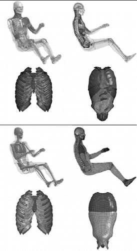
Methods: The simplified occupant model (M50-OS) was developed using the same source geometry as the M50-O. Though some meshed components were preserved, the total element count was reduced by remeshing, homogenizing, or in some cases omitting structures that are explicitly contained in the M50-O. Bones are included as rigid bodies, with the exception of the ribs, which are deformable but were remeshed to a coarser element density than the M50-O. Material models for all deformable components were drawn from the biomechanics literature. Kinematic joints were implemented at major articulations (shoulder, elbow, wrist, hip, knee, and ankle) with moment vs. angle relationships from the literature included for the knee and ankle. The brain of the detailed model was inserted within the skull of the simplified model, and kinematics and strain patterns are compared.
Results: The M50-OS model has 11 contacts and 354,000 elements; in contrast, the M50-O model has 447 contacts and 2.2 million elements. The model can be repositioned without requiring simulation. Thirteen validation and robustness simulations were completed. This included denuded rib compression at 7 discrete sites, 5 rigid body impacts, and one sled simulation. Denuded tests showed a good match to the experimental data of force vs. deflection slopes. The frontal rigid chest impact simulation produced a peak force and deflection within the corridor of 4.63 kN and 31.2%, respectively. Similar results vs. experimental data (peak forces of 5.19 and 8.71 kN) were found for an abdominal bar impact and lateral sled test, respectively. A lateral plate impact at 12 m/s exhibited a peak of roughly 20 kN (due to stiff foam used around the shoulder) but a more biofidelic response immediately afterward, plateauing at 9 kN at 12 ms. Results from a frontal sled simulation showed that reaction forces and kinematic trends matched experimental results well. The robustness test demonstrated that peak femur loads were nearly identical to the M50-O model. Use of the detailed model brain within the simplified model demonstrated a paradigm for using the M50-OS to leverage aspects of the M50-O. Strain patterns for the 2 models showed consistent patterns but greater strains in the detailed model, with deviations thought to be the result of slightly different kinematics between models. The M50-OS with the deformable skull and brain exhibited a run time 4.75 faster than the M50-O on the same hardware.
Conclusions: The simplified GHBMC model is intended to complement rather than replace the detailed M50-O model. It exhibited, on average, a 35-fold reduction in run time for a set of rigid impacts. The model can be used in a modular fashion with the M50-O and more broadly can be used as a platform for parametric studies or studies focused on specific body regions.
Introduction
Approximately 1.24 million people die each year worldwide from motor vehicle crashes, and an additional 20 to 50 million more suffer nonfatal injuries (World Health Organization Citation2009). Improving vehicular safety requires knowledge of the underlying occupant biomechanics. Computer simulations of the human body using finite element analysis (FEA) are gaining in popularity and have the potential to enhance the volume of biomechanically relevant data obtained from crash simulations. Computational models of anthropometric test devices (ATDs) can also be used to this end; however, these models are developed to match specific ATD geometry and construction, which, by necessity, deviates from the human body. The increased complexity of computational human body models in recent years has resulted in significantly increased computational expense. Taken together, these observations signal the need for a complementary model that provides the biofidelic aspects of the human body model with reduced run time and rapid positioning capabilities of ATD models. The development of this type of human body model is the focus of this work.
The Global Human Body Models Consortium (GHBMC) is an international consortium of industry, government, and academic research groups that have consolidated the development of human body finite element models for injury prediction and prevention. The GHBMC 50th percentile male seated occupant model v4.3 (M50-O) has 2.2 million elements. The geometry of the M50-O was developed to represent an average male anthropometry (Gayzik et al. Citation2011) and was validated at the body region and full-body model levels.
The head was validated with 35 loading cases demonstrating that the head responses were comparable to experimental measurements in terms of pattern, peak values, or time histories (Mao et al. Citation2013). It has also been used in the development of advanced brain injury criteria (Takhounts et al. Citation2013). The neck response was investigated by assessing rear impact scenarios, individual loading of craniovertebral ligaments, and progressive failure of the ligaments (DeWit et al. 2012; Fice et al. Citation2011; Mattucci et al. Citation2012, 2013). Cross-sectional planes were defined to investigate neck forces (White et al. 2015). The ribs were tested in a hierarchical fashion (Kindig et al. 2015; Poulard et al. Citation2014) under anterior–posterior bending loads, and the effect of cortical thickness and material properties were evaluated (Li, Kindig, Kerrigan, et al. 2010; Li, Kindig, Subit, and Kent 2010). A new cortical thickness mapping technique was applied to the coxal bone from CT images and the effects of posture on hip injury tolerance were analyzed (Kim et al. 2012; Yue et al. 2014). The lower limbs were also tested under complex loading resulting from blunt impacts (Shin and Untaroiu Citation2013; Shin et al. Citation2012; Untaroiu et al. Citation2013). The full-body M50-O was validated by mass distribution (Vavalle et al. 2014) and in a variety of hub and lateral impacts (Hayes et al. Citation2014; Park et al. Citation2013; Vavalle et al. Citation2015; Vavalle et al. Citation2012).
Though biomechanical data for select crash-induced injuries can be obtained from the M50-O model, other uses such as parametric studies or the simulation of longer events (>250 ms) are impeded by the run time. The M50-O completes a 250 ms frontal sled simulation in approximately 24 h on 48 CPU computational cluster. It is feasible to envision a scenario when only a certain aspects of the response are of interest to the researcher. Therefore, the goal of the current study is to develop a 50th percentile male occupant simplified model (M50-OS) that is less computationally expensive than the M50-O but is reasonably biofidelic and can serve as a platform for modular use of components of the M50-O. As a tradeoff, the M50-OS is not intended to predict crash induced injuries, unlike its more detailed counterpart.
Methods
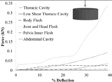
The M50-OS was developed using the same source geometry as the GHBMC 50th percentile male seated occupant model ver. 4.3 (Gayzik et al. Citation2011). The M50-OS uses a comparatively coarser mesh (8–10 mm) for deformable elements than the M50-O (2–4 mm). As large regions of the M50-O model were remeshed, the mass distribution was verified using segment planes and data from McConville et al. (1980; Vavalle et al. 2013). The materials used in the model are found in Table A3 (see online supplement). All deformable structures representing soft tissues were simulated using LS DYNA in a uniaxial compressive simulation, using a 10 mm × 25 mm diameter hexahedral cylinder at 0.1 s−1 to 40% deformation, a tissue sample sized used in previous studies (Kemper et al. Citation2012). The soft tissue, including the flesh components and internal cavity compartments, required stable and computationally inexpensive material models for which parameters were not always available in the literature. Instead, material models obtained in the literature were utilized in the compression simulation to obtain force versus deflection curves that were then used as inputs into material models that demonstrated the desired characteristics. Specific data on each material are discussed below.
The head of the M50-OS utilizes the same skull, face, and scalp meshes as the M50-O. The skull is modeled as a rigid body but the skin and outer flesh was kept deformable. The brain was replaced with a single mass node at the center of gravity of the brain and constrained to the skull. The deformable skin material definition was preserved from the M50-O (Mao et al. Citation2013). The head flesh is modeled to represent charcoal polyester used in previous dummy models (Canha et al. Citation2000). This material model is commonly used, because it is stable and can be adjusted by a nominal stress versus strain curve and a viscous coefficient (Croop and Lobo Citation2009). The cervical spine of the M50-OS was modeled using the vertebral body geometry from the M50-O and the definitions from Dibb et al. (2013) for the osteo-ligamentous spine. Briefly recapping their approach, the vertebral bodies in the neck were modeled as rigid with constraints to nodes at the centers of rotation (COR) between neighboring bodies. At each COR there were 3 translational and 3 rotational dampers implemented. One-dimensional muscle elements utilizing the definitions from Dibb et al. (2013) were added to constrain the scapula.
The deformable flesh components are majority hexahedral elements with a hemispherical tetrahedral element region used locally around major articulations (for positioning purposes). In the M50-O, the flesh was modeled using a hyperelastic material that can incorporate strain stiffening via a uniaxial load curve (Yamada and Evans Citation1970) and is based on the Ogden rubber model. Because this material model (*MAT_SIMPLIFIED_RUBBER) was relatively computationally expensive, the M50-OS utilizes a direct implementation of Ogden rubber (*MAT_OGDEN_RUBBER) that was determined to be well suited for white adipose tissue (Egelbrektsson Citation2011). The parameters for this material were determined by running the uniaxial compressive simulation using the parameters provided by Egelbrektsson (Citation2011) to obtain a force–deflection curve. This curve was subsequently stiffened at high strains (>35%) by increasing the corresponding force by 50% for stability.
The viscera in the thoraco-abdominal cavity were modeled as a 2-layered homogeneous component separated at the location of the diaphragm (). The upper section was subsequently split into 2 additional components with a low-shear region at the center of the chest cavity to mimic the presence of fluid and to tune the response in frontal impacts. These 3 sections were modeled as a highly compressible low-density foam that is commonly used to model ATDs (Canha et al. Citation2000), but the load curve defining the nominal stress versus strain and elastic modulus was specific to each region and obtained using the methods described previously. The outer component of the thorax cavity was modeled using the compressive properties of the lung (Yuen et al. Citation2008). The inner, low-shear region of the thorax was consistent with the compressive behavior of a heart tissue model (Deng et al. Citation1999) and the abdomen section utilized the compressive behavior of the liver (Lee and Yang Citation2001).
The rib cage retained the same properties as the M50-O but utilized a coarser mesh. A uniform thickness of 0.75 mm was applied to the cortical bone layer, shown in . Because of the mesh difference, a study on the force deflection response of the rib cage at localized areas was conducted and compared against literature as well as the M50-O. A segment-based contact around the clavicle was defined to increase stability and to improve the response to seat belt loading. A tied contact was defined to prevent separation of the rib cage and the thoracic flesh. The thoracic spine, lumbar spine, costovertebral joints, and upper extremity joints all retained the same definitions as in the M50-O. The constraints in the thoracic spine and costo-vertebral junction utilize a generalized constrained joint that was retained from the M50-O occupant model and are based biomechanical tests of these locations (Duprey et al. Citation2010). The lumbar intervertebral discs were also retained from the M50-O, incorporating 1D elements to allow for motion between vertebral bodies.
The pelvic bones utilized the same surface mesh from the M50-O (quadrilateral shell) but are modeled as rigid shells. Due to the organic shape of the pelvis, a tetrahedral region of elements was used between the outer flesh and the pelvis. This inner flesh layer is defined as *MAT_FU_CHANG_FOAM, which has proven to be stable in high lateral force impacts. This material model performs well in rate-dependent impacts and has been used as an alternative to low-density foam (Serifi et al. Citation2003). A segment-based contact was defined between the 2 flesh layers (stiffer inner and more flexible outer) in the pelvis to increase stability in large deformation simulations.
The flesh mesh at the wrist, elbow, shoulder, hip, knee, and ankle used tetrahedral elements in a hemisphereical configuration to allow for rotation about the joints. This was to provide reasonble external contours depending on the postion of the model. The material model at the joints required the use of stiffer foam due to the large compressive loads on flesh that was in close proximity to rigid bones. This was similar to the head flesh (Croop et al. Citation2009). Revolute joints were used at the wrists, elbows, ankles, and knees, and spherical joints were used at the glenohumeral joint and acetabulum. Passive elastic joint moments were included in the lower extremity joints (knee and ankle) to provide accurate moment versus angle relationships (Riener and Edrich Citation1999). Riener and Edrich (Citation1999) performed a study on 10 healthy male subjects instrumented with strain gauge–based monoaxial load cells and performed rotations about the knee and ankle. The data were incorporated into the M50-OS by utilizing the *CONSTRAINED_JOINT_STIFFNESS cards to provide moment versus joint angle relationships. The cross-joint meshes are constrained to the underlying bone and do not have contacts defined against each other (). All such limb caps were included in contact definitions with the surrounding structures of the body or its simulation environment. Automatic single surface contacts and an interior contact were used to define the interactions among all of the components of the M50-OS.
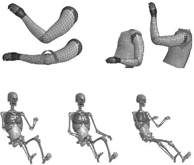
Positioning can be applied in a preprocessor without the need to run positioning simulations with the M50-OS. A code to position the M50-OS model was written in the open source software LS-PrePost (v4.1, LSTC, Livermore, CA), shown in . The joints defined in the positioning tree include the C7/T1 facet joint, glenohumeral joints, humeroulnar joints, radiocarpal joints, acetabulofemoral joints, tibiofemoral joints, and talocrural joints. A local coordinate system for each point of rotation was defined and utilizes nodes defined in the spherical or revolute joints in order to reposition the model. This positioning includes maximum permissible angles of rotation by axis and limb. The upper arms, lower arms, hands, upper legs, lower legs, feet, and neck (small changes only) can be adjusted to desired positions.
The M50-OS was programmed to output data for comparisons to the literature and the M50-O. In this study, we use a constrained node at the head center of gravity for the head accelerometer data, a femur load sensor, and several locations on the model to provide kinematic outputs. The femur axial force outputs use a beam element that includes 2 coincident nodes; one node is constrained to the distal rigid bone and the other node is constrained to the proximal rigid bone. The beam itself is designed to act like a load cell to output the axial force. A similar approach was used to implement a tibia load cell.
All simulations were run using LS-DYNA R. 6.1.1. MPP on the DEAC cluster at Wake Forest University, using 48 CPUs and Infiniband interconnects. The simplified model rib cage was subjected to denuded rib point-loading simulations at 7 locations on the rib cage. The methods follow those of Kindig et al. (2013) and Poulard et al. (Citation2014), who performed a similar validation of the M50-O. Because the rib meshes were altered, this work was completed on the M50-OS to ensure biofidelity and demonstrate a consistent response, despite the change in mesh density to a coarser mesh. An elastic sphere and rigid plate setup was used as shown in the bottom right of Figure A1 (see online supplement; Poulard et al. Citation2014). The impactor's normal direction was such that it was perpendicular to the sphere and it translated inward, causing rib deformation at a constant, quasistatic rate throughout the simulations.
The M50-OS was simulated in one robustness scenario, 4 rigid impacts, and a frontal sled pulse. The robustness simulation was a knee bolster–type impact with a 4.9 m/s initial velocity prescribed to the human body model and designed to be compared only to the same output from the M50-O. Rigid impacts included a 23 kg hub impact to the chest with an initial velocity of 6.7 m/s (Kroell et al. Citation1974; Lebarbé and Petit Citation2012) and a 48 kg bar impact to the abdomen with an initial velocity of 6 m/s (Hardy et al. Citation2001). The lateral impacts included a 23.4 kg plate impact at 12 m/s to the right arm (Kemper et al. Citation2008) and a lateral sled simulation with a model initial velocity of 6.7 m/s impacting a rigid wall (Cavanaugh et al. Citation1990). The M50-OS was repositioned so that the right humerus was parallel with the impactor in the 12 m/s impact to more closely match the posture defined in the experiment. In the 6.7 m/s lateral sled impact, the M50-OS was repositioned so that the left arm was placed over the left leg and gravity settled for 100 ms prior to initiation of the impact. The frontal sled simulation is based on work by Shaw et al. (Citation2009) utilizing an 11.1 m/s frontal pulse. The arms are positioned to be near the legs of the model, which is first belted and then allowed to gravity settle for 100 ms, followed by a 150 ms event. This is done to allow for the model to be in good contact with the seat and lower extremity restraints. The results from dynamic simulations were quantitatively evaluated for magnitude and phase agreement with the experimental data using an objective comparison software package, CORrelation and Analysis (CORA v. 3.5.1; Correlation and Analysis software; Gaimersheim, Germany). CORA was not run for the denuded rib test and or frontal thorax hub impact because time-valued data were not available (both are cross-plots of force vs. deflection).
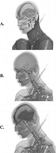
To demonstrate the modular use of the M50-OS, all parenchymal structures, meninges, cerebrospinal fluid, ventricles, and sinuses of the M50-O brain (n = 27 parts) were imported. Deformable properties for the inner and outer tables of the skull and diploe layer were imported into the M50-OS to demonstrate its modular use with the M50-O (Mao et al. Citation2013). All modeling considerations were identical between the modified M50-OS (with brain) and M50-O in this region. The mass node that represented the brain was removed from the model. The model was simulated in the Shaw et al. (Citation2009) frontal sled test for 100 ms of settling and the first 100 ms of the event. Kinematics and deformation of the brain model were investigated.
Results
The M50-OS contains a total of 11 contacts: 4 automatic single surface, 5 automatic surface-to-surface contacts, a tied nodes to surface, and a contact interior. The M50-O, in contrast, utilizes a total of 447 contacts. The total number of elements was reduced to 354,000, including 205,000 rigid elements and 149,000 deformable elements, compared to the detailed M50-O, which has 2.2 million. The resulting components of the model are shown in Table A1 (see online supplement), organized by body structure, and the uniaxial compression tests performed to evaluate the soft tissue components are shown in . The smallest time step of the M50-OS was 2.8 μs, 28 times larger than the detailed M50-O, 0.1 μs. The measured section masses of the M50-OS were within one standard deviation of the reported masses in McConville et al. (Citation1980) for 8 of 10 given body regions. The upper arm and foot of the M50-OS are 1.05 and 1.17 standard deviations from the experimental mean masses. This discrepancy is due to the overlapping elements around moveable joints at the shoulder and ankle.
The simplified model denuded rib loading simulations were completed on the M50-OS, compared to experimental data (Kindig et al. 2013; Poulard et al. Citation2014) and the M50-O. The overall response showed good agreement with the experimental corridors and closely followed the response of the M50-O (see Figure A1). The point loading test on the ribs demonstrated small oscillations at lower displacements most prominently at rib 9. The oscillations decreased as the rib was loaded and the response became more linear as predicted in the experimental data and the M50-O. Rib 9 showed one of the poorer correlations, which is likely influenced by the presence of costal cartilage, which can vary significantly between specimens.
The knee bolster impact provided axial forces along the left and right femur. The maximum loads were 3.82 and 3.80 kN for the left and right femur; images from the simulation are shown in Figure A4 (see online supplement). The M50-O, in the same boundary condition, resulted in similar peak loads for the left and right femur of 3.73 and 3.71 kN, respectively. The kinematic and kinetic results from the remaining 4 rigid impacts are shown in . In the frontal thorax hub impact the peak thorax deflection and peak force were 31.2% and 4.6 kN, respectively. The frontal abdominal bar impact response remained in the corridors during the loading phase and produced a peak abdominal deflection and peak force of 76.2% (anterior abdominal deflection is considered the distance from the outermost flesh of the model to the anterior surface of L3) and 5.2 kN, respectively, with a CORA score of 0.69. In the lateral rigid sled simulation, forces for the thorax, abdomen, and shoulder were averaged to represent the torso force with a peak of 8.71 kN. The CORA score for the torso was 0.72. The thorax lateral impact force resulted in a peak of 20.8 kN corresponding to the impact of stiffer material at the shoulder; however, the plateau occurring after these initial spikes matched the data well and the CORA score was 0.58.
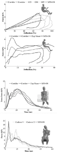
The frontal sled outputs considered in this study included head kinematics, sternum deflection, pelvic motion, and reaction loads on the buck. The results are shown by using the SAE J211 sign convention. The knee and foot pan used a local coordinate systems and the loads perpendicular and parallel to the plane are reported. The results, shown in Figure A2 (see online supplement), were selected to demonstrate the global response of the M50-OS. The head vertical displacement was 12 cm greater than the experimental mean with a CORA score of 0.55. The maximum sternum frontal displacement matched the experimental mean with a CORA score of 0.83. The left and right knee forces in the local X direction matched the experimental results with CORA scores of both 0.61. Taken together, the knee and feet reaction loads reasonably follow the contours of the experiment, and the gross motion of the body (Figure A4) suggests that the joint modeling approach is reasonable. Some aspects of the response were not captured, including the shearing force on the seat pan and the upward motion of the pelvis. The mean CORA values between models are nearly identical for the data presented herein (0.63 for M50-O vs. 0.59 for M50-OS).
The modular addition of the M50-O brain in the M50-OS is shown in . Kinematics of the baseline M50-OS and M50-OS with brain appeared unchanged. Based on the simulation pace of the M50-O (minutes of real-time for each millisecond of simulation time), the M50-OS demonstrated a 4.75-fold decrease in run time for this simulation, which utilized a deformable skull and brain. Kinematics of the M50-O are shown in Figure A2 and indicate some differences. The M50-O exhibited less forward flexion, slightly greater lateral displacement, and slightly less vertical displacement. Instantaneous strain fields at 75 ms (time of peak change in X velocity) are shown in Figure A3 (see online supplement). Though the strain data differ in magnitude (which is suspected to be a function of the differences in kinematics), similar patterns emerge. Larger strains are found in the cerebrum, with lower values in the deep structures such as the brainstem. Cross-sectional images show lower strains in the frontal and occipital portions of the brain, with larger strains occurring in the superior parietal lobe. The comparisons are not intended to comment on injury probability (there was no head strike in the simulation) but merely to show that similar trends are found when using the M50-OS in this modular capacity.
Discussion
Using the same source geometry of the GHBMC average male detailed occupant model, the M50-OS provides comparable gross kinematic and kinetic outputs. The total run time was closely monitored throughout the development process. The same hardware was used for both the M50-O and M50-OS, 48 central processing units on Infiniband high-speed interconnects, in order to quantify the run time. The rigid impacts demonstrated a run time reduction of 35 times on average, and the sled impact was reduced by 23 times (see Table A2, online supplement). The model was 5 times faster when including deformable structures of the head and brain. At the same time, biofidelity was only marginally affected when compared to the M50-O using CORA.
The reductions in run time provide a substantial opportunity for parametric modeling with the GHBMC M50-OS. To put it in perspective, nearly 35 parameter variations could be run with the M50-OS in the time required to run a single study with the M50-O. As discussed in the Introduction, some of the attributes of the M50-OS mimic those of traditional ATD models (run time, positioning, and virtual instrumentation). The key difference in relation to traditional ATD models is the use of the same body habitus as the GHBMC M50-O, which, as demonstrated, opens the opportunity to modularly include aspects of the detailed human body within it. Due to geometrical differences, this would not be possible with an ATD model. Though a single modular simulation was created, the approach outlined could be used to insert other parts of the model, such the lower extremity or thoraco-abdominal cavity. Even the inclusion of the brain and deformable skull, which demonstrated a more modest 4.75-fold run time reduction, would translate into a researcher having simulations results within a typical workday rather than having to run simulations overnight.
The applications of the M50-OS include parametric studies of either an aspect of the model itself (i.e., studying the effects of various techniques to model the spine) or the environment (e.g., airbag system interaction). The M50-OS is ideally suited to study and develop seat belt systems because the body contours match those of human body. This simplified version of a more complex model is not designed to be mutually exclusive but, rather, to be used together. For example, a test matrix can be explored with the simplified model to narrow down to a set of critical cases, which could then be further explored with the detailed model.
The global responses compared favorably with the literature force and displacement curves, with peaks occurring at the correct phase in time and of an appropriate magnitude in general. The frontal thorax hub and abdominal impacts provided peak deflections comparable to the corridors. However, in both cases the peak force was toward the upper bound due to the rubber and foam materials selected in these regions. It was necessary to utilize the given materials in order to provide stability to the M50-OS such that the model can be simulated in high-impact scenarios. In the 12 m/s lateral impact one peak occurred early in the simulation, which was due to the rigid plate contacting the shoulder of the right upper extremity. The joints required a stiffer material due to noted instabilities during the development process and therefore exhibit an initial peak. Following the peak, the force leveled off and decreased as the model lost contact with the impactor. The lateral sled impact simulation provided similar results in the thoracic response in a 6.7 m/s sled impact, again from the joint mesh. Both of these lateral impacts are highly dependent on the initial placement of the upper extremity being impacted. With the simplified model we were able to easily test arm configurations. In addition, it was demonstrated that the M50-OS has the capabilities to output accurate femur forces.
The denuded rib cage simulations showed that despite using a coarser mesh, the response closely matched the literature and the M50-O. By completing these isolated simulations, it suggests that the mesh adjustments in the rib cage does not alter the gross force vs. deflection of the M50-OS thorax. It should be noted that the intended use of this model does not include injury prediction and, as such, stress and strain values within the ribs were not analyzed. Researchers interested in such data should use the M50-O rather than M50-OS in that case.
It should be noted that this article focuses on the development process of this model, initial validation, and demonstration of the modular use of the model. Kinematics of the model are limited to the frontal crash mode. Further work should be undertaken on lateral kinematic validation and specific work for the pelvis and lower extremity, which will be considered in future studies. For example, the pelvic motion in the frontal sled impact failed to capture the upward motion of the pelvis.
This simplified model is an additional computational tool for the study of injury biomechanics. We have demonstrated reasonable biofidelity and significant runtime reduction in comparison to the 50th percentile male FBM v4.3 GHBMC model. The simplified model does not require pre-event simulations for repositioning, and modular use of the model was demonstrated with the M50-O brain. It is ideally suited for parametric studies, studies toward the design of belt systems, fit within an occupant compartment, or studies that require a long run time.
Acknowledgments
The authors thank Dr. Matt Panzer and Dr. David Poulard of the Center for Applied Biomechanics for providing digitized data from Kindig et al. (2015).
Funding
Funding for this project has been provided by the Global Human Body Models Consortium, LLC. All simulations were run on the DEAC cluster at Wake Forest University, with support provided by Drs. David Chin and Timothy Miller.
Supplemental Materials
Supplemental data for this article can be accessed on the publisher's website
Appendix
Download Zip (3.5 MB)References
- Canha J, DiMasi F, Tang Y, Haffner M, Shams T. Development of a finite element model of the THOR crash test dummy. Warrendale, PA: SAE; 2000. SAE Technical Paper No. 2000-01-0159.
- Cavanaugh JM, Walilko TJ, Malhotra A, Zhu Y, King AI. Biomechanical response and injury tolerance of the pelvis in twelve sled side impacts. Warrendale, PA: SAE; 1990. SAE Technical Paper No. 902305.
- Croop B, Lobo H. Selecting material models for the simulation of foams in LS-DYNA. In: Proceedings of the 7th European LS-DYNA Conference, Salzburg, Germany, 2009.
- Deng Y-C, Kong W, Ho H. Development of a finite element human thorax model for impact injury studies. Warrendale, PA: SAE; 1999. SAE Technical Paper No. 1999-01-0715.
- DeWit JA, Cronin DS. Cervical spine segment finite element model for traumatic injury prediction. J Mech Behav Biomed Mater. 2012;10:138–150.
- Dibb AT, Cox CA, Nightingale RW. Importance of muscle activations for biofidelic pediatric neck response in computational models. Traffic Inj Prev. 2013;14(Suppl):S116–S127.
- Duprey S, Subit D, Guillemot H, Kent RW. Biomechanical properties of the costovertebral joint. Med Eng Phys. 2010;32:222–227.
- Egelbrektsson K. Evaluation of Material Models in LS-DYNA for Impact Simulation of White Adipose Tissue [Master's thesis]. Gothenburg, Sweden: Chalmers University of Technology; 2011.
- Fice JB, Cronin DS, Panzer MB. Cervical spine model to predict capsular ligament response in rear impact. Ann Biomed Eng. 2011;39:2152–2162.
- Gayzik FS, Moreno DM, Geer CP, Wuertzer SD, Martin RS, Stitzel JD. Development of a full body CAD dataset for computational modeling: a multi-modality approach. Ann Biomed Eng. 2011;39:2568–2583.
- Hardy WN, Schneider LW, Rouhana SW. Abdominal impact response to rigid-bar, seatbelt, and airbag loading. Stapp Car Crash J. 2001;45:1–32.
- Hayes AR, Vavalle NA, Moreno DP, Stitzel JD, Gayzik FS. Validation of simulated chestband data in frontal and lateral loading using a human body finite element model. Traffic Inj Prev. 2014;15(2):181–186.
- Iwamoto M, Kisanuki Y, Wantanabe I, Furusu K, Miki K, Hasegawa J. Development of a finite element model of the Total Human Model for Safety (THUMS) and application to injury reconstruction. Paper presented at: IRCOBI; 2002; Munich, Germany.
- Kemper AR, McNally C, Kennedy EA, Manoogian SJ, Duma SM. The influence of arm position on thoracic response in side impacts. Stapp Car Crash J. 2008;52:379–420.
- Kemper AR, Santago AC, Stitzel JD, Sparks JL, Duma SM. Biomechanical response of human spleen in tensile loading. J Biomech. 2012;45:348–355.
- Kim YH, Kim JE, Eberhardt AW. A new cortical thickness mapping method with application to an in vivo finite element model. Comput Methods Biomech Biomed Eng. 2012;17:997–1001.
- Kindig M, Li Z, Kent R, Subit D. Effect of intercostal muscle and costovertebral joint material properties on human ribcage stiffness and kinematics. Comput Methods Biomech Biomed Eng. 2015;18:556–570.
- Kroell CK, Schneider DC, Nahum AM. Impact tolerance and response of the human thorax II. SAE Technical Paper. Warrendale, PA: SAE; 1974.
- Lebarbé M, Petit P. New biofidelity targets for the thorax of a 50th percentile adult male in frontal impact. In: Proceedings of the 2012 IRCOBI Conference, Dublin, Ireland, 2012.
- Lee JB, Yang KH. Development of a finite element model of the human abdomen. Stapp Car Crash J. 2001;45:79–100.
- Li Z, Kindig MW, Kerrigan JR, et al. Rib fractures under anterior–posterior dynamic loads: experimental and finite-element study. J Biomech. 2010;43:228–234.
- Li Z, Kindig MW, Subit D, Kent RW. Influence of mesh density, cortical thickness and material properties on human rib fracture prediction. Med Eng Phys. 2010;32:998–1008.
- Mao H, Zhang L, Jiang B, et al. Development of a finite element human head model partially validated with thirty five experimental cases. J Biomech Eng. 2013;135:111002.
- Mattucci SF, Moulton JA, Chandrashekar N, Cronin DS. Strain rate dependent properties of younger human cervical spine ligaments. J Mech Behav Biomed Mater. 2012;10:216–226.
- Mattucci SF, Moulton JA, Chandrashekar N, Cronin DS. Strain rate dependent properties of human craniovertebral ligaments. J Mech Behav Biomed Mater. 2013;23:71–79.
- McConville J, Churchill T, Kaleps I, Clauser C, Cuzzi J. Anthropometry Relationships of Body and Body Segment Moments of Inertia. Yellow Springs, OH: Anthropology Research Project Inc; 1980.
- Park G, Kim T, Crandall JR, Arregui-Dalmases C, Luzon-Narro J. Comparison of kinematics of GHBMC to PMHS on the side impact condition. Paper presented at: IRCOBI; 2013; Gothenburg, Sweden.
- Poulard D, Kent R, Kindig M, Li Z, Subit D. Thoracic response targets for a computational model: a hierarchical approach to assess the biofidelity of a 50th-percentile occupant male finite element model. J Mech Behav Biomed Mater. 2014;45:45–64.
- Riener R, Edrich T. Identification of passive elastic joint moments in the lower extremities. J Biomech. 1999;32:539–544.
- Serifi E, Hirth A, Matthaei S, Mullerschon H. Modeling of foams using Mat83—preparation and evaluation of experimental data. Paper presented at: 4th European LS-DYNA Users Conference; 2003; Ulm, Germany.
- Shaw G, Parent D, Purtsezov S, et al. Impact response of restrained PMHS in frontal sled tests: skeletal deformation patterns under seat belt loading. Stapp Car Crash J. 2009;53:1–48.
- Shin J, Untaroiu C. Biomechanical and injury response of human foot and ankle under complex loading. J Biomech Eng. 2013
- Shin J, Yue N, Untaroiu CD. A finite element model of the foot and ankle for automotive impact applications. Ann Biomed Eng. 2012;40:2519–2531.
- Takhounts EG, Craig MJ, Moorhouse K, McFadden J, Hasija V. Development of brain injury criteria (BrIC). Stapp Car Crash J. 2013;57:243–266.
- Untaroiu CD, Yue N, Shin J. A finite element model of the lower limb for simulating automotive impacts. Ann Biomed Eng. 2013;41:513–526.
- Vavalle N, Davis M, Stitzel J, Gayzik F. Quantitative validation of a human body finite element model in dynamic pendulum impacts. Ann Biomed Eng. 2015
- Vavalle NA, Moreno DP, Rhyne AC, Stitzel JD, Gayzik FS. Lateral impact validation of a geometrically accurate full body finite element model for blunt injury prediction. Ann Biomed Eng. 2012;41:497–512.
- Vavalle NA, Thompson AB, Hayes AR, Moreno DP, Stitzel JD, Gayzik FS. Investigation of the mass distribution of a detailed seated male finite element model. J Appl Biomech. 2014;30:471–476.
- White NA, Moreno DP, Gayzik FS, Stitzel JD. Cross-sectional neck response of a total human body FE model during simulated frontal and side automobile impacts. Comput Methods Biomech Biomed Eng. 2015;18:293–315.
- World Health Organization. Global Status Report on Road Safety. 1st ed. Geneva, Switzerland: Author; 2009.
- Yamada H, Evans FG. Strength of Biological Materials. Williams & Wilkins; 1970.
- Yue N, Untaroiu CD. A numerical investigation on the variation in hip injury tolerance with occupant posture during frontal collisions. Traffic Inj Prev. 2014;15:513–522.
- Yuen K, Cronin DS, Deng Y. Lung response and injury in side impact conditions. In: Proceedings of the International Research Council on the Biomechanics of Injury Conference. Vol. 16. 2008.