Abstract
Osteoporosis is a non-pulmonary manifestation whose true prevalence is uncertain in severe chronic obstructive pulmonary disease (COPD). We describe the prevalence and risk factors for osteoporosis in a large, well characterized COPD cohort. Dual energy x-ray absorptiometry of the lumbar spine and hip, full pulmonary function testing, cardiopulmonary exercise test, 6 minute walk distance and demographics were performed in 179 non-selected COPD patients. Patients were 59 ± 7 years old, smoked 53 ± 32 pack years, FEV1 26% ± 9.8, and 45% were currently taking prednisone. Bone mineral density measurements were abnormal in 97%; 66% had dual energy X-ray absorptiometry defined osteoporosis, while 31% had osteopenia. The prevalence of osteoporosis in males versus females was 70% versus 62% (p = 0.33); both groups had similar fracture rates. The prevalence of osteoporosis in African Americans versus Caucasians was 69% versus 65% (p = 0.78). Caucasians had a significantly lower Ward's Triangle T score than African Americans (−2.52 ± −0.96 vs. −2.16 ± −0.91, p = 0.04). Those with bone fractures took higher doses of prednisone than those without fractures. Univariate analysis identified BMI and FVC% as predictors for osteoporosis (p = 0.03 OR 0.934 p = 0.006 OR 0.974). Multivariate analysis revealed only FVC% as a significant predictor (p = 0.006, OR 0.974). Osteoporosis is highly prevalent in severe COPD, and affects males and African Americans to a similar degree as females and Whites. Osteoporosis should be considered in severe COPD regardless of race or gender.
Key words: :
INTRODUCTION
Osteoporosis has recently been reported to be an important problem in COPD. COPD patients have several risk factors that predispose them to accelerated bone loss such as prolonged steroid use, tobacco smoking, decreased mobility, and poor nutritional status (Citation[1], Citation[2]). Although osteoporosis may be asymptomatic, reduced bone mass increases the risk for vertebral, rib and hip fractures, factors that can add significantly to COPD morbidity (Citation[3]). Previous small cross-sectional studies have demonstrated an association between chronic systemic corticosteroid use and reduced bone mineral density, primarily in regions with high trabecular bone content such as the ribs and vertebrae (Citation[4]). In the general population, women, especially post-menopausal women, have a greater prevalence of osteoporosis than males. Moreover, osteoporosis is more prevalent among Caucasians than African Americans (AA). No studies to date have examined the occurrence of osteoporosis in severe COPD patients with attention to the influence of race and gender on the prevalence and presentation of osteoporosis.
Herein, in a large well characterized COPD cohort, we report the effects of race, gender and severity of airflow obstruction on the prevalence of osteopenia, osteoporosis and bone fracture rate.
MATERIALS AND METHODS
Study patients
We retrospectively analyzed the charts and database of all COPD patients seen at the Temple Lung Center between 1996 and February 2006 who had dual energy X-ray absorptiometry (DEXA) as part of standard testing for lung transplant or lung volume reductive surgery program (n = 179). DEXA scans was performed in all COPD patient candidates who presented for the two programs in an unselected manner. We also recorded demographic factors, smoking history, current use of steroids and other medications, full pulmonary function data, symptom limited cardiopulmonary exercise, and 6-minute walk distance (6 MWD). Patients were further stratified by GOLD class (Citation[5]) and modified BODE scores (Citation[6]). A diagnosis of COPD was based on pulmonary function testing according to American Thoracic Society guidelines. The study was approved by our institutional review board for human research.
Bone mineral density measurements
The Bone mineral density (BMD) of the lumbar spine (LS), Ward's Triangle (WT), and total hip were measured using DEXA (Hologic ADR 4500 SL). Measurements were expressed as grams of bone mineral per square centimeter of bone. The results were also reported as a T-score (a standard deviation measure related to peak bone mass for a young adult). Osteoporosis was defined according to the World Health Organization study group criteria for osteoporosis as a T-score ≤ −2.5 standard deviations below peak bone mass, and osteopenia as a T-score between −1 to −2.5 standard deviations below peak bone mass.
Fractures
The number of fractures was determined based on chart review to identify x-ray reports that were positive for fractures (n = 27). X-rays were ordered by the patients' primary physician and based on clinical symptoms. There was no routine bony X-ray screening conducted for asymptomatic fractures.
Pulmonary function and 6 MWD
Pulmonary function testing was done following ATS standards (Citation[7], Citation[8], Citation[9]). Spirometry, lung volumes and diffusion capacity were measured post bronchodilator administration using predicted standards as reported by Crapo et al. (Citation[10]). 6 MWD was done in a 100 foot linear corridor as previously reported (Citation[11]).
Modified BODE score
We modified the BODE index as reported by Celli et al. (Citation[6]) by substituting a modified version of the BORG score (Citation[12]) for the modified medical research council (MMRC) dyspnea scale ().
Table 1 Variables and point values used for the computation of the modified BODE score
Statistical methods
All data are presented as mean ± standard deviation. Student's t-test was used to analyze continuous variables and Fisher's Exact test were used to compare categorical variables between groups. Pearson and a Spearman Correlation were used to analyze relationships among patient characteristics. Univariate and multivariate logistic regression analysis was used to estimate the effects of potential predictors (risk factors) on the occurrence of osteoporosis. Predictors were expressed as odds ratios with 95% confidence intervals. For purposes of the analysis, osteoporosis was defined as a dexascan peripheral or central t score of −2.5 or less. All analyses were carried out SAS software V9.1. Statistical significance was defined as p ≤ 0.05.
RESULTS
Demographics
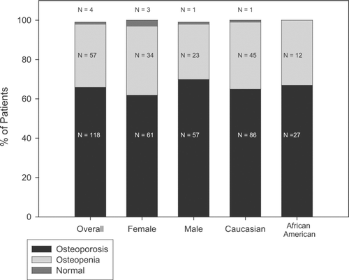
Patient demographics are listed in . Males and females (55%) were statistically similar in age, BMI, pack-year smoking history, FEV1%, TLC%, M-BODE, and BMD. 6 MWD and DLCO% were significantly lower in females compared to males. The prevalence of osteoporosis was similar among males and females () and fracture rate was not significantly different.
Table 2 Characteristics of study subjects by race and gender
Figure 1 The prevalence of osteoporosis was similar among males and females and fracture rate was not significantly different. The prevalence of osteoporosis was similar among African Americans and Caucasians (69.2% vs. 65.2%, p = 0.78).
Our study included 132 Caucasians and 39 AA (). The remaining 8 patients were Hispanic and Asian. Caucasians and AA were statistically similar with regards to age, weight, BMI, current prednisone use, pulmonary function, 6 MWD, and modified BODE. However, AA had smoked significantly less than Caucasians (37 ± 23 vs. 58 ± 33, p < 0.001).
Bone mineral density
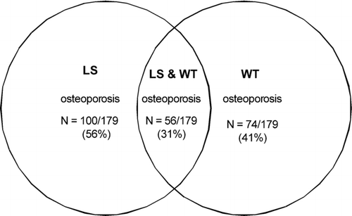
Osteoporosis and osteopenia were almost universal in our study population. Only 4 (3%) of severe COPD patients had a normal BMD at both LS and WT sites while 118 (66%) had osteoporosis and 57 (31%) had osteopenia. shows the occurrence of osteoporosis according to involved site. 59 (33%) patients had osteopenia at LS, and 95 (53%) had osteopenia at WT.
Figure 2 Distribution of osteoporosis & osteopenia by site involved in all COPD patients (n = 179).
The average BMD for all COPD patients, as well as divided by gender and race are listed in . There were no significant differences in the prevalence of osteoporosis or osteopenia between males and females (), nor any significant difference in the location where osteoporosis was identified.
Similarly, the frequency of osteoporosis was similar among AA and Caucasians (69% vs. 65%, p = 0.78) (). There was no significant difference in the presence of osteopenia between AA and Caucasians. Among AA the prevalence of osteopenia and osteoporosis at LS were 11 (28%) and 25 (64%), and in Caucasians 44 (33%) and 71 (54%), while at WT the prevalence of osteopenia and osteoporosis in Caucasians was 67 (51%) and 59 (45%), and in AA 24 (62%) and 13 (33%). Caucasians had significantly lower WT T scores and lower LS T scores that trended toward significance when compared to African Americans ().
Table 3 Bone mineral density of subjects studied in all group and gender and race
Bone fracture results
There was no significant difference in fracture rate based on gender or race. Fractures were reported on radiographs in 8 (13%) osteopenic patients, and 19 (16%) patients with osteoporosis. In the total group, 27 (15%) COPD patients had fractures; 15 (56%) females and 12 (44%) males. The fracture group had a lower WT BMD than the group without fractures (−2.77 ± −1.03 vs. −2.35 ± −0.95, p = 0.039). The most common fracture site was vertebral compression fractures (n = 14), then rib fractures (n = 8), followed by the knee (n = 2), wrist (n = 2) and hip (n = 1) fractures.
Risk factors and correlations
In the entire study population, only 22 patients (12%) were currently prescribed bisphosphonates and 20 patients (11%) were on calcium with or without vitamin D supplementation. Moreover, COPD patients not prescribed a statin had lower WT T scores than those who used statins (−2.47 vs. −1.99, p = 0.04). Prednisone users tended to have lower WT T scores compared to patients not currently using prednisone, but this did not reach statistical significance. In patients with identified fractures, 4% (1/27) were on a statin agent, compared with 12% (18/152) of the non-fracture group. Smoking pack year trended to be higher in patients with fractures than in those without fractures (63 ± 33 vs. 51 ± 32, p = 0.081).
Among the all COPD patients, there was a correlation between BMI and WT T scores (). There was also a weak correlation between FEV1% and LS T-scores (R = 0.18, p = 0.02). Univariate analysis identified only BMI and FVC% as being significant in predicting the development of osteoporosis (), and in subsequent multivariate analysis only FVC% was a significant predictor of osteoporosis based on LS & WT T score (), indicating that as FVC% increased, the bone mineral density (T-score) improved. No other factors were found to be significant predictors of osteoporosis.
Figure 3 Correlations between WT T score and BMI in all COPD patients (n = 179).
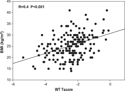
Table 4 Univariate and multivariate analysis of risk factors by logistic regression among all COPD (N = 179) who were identified as having osteoporosis (T-score ≤ –2.5)
GOLD staging and osteoporosis
We classified patients according to GOLD Stage (). The majority of patients were GOLD stage 3 or 4. The prevalence of osteoporosis was greater than 50% regardless of GOLD stage (Stage 2–57%, stage 3–54% and Stage 4–68%). No fractures where found in Stage 2 patients, while 20% of patients in Stage 3, and 14% patients in stage 4 had fractures. Six-minute walk distance was significantly decreased in Stage 4 compared to Stage 2 and 3.
Table 5 GOLD stage
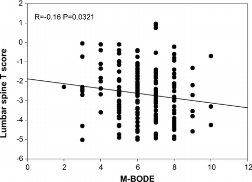
The mean modified BODE score in patients with osteoporosis was higher than patients without osteoporosis (6.41 ± 1.41 vs. 5.91 ± 1.70, p = 0.07), and modified BODE score correlated with lumbar spine T score ().
Figure 4 Correlation between LS T score and modified M-BODE score.
DISCUSSION
Our study shows a high prevalence of osteoporosis and fractures among patients with severe COPD regardless of sex or race. Lower BMI and FVC% are significant risk factors for osteoporosis in COPD patients. Overall, a T-score < −1.0 was present in 97% of the 179 patients. In our study 70% of men and 69% of AA with COPD were identified as having osteoporosis. While BMD measurements were similar among males and females, AA had a significantly higher WT BMD compared to Caucasians.
In the general population higher age, female gender, and low body weight are accepted risk factors for the development of osteoporosis. The National Osteoporosis Foundation estimates 80% of women and 20% of men over the age of 50 are thought to have osteoporosis. Among patients with COPD, 36 to 60% have been identified as having osteoporosis (Citation[1], Citation[13]). The mechanisms responsible for osteoporosis in patients with COPD appear to be multi-factorial and include corticosteroid use, reduced physical activity, poor nutrition, low body mass index, tobacco use, decreased exposure to sunlight, nutritional deficiencies, and hyper-catabolic effects of ongoing inflammatory processes (Citation[14]). To date the focus has been on exposure to corticosteroids, both systemic and inhaled. Systemic corticosteroids have been shown to adversely affect bone mineral density by decreasing new bone formation while increasing bone catabolism and accelerating bone loss. The rate of bone loss correlated loosely with the daily dose and duration of steroid therapy (Citation[1], Citation[15]). Most studies of BMD in advanced lung disease have found corticosteroid use to be an independent risk factor for osteoporosis (Citation[13], Citation[16]–19).
Patients with severe COPD had an equal incidence of osteoporosis among males and females. This differs from expected rates in the general population where females, especially post-menopausal females are at higher risk. The increased occurrence of osteoporosis among males could not be accounted for by differences in BMI, smoking history, or steroid use. McEvoy et al. reported that vertebral fractures were common in older men with COPD (Citation[20]) and the likelihood of bony fractures was greatest in men on continuous systemic corticosteroids.
In the general population, race is also a factor affecting the prevalence of osteoporosis with AAs having a lower incidence than Caucasians independent of gender. Studies in young adults show BMD is higher in black men and women than in white men and women, but there was no difference in women and men of the same race (Citation[12], Citation[13]). Looker et al reported 3–6% (1–2 million) of men have osteoporosis and 28–47% (8–13 million) have osteopenia; Most of the older U.S. adults with low femur BMD are women, but the number of men is substantial (Citation[21]). In contrast in our study, AA COPD patients had a 69% incidence of osteoporosis compared to 65% of Caucasians. We also found that AA COPD patients had significantly higher WT BMD and WT T scores than Caucasians. Demographic factors were similar between AA and Caucasians, except for the significant decrease in pack-years smoked.
We did not find the same association between corticosteroid use and BMD in severe COPD. This may have been due to the fact that we were only able to determine if patients were currently on steroids at the time of evaluation and not the total number of years or cumulative steroid dose.
Our study found lower BMI and FVC% independently associated with development of low T-score. In a recent study by Vrieze et al., BMI and FEV1 were risk factors for lower T scores in COPD patients (Citation[22]). Another study suggests that weight loss is associated with loss of BMD (Citation[23]). Katsura also found that in females with COPD there was a positive correlation between BMI and BMD (Citation[24]). Schliah et al. showed that FVC% and FEV1 were significantly decreased in patients with spinal osteoporotic fractures as compared to patients with chronic lower back pain without osteoporosis (Citation[25]). The low FEV1 and FVC in our patient group are most likely due to the severity of COPD in our patient population. In our study multivariate analysis revealed FVC% to be the only factor associated with osteoporosis in other words, patients with greater airflow obstruction had more negative T scores.
The overall fracture rate among all COPD patients in our study was 15%, this is lower than the fracture rate of 26.8% reported by Papaioannou (Citation[26]) and the fracture rates reported by McEvoy (Citation[20]). This discrepancy is most likely due to the fact that our fracture rate was based on radiographs done by the treating physician in response to patient's symptoms and therefore underestimates the true prevalence.
In conclusion, among patients with severe COPD not only Caucasian and female have significantly decreased bone mass, but also AA and males. Therefore, any patient with severe COPD, regardless of race or gender is likely to have osteoporosis or osteopenia. Clinicians treating COPD need to have a high index of suspicion for osteoporosis or osteopenia in order to treat patients and hopefully avoid the development of bone fractures.
This paper was funded by Pennsylvania Dept. of Health RFA 02-07-20.
REFERENCES
- Biskobing D M. COPD and osteoporosis. Chest 2002; 121: 609–620
- Goldstein M F, Fallon J J, Jr, Harning R. Chronic glucocorticoid therapy-induce osteoporosis in patients with obstructive lung disease. Chest 1999; 116: 1733–1749
- National Institues of Health Consensus Development Panel on Osteoporosis. Prevention, diagnosis, and therapy. JAMA 2001; 285: 785–795
- Seeman E., Wagner H, Offord K, Kumar R, Johnson W, Riggs B. Differential effects of endocrine dysfunction on the axial and appendicular skeleton. J Clin Invest 1982; 69: 1302–1309
- NHLBI/WHO. Global Initiative for Chronic Obstructive Lung Disease (GOLD) workshop report: global strategy for the diagnosis, management, and prevention of chronic obstructive pulmonary disease. 2005, Available at: http://www.goldcopd.com Accessed November 11, 2005
- Celli B R, Cote C G, Marin J M, Casanova C, Montes de Oca M, Mendez R A, Plata V P, Cabral H J. The body-mass index, airflow obstruction, dyspnea, and exercise capacity index in Chronic Obstructive Pulmonary Disease. N Engl J Med 2004; 350: 1005–1012
- American Thoracic Society. Lung function testing: selection of reference values and interpretative strategies. Am Rev Respir Dis 1991; 144: 1202–1218
- American Thoracic Society. Standardization of spirometry, 1994 update. Am J Respir Crit Care Med 1995; 152: 1107–1136
- American Thoracic Society. Single-breath carbon monoxide diffusing capacity (transfer factor): recommendations for a standard technique–1995 update. Am J Respir Crit Care Med 1995; 152: 2185–2198
- Crapo R O, Morris A H, Gardner R M. Reference spirometric values using techniques and equipment that meets ATS recommendations. Am Rev Respir Dis 1981; 123(6)659–664
- American Thoracic Society. ATS Statement: Guidelines for the Six-Minute Walk Test. Am J Respir Crit Care Med 2002; 166: 111–117
- Weinstein M S, Martin U J, Crookshank A D, Chatila W, Vance G B, Gaughan J P, Furukawa S, Criner G J. Mortality and functional performance in severe emphysema after lung volume reduction or transplant. COPD, (In press)
- Iqbal F, Michaelson J, Thaler L, et al. Declining bone mass in men with chronic pulmonary disease: contribution of glucocorticoid treatment, body mass index, and gonadal function. Chest 1999; 116: 1616–1624
- Eastell R. Treatment of postmenopausal osteoporosis. N Engl J Med 1998; 338: 736–746
- Ionescu A A, Schoon E. Osteoporosis in chronic obstructive pulmonary disease. Eur Respir J Suppl Nov, 2003; 46: 64s–75s
- Tschopp O, Boehler A, Speich R, Weder W, Seifert B, Russ E W, Schmid C. Osteoporosis before lung transplantation: association with low body mass index, but not with underlying disease. Am J Transplant Feb, 2002; 2(2)167–172
- Shane E, Silverberg S J, Donovan D, Papadopoulos A, Staron R B, Addesso V, Jorgesen B, McGregor C, Schulman L. Osteoporosis in lung transplantation candidates with end-stage pulmonary disease. Amer J Med 1996; 101(3)262–269
- National Institues of Health Consensus Development Panel on Osteoporosis. Prevention, diagnosis, and therapy. JAMA 2001; 285: 785–795
- Sin D. The risk of osteoporosis in caucasian men and women with Obstructive Airways Disease. Amer J Med 2003; 114: 10–14
- Mc Evoy C E, Ensrud K E, Bender E, Harry K. Association between corticosteroid use and vertebral fractures in older men with Chronic Obstructive Pulmonary Disease. Am J Respir Crit Care Med 1998; 157(3)704–709
- Looker A C, Orwoll E S, Johnston C C, Jr, Lindsay R L, Wahner H W, Dunn W L, Calvo M S, Harris T B, Heyse S P. Prevalence of low femoral bone density in older U.S. adults from NHANES III. J Bone Miner Res 1997; 12: 1761–1768
- Vrieze A, de Greef M H, Wýkstra P J, Wempe J B. Low bone mineral density in COPD patients related to worse lung function, low weight and decreased fat-free mass. Osteoporosis Int 2007, March 9
- Margolis K L, Ensrud K E, Schreiner P J, Tabor H K. Body size and risk for clinical fractures in old women. Study of Osteoporotic Fractures Research Group. Ann Intern Med 2000; 133: 123–127
- Katsura H, Kida K. A comparison of bone mineral density in elderly female patients with COPD and bronchial asthma. Chest 2002; 122: 1949–1955
- Schlaich C, Minne H W, Bruckner T. Reduced pulmonary function in patients with spinal osteoporotic fractures. Osteoporos Int 1998; 8(3)261–267
- Papaioannou A, Parkinson W, Ferko N, Probyn L. Prevalence of vertebral fractures among patients with chronic obstructive pulmonary disease in Canada. Osteoporos Int 2003; 14(11)913–917