
ABSTRACT
Isokinetic dynamometry represents the clinical gold standard for strength assessment but testing lack consensus. Elite youth male football players (n = 28) completed 20 repetitions (analysed as four epochs) of eccentric knee flexor (eccKF) and concentric knee extensor (conKE) trials at 60, 180 and 270°∙s−1, quantifying peak torque (PT) and functional range (FR). There was a significant (P < 0.001) main effect for fatigue and angular velocity in conKE PT; eccKF PT was not significant across epoch (P = 0.35) and velocity (P = 0.12) and a velocity x epoch interaction highlighted more repetitions were required to elicit fatigue as velocity increased. FR decreased with fatigue (P < 0.001) and velocity (P < 0.01) in conKE and eccKF, indicative of a narrowing of the strength curve. Clinical interpretation advocates an isokinetic test comprising at least 15 reps at a velocity ≥ 180°∙s−1 and analysis beyond the peak of the strength curve (PT) to inform clinical reasoning and individualized exercise prescription.
Introduction
The primary concern for a sports medicine practitioner working in a professional football club academy is the medical welfare of the player. A philosophy of evidence-based-practice dictates that research is conducted in such a way as to optimize the practical implementation of sports medicine support for each player. However, the demands of the professional context dictate that conducting research is time limited, and data collection has to deliver maximal gains from minimal time investment from the player. Efficacy, therefore, becomes a primary concern, and isokinetic dynamometry for the assessment of strength as a primary and modifiable risk factor for muscular strain injury is one such example, and the focus of this study.
Musculature injuries are a significant problem in professional football (Ekstrand et al., Citation2013), with knee extensor (quadriceps) and knee flexor (hamstring) injury incidence ranging from 19 to 32% (Ekstrand et al., Citation2011) and 12 to 37% (Opar et al., Citation2012), respectively, with implications for senior professional (Ekstrand et al., Citation2011; Woods et al., Citation2004) and elite youth players (Cloke et al., Citation2012). This is exacerbated by a high (~ 17%, Ekstrand et al., Citation2011) rate of injury recurrence for both hamstring and quadricep injuries in elite senior and youth players (Brooks et al., Citation2006; Ekstrand et al., Citation2016; Le Gall et al., Citation2006). Injury recurrence is typically associated with lengthier absences (Liu et al., Citation2012) and impacts upon the development of the young player. Muscular strength deficits (Askling et al., Citation2013; Croisier et al., Citation2008) and particularly reduced eccentric muscle strength (Nielsen & Yde, Citation1989; Van Dyk et al., Citation2016), of the quadriceps and hamstrings, are acknowledged as modifiable risk factors to reduce risk of myofascial, muscular or myotendinous junction and tendinous injuries.
Isokinetic dynamometry is considered to be the gold standard clinical assessment of muscular strength (Stark et al., Citation2011) but the literature highlights varied and often arbitrary selection of repetitions and testing velocities (Askling et al., Citation2003; Small et al., Citation2010; Van Dyk et al., Citation2017), at times offering little functional relevance to the mechanism of injury (Greig & Siegler, Citation2009). Previous studies have used three repetitions (Eustace et al., Citation2017), ten (Van Dyk et al., Citation2017), fifty (Sangier & Tourny, Citation2007), and in excess of one-hundred repetitions (Souron et al., Citation2018). Thigh musculature strength assessments are often completed with isokinetic velocities ranging from 60°·s−1 to 180°·s−1 (Croisier et al., Citation2008; Small et al., Citation2010; Van Dyk et al., Citation2016), with other studies using 300°·s−1 (Greig, Citation2008) to better reflect sport-specific functional demands acknowledging angular velocities of ~ 400°·s−1 during change of direction tasks (Nedergaard et al., Citation2014). The previous literature also highlights a lack of consensus in data analysis and the definition of outcome measures. Anterior and posterior thigh musculature imbalances have previously been quantified using a Hamstring:Quadriceps strength ratio (Aagard et al., Citation1998; Holcomb et al., Citation2007) defined as the ratio between peak concentric torque (PT) of hamstring and quadriceps. The functional strength ratio more appropriately considers the eccentric hamstring:concentric quadrideps strength ratio (Willigenburg et al., Citation2018), with a value closer to 1.0 considered to reduce the risk of hamstring injury (Holcomb et al., Citation2007; Orchard et al., Citation1997), but is also quantified using PT. The traditional use of peak torques and control ratios has more recently been usurped by a broader consideration of the strength curve, including angle-specific torque and functional range (Eustace et al., Citation2017).
The clinical gold standard must be applicable in a way that suits the demands of a professional football club. Anecdotally practitioners have expressed concern over the time taken, the risk of injury, and the clinical value in isokinetic assessments. This helped to shape a desire for a single data collection trial, and a data analysis process that would directly inform training interventions and would be transferable across medical departments (physiotherapy, sports science, strength and conditioning). The aim of this study was to utilize a battery of isokinetic testing to inform prescription of a single trial for subsequent player profiling within a professional football club. Whilst previous studies highlight the lack of power in predicting injury (Green et al., Citation2017; Van Dyk et al., Citation2016, Citation2017), this negates the purpose of assessment which is to inform (p)rehabilitation rather than to predict injury. The present study considers elite youth players, with age acknowledged as a risk factor for thigh musculature injuries. It has been suggested that elite adolescent athletes between the ages of 16 and 18, as well as periods after adolescent growth demonstrate a greater risk of thigh musculature injury (Cloke et al., Citation2012; Van der Sluis et al., Citation2015).
Methods
Participants
Twenty-eight elite youth male players (Age: 16.7 ± 1.3 years; height: 179.2 ± 11.2 cm; Mass: 72.4 ± 8.6 kg) from the same club in the English Football League Championship were recruited to participate in this study. Inclusion criteria required the participants to have received no quadriceps or hamstrings injuries within the previous 12 weeks, be contracted to the football club, be aged between 16 and 19, and be completing a weekly training volume of ~14 hour week−1. Prior to each experimental condition, all participants were required to complete a health screening procedure comprising a health, physical activity and pre-exercise control questionnaire. All players provided written consent and were made aware that data would remain anonymized and would not affect their standing within the team. Parent/guardian consent was also obtained for the youth players aged below 18 years. Ethical consent was provided by Edge Hill University’s research ethics committee and in accordance with the Helsinki declaration.
Experimental procedures
Players completed a single experimental trial, and in attempt to reduce the influence of residual fatigue, as identified in the PPI consultations, a 48-h rest-period from exercise was factored into the player’s weekly macrocycle. Pre-testing the dynamometer was calibrated in accordance with manufacturer guidelines, whilst participants completed a standardized 5-min warm up on a stationary cycle ergometer (Monark, 824E, Sweden) at 60 W (Eustace et al., Citation2017) and were given opportunity to complete self-directed mobilizations and stretching. Three trial repetitions were completed prior to each experimental trial, with an experimental trial comprising 20 maximal effort repetitions of dominant limb isokinetic (Biodex Medical Systems, System 2, Shirley, New York, USA) strength assessments at angular velocities of 180, 270, and 60°∙s−1. This testing order was designed to minimize the impact of the slowest 60°∙s−1 trial, associated with greater time under tension, on subsequent trials (Greig, Citation2008). Concentric knee extensor (conKE) and eccentric knee flexor (eccKF) trials were completed in randomized order. Passive knee flexion performed at 60°·s−1 separated each repetition, with a rest period of 10 min interspersing each set. Verbal feedback was providing solely during the familiarization trial with no feedback provided during the experimental trial. The isokinetic dynamometer was set up specific to each individual with the crank axis aligned with the axis of rotation of the knee and anatomically referenced to 90° of knee flexion. Data collection reflected the full range of motion for each player, with a gravity correction applied with 0° of knee flexion. The lever arm cuff securely fastened around the ankle, approximately 5 cm proximal to the malleolus (Eustace et al., Citation2017; Greig, Citation2008).
Data analysis
Each 20-repetition set was sub-divided into four epochs of five repetitions and the peak repetition of each epoch was identified and used for data analysis (Van Dyk et al., Citation2016). Analysis was restricted to isokinetic data, defined by applying a 1% cut-off to the predetermined angular velocity. The peak torque (PT) was identified for each rep, with functional range (FR) defined as the range over which 85% of PT was maintained (Eustace et al., Citation2017). PT values recorded from the eccKF and conKE assessments were used to calculate the traditional dynamic control ratio (DCR) at each testing velocity.
Statistical analyses
Statistical analysis was conducted using PASW Statistics Editor 24.0 for windows (SPSS Inc., Chicago, IL USA). Before parametric analysis, the assumptions of normality of the residual values were assessed using a Shapiro–Wilk test, with significance set at P ≤ 0.05. Subsequently, a repeated measures general linear model was used to investigate main effects for testing velocity, epoch and velocity x epoch interactions in each parameter. Where appropriate post-hoc pairwise comparisons with a Bonferonni correction were applied. For all significant interactions, 95% confidence intervals (CI) and observed power are reported in conjunction with the frequentist statistical approach. All data were reported as mean ± SD.
Results
Peak torque
summarizes the influence of angular velocity and epoch on peak torque. There was a significant (P < 0.001; Observed Power = 0.99) main effect for velocity in conKE, with PT decreasing as velocity increased. There was no significant (P = 0.12) main effect for velocity in EccKF, with PT maintained at ~126 Nm.
Figure 1. The influence of (a) angular velocity and (b) epoch on conKE and eccKF PT.
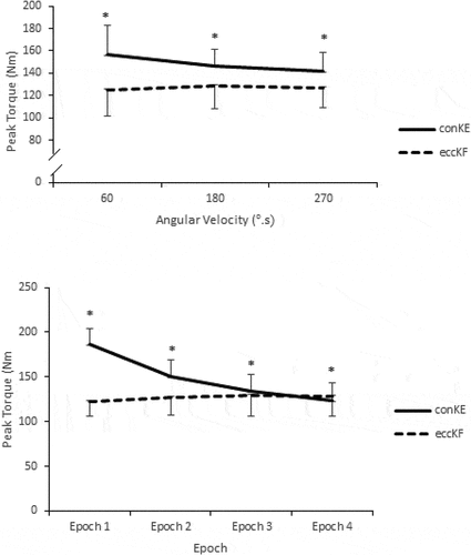
There was also a significant (P < 0.001; Observed Power = 0.98) main effect for epoch in conKE with PT decreasing with prolonged exercise duration. There was no significant (P = 0.35) main effect for epoch in eccKF PT.
summarizes the influence of contraction modality, angular velocity and epoch on PT. The general linear model (GLM) identified a significant epoch x velocity conKE interaction (P < 0.001; Observed Power = 0.97) with significantly (P ≤ 0.01) higher values recorded for 60°·s−1 when compared to all other velocities in each epoch, whilst 180°·s−1 displayed significantly higher values (P < 0.01) when compared to 270°·s−1. Furthermore, an epoch x velocity interaction was also identified for eccKF PT (P < 0.001; Observed Power = 0.75) with significant differences highlighted in epoch 2 (P ≤ 0.01), epoch 3 (P ≤ 0.01) and epoch 4 (P ≤ 0.01).
Table 1. The influence of angular velocity, contraction type and epoch on PT.
Functional range
summarizes the influence of angular velocity and epoch on functional range. There was a significant main effect for angular velocity in conKE FR (P < 0.001; Observed Power = 0.99) with a significantly lower FR at 270°·s−1 (18.09 ± 4.25°) when compared to 60°·s−1 (20.20 ± 5.67°; P = 0.01; 95% CI = 0.40 to 3.81). A significant (P < 0.01; Observed Power = 0.99) main effect for velocity in eccKF FR was also identified, with significantly lower values at 270°·s−1 (18.48 ± 9.15°) compared to 60°·s−1 (24.23 ± 7.14°; P = 0.03; 95% CI = −10.80 to −0.69) and 180°·s−1 (21.66 ± 10.25°; P = 0.01; 95% CI = −5.95 to −0.42).
Figure 2. The influence of (a) angular velocity and (b) epoch on conKE and eccKF FR.
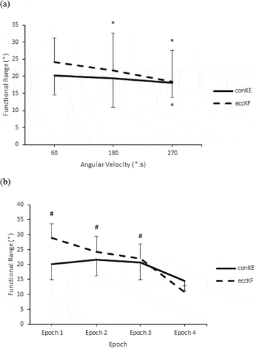
There was also a significant main effect (P < 0.001; Observed Power = 0.85) for epoch in conKE with significantly lower functional range during epoch 4 (14.53 ± 3.29°) when compared to epoch 1 (20.21 ± 5.23°; 95% CI = −0.17 to – 2.20), epoch 2 (21.58 ± 5.33; 95% CI = −11.89 to – 2.22) and epoch 3 (20.72 ± 5.89; 95% CI = −10.48 to −1.90). Significant velocity x epoch interactions are identified (P < 0.001; Observed Power = 0.98) for conKE FR in epoch 3 only, with significantly lower functional ranges highlighted at 270°∙s−1 (13.05 ± 2.94°) when compared to 60°∙s−1 (24.74 ± 6.35°; P < 0.001; 95% CI = – 17.81 to – 5.56) and 180°∙s−1 (24.37 ± 9.97°; P < 0.001; 95% CI = – 16.60 to −6.03).
A similar trend was observed for eccKF FR, with a significant main effect for epoch (P < 0.001; Observed Power = 0.98) highlighting significantly lower values at epoch 4 (10.72 ± 2.07°) when compared to epoch 1 (28.86 ± 4.71°; P = <0.001; 95% CI = −25.98 to −10.30), epoch 2 (24.33 ± 5.21°; P = 0.01; 95% CI = −24.44 to −2.79) and epoch 3 (21.92 ± 4.98°; P = 0.02; 95% CI = −21.09 to −1.30). No significant (P = 0.56) eccKF FR epoch x velocity interactions were identified.
Dynamic control ratio (DCR)
highlights a significant main effect for DCR PT60 (P < 0.001; Observed Power = 0.98) and epoch, with post-hoc Bonferonni correction factors highlighting a temporal fatigue effect (P < 0.001; Observed Power = 0.91) with significantly increased values observed during epoch 1 (1.61 ± 0.34), when compared to all subsequent variables. No significant epoch main effects were identified for DCR PT180 (P = 0.31) nor DCR PT270 (P = 0.64).
Table 2. The influence of epoch and angular velocity on dynamic control ratio PT (DCR PT).
Discussion
The primary aim of this study was to prospectively develop an optimal isokinetic screening trial based on analysis of an isokinetic test on elite youth football players. In the clinical context, the optimal screening trial would be time and resource effective whilst offering the necessary analysis to inform interventions.
Practical implications for isokinetic data collection
A primary consideration in isokinetic assessment is the testing velocity (Greig, Citation2008). There was a significant main effect for velocity in conKE peak torque, as expected given the classic force–velocity relationship of muscle and supporting previous observations of reduced conKE strength with increasing velocity (Eustace et al., Citation2017; Greig, Citation2008). In contrast, eccKF peak torque is maintained with increasing velocity, and therefore as angular velocity increased the quadriceps dominance apparent at slow velocities was reduced. The conKE and eccKF torque-velocity curves did not cross as observed by Eustace et al. (Citation2019) in senior professional players, where the eccentric knee flexors became dominant at velocities greater than approximately 200°∙s−1. This might reflect the relative training status of the samples, with the youth players used in the current study not generating the same magnitude of peak torques observed by Eustace et al. (Citation2017). The lack of a main effect for velocity in eccKF would suggest that angular velocity is not absolutely paramount when developing a screening trial, where perceived safety of slower velocities might be preferable to the arguably more functionally relevant velocities approaching the limits of isokinetic dynamometers. However, testing velocity is only part of the clinical reasoning process in data collection and research design.
In the current study, data collection included 20-repetitions, subsequently sub-divided into four epochs. There was a significant main effect for epoch (with the data pooled for all velocities) in conKE but not in eccKF, suggesting that the concentric knee flexors exhibited an acute within-set fatigue effect. This observation might reflect a specific response to modality, differentiating the mechanism of concentric and eccentric muscular contractions. Alternatively, this finding might be indicative of a lack of strength endurance in concentric knee flexors, which is not apparent in the eccentric knee flexors. Relative adaptations in conKE strength and eccKF strength endurance in these youth players might reflect their training history, but might also identify specific training needs. Whilst strength is widely acknowledged as a primary risk factor for muscular strain injury (Croisier et al., Citation2008; Van Dyk et al., Citation2016), the physical demands of football and epidemiological observations of increased injury risk during the latter stages of match-play also stress the importance of strength endurance. The resistance of eccKF strength to a 20-repetition set might reflect a history of adaptation to intermittent running, whereas the decline over 20 repetitions in conKE might reflect a gym-based strength emphasis defined by lower repetitions.
With the data considered at each testing velocity discretely, the velocity x epoch interaction in eccKF peak torque highlighted that a fatigue-induced inflection point in peak torque shifted towards the latter parts of the set as angular velocity increased. This might represent the muscular work done per repetition, with the increase in angular velocity over the same range eliciting a lower demand and therefore enabling more repetitions before the onset of acute fatigue. Therefore, whilst testing velocity does not influence the magnitude of peak torque in eccentric hamstrings, there is a velocity x rep interaction that should inform data collection. In this respect, we advocate a higher testing velocity within the constraints of the equipment, since a higher velocity would offer greater functional specificity to the mechanism of injury (Eustace et al., Citation2017; Greig & Siegler, Citation2009), without increasing the risk of injury perceived by some practitioners. At this higher angular velocity, we would advocate 15–20 repetitions in order to induce a fatigue effect. Differentiating deficits in strength and/or strength endurance would better inform personalized interventions and exercise prescription.
Practical implications for isokinetic data analysis
The 20-repetition set would enable the calculation of a fatigue index, further informing the training needs of the player beyond maximum strength. The inclusion of functional range in data analysis is designed to extend consideration of the strength curve beyond the single maxima in peak torque that is typically presented in the literature. Considering strength as a function of velocity and joint angle helps to inform individualized training interventions, particularly when considering strength deficits are the aetiological risk (Croisier et al., Citation2008; Van Dyk et al., Citation2016). The main effect for velocity observed in functional range is partly influenced by a narrowing of the isokinetic range and should, therefore, be treated with caution, but the influence of epoch also suggests a narrowing of the strength curve. This has implications for injury, since the demands of the sport require knee range of motion beyond the angle of peak torque, and the mechanism of injury can be diverse.
The dissociation between peak torque and functional range in their sensitivity to testing velocity and repetition volume adds value to this metric, in contrast to work done for example, which is highly correlated with peak torque (Dvir, Citation2014). Whilst in conKE the player reduced peak torque but was able to retain functional range for more reps, in eccKF the player maintained peak torque but suffered a progressive reduction in functional range. This differential between peak and width of the curve has implications for injury risk, and subsequently for exercise prescription. Angle-match torque data are additional means of considering specifics of the strength curve (Eustace et al., Citation2017), but at the higher testing velocities the isokinetic phase is reduced. In the current study, the isokinetic phase at the highest angular velocities did not offer data beyond approximately 35° of knee flexion in this sample of elite youth male footballers. We would, therefore, advocate angle-matched data where there was a specific rationale, for example, when monitoring rehabilitation of an injured player. The lack of isokinetic torque data at high velocities might be supplemented by isometric testing where appropriate.
Care should be taken in generalizing these findings beyond the specific sample and testing protocol used. The players were elite male youth footballers with implications for their training status and history, injury free, and tested in a non-fatigued state. The optimum isokinetic screen might, therefore, differ in other samples, such as senior or female players, and in other sports reflecting specificity in adaptation. The influence of previous injury on the response to a high velocity and high volume test also warrants consideration. Similarly, the testing protocol of 20 repetitions was used to reflect the strength endurance training regime of these players, and future research might consider more repetitions and/or more sets, acknowledging a potential lag in response to fatigue.
Conclusion
Isokinetic profiling is widely acknowledged as the gold standard measure of strength (Buckthorpe et al., Citation2018; Eustace et al., Citation2017; Van Dyk et al., Citation2017) but should be considered as a means of informing needs-based interventions, and not as a predictor of injury. Strength imbalances should be considered as an internal, modifiable risk factor, but many complex interactions may then precede an injurious event. In data collection, we advocate high velocity testing ≥ 180°∙s−1 to provide greater functional relevance to the common mechanisms of injury, to reflect the potential cross-over defined by ipsi-lateral strength symmetry, and to reduce the total amount of work done per repetition. Correspondingly, we advocate a high number ≥ 15 reps at this velocity to induce an acute inflection and to enable calculation of fatigue indices and needs in strength endurance. In analysis, we advocate investigation away from the single maxima of peak torque and towards a broader consideration of the strength curve. Identifying personalized needs defining strength deficits as a function of velocity, joint angle and repetition volume provides a more comprehensive profile upon which to develop evidence-based clinical exercise prescription.
Acknowledgments
The authors wish to thank all of the players at Preston North End Football Club Academy who participated in the study as well their parents and all of the Academy Medical Staff with additional thanks to Nick Harrison (Academy Manager) and Mathew Jackson (Head of 1st Team Medicine) in enabling this project to be completed.
Disclosure statement
No competing interests declared.
References
- Aagard, P., Simonsen, E. B., Magnusson, S. P., Larsson, B., & Dhyre-Poulsen, P. (1998). A new concept for isokinetic hamstring: Quadricepsmuscle strength ratio. American Journal of Sports Medicine, 26(2), 231–237. https://doi.org/10.1177/03635465980260021201
- Askling, C., Karlsson, J., & Thorstensson, A. (2003). Hamstring injury occurrence in elite soccer players after preseason strength training with eccentric overload. Scandinavian Journal of Medicine & Science in Sports, 13(4), 244–250. https://doi.org/10.1034/j.1600-0838.2003.00312.x
- Askling, C., Tengvar, M., & Thorstensson, A. (2013). Acute hamstring injuries in Swedish elite soccer: A prospective randomised controlled clinical trial comparing two rehabilitation protocols. British Journal of Sports Medicine, 47(15), 953–959. https://doi.org/10.1136/bjsports-2013-092165
- Brooks, J., Fuller, C., Kemp, S., & Reddin, D. (2006). Incidence, risk, and prevention of hamstring muscle injuries in professional rugby union. The American Journal of Sports Medicine, 34(8), 1297–1306. https://doi.org/10.1177/0363546505286022
- Buckthorpe, M., Wright, S., Bruce-Low, S., Nanni, G., Sturdy, T., Gross, A., Bowen, L., Styles, B., Della Villa, S., Davison, M., & Gimpel, M. (2018). Recommendations for hamstring injury prevention in elite soccer: Translating research into practice. British Journal of Sports Medicine, 53(7), 449–456. https://doi.org/10.1136/bjsports-2018-099616
- Cloke, D., Moore, O., Shab, T., Rushton, S., Shirley, M., & Deehan, D. (2012). Thigh muscle injuries in youth soccer. The American Journal of Sports Medicine, 40(2), 433–439. https://doi.org/10.1177/0363546511428800
- Croisier, J., Ganteaume, S., Binet, J., Genty, M., & Ferret, J. (2008). Strength imbalances and prevention of hamstring injury in professional soccer players. The American Journal of Sports Medicine, 36(8), 1469–1475. https://doi.org/10.1177/0363546508316764
- Dvir, Z. (2014). Relevant, less relevant and irrelevant isokinetic strength test parameters: Some critical comments. Movement & Sport Sciences, 85, 15–21. https://doi.org/10.1051/sm/2013088
- Ekstrand, J., Askling, C., Magnusson, H., & Mithoefer, K. (2013). Return to play after thigh muscle injury in elite soccer players: Implementation and validation of the Munich muscle injury classification. British Journal of Sports Medicine, 47(12), 769–774. https://doi.org/10.1136/bjsports-2012-092092
- Ekstrand, J., Hägglund, M., & Waldén, M. (2011). Epidemiology of muscle injuries in professional soccer. The American Journal of Sports Medicine, 39(6), 1226–1232. https://doi.org/10.1177/0363546510395879
- Ekstrand, J., Waldén, M., & Hägglund, M. (2016). Hamstring injuries have increased by 4% annually in men’s professional soccer, since 2001: A 13-year longitudinal analysis of the UEFA Elite Club injury study. British Journal of Sports Medicine, 50(12), 731–737. https://doi.org/10.1136/bjsports-2015-095359
- Eustace, S. J., Page, R., & Greig, M. (2019). Isokinetic strength differences between elite senior and youth female soccer players identifies training requirements. Physical Therapy in Sport, 39, 45–51. https://doi.org/10.1016/j.ptsp.2019.06.008
- Eustace, S. J., Page, R. M., & Greig, M. (2017). Contemporary approaches to isokinetic strength assessments in professional soccer players. Science and Medicine in Soccer, 1(3), 251–257. https://doi.org/10.1080/24733938.2017.1371851
- Green, B., Bourne, M., & Pizzari, T. (2017). Isokinetic strength assessment offers limited predictive validity for detecting risk of future hamstring strain in sport: A systematic review and meta-analysis. British Journal of Sports Medicine, 52(5), 329–336. https://doi.org/10.1136/bjsports-2017-098101
- Greig, M. (2008). The influence of soccer soccer-specific fatigue on peak isokinetic torque production of the knee flexors and extensors. American Journal of Sports Medicine, 36(7), 9–16. https://doi.org/10.1177/0363546508314413
- Greig, M., & Siegler, J. (2009). Soccer-specific fatigue and eccentric hamstrings muscle strength. Journal of Athletic Training, 44(2), 180–184. https://doi.org/10.4085/1062-6050-44.2.180
- Holcomb, W. R., Rubley, M. D., Lee, H. J., & Guadagnoli, M. A. (2007). Effect of hamstring-emphasized resistance training on hamstring: Quadricepsstrength ratios. Journal of Strength and Conditioning Research, 21(1), 41–47. https://doi.org/10.1519/R-18795.1
- Le Gall, F., Carling, C., Reilly, T., Vandewalle, H., Church, J., & Rochcongar, P. (2006). Incidence of injuries in elite french youth soccer players. The American Journal of Sports Medicine, 34(6), 928–938. https://doi.org/10.1177/0363546505283271
- Liu, H., Garrett, W., Moorman, C., & Yu, B. (2012). Injury rate, mechanism, and risk factors of hamstring strain injuries in sports: A review of the literature. Journal of Sport and Health Science, 1(2), 92–101. https://doi.org/10.1016/j.jshs.2012.07.003
- Nedergaard, N. J., Kersting, U., & Lake, M. (2014). Using accelerometry to quantify deceleration during a high-intensity soccer turning manoeuvre. Journal of Sports Science, 32(20), 1897–1905. https://doi.org/10.1080/02640414.2014.965190
- Nielsen, A., & Yde, J. (1989). Epidemiology and traumatology of injuries in soccer. The American Journal of Sports Medicine, 17(6), 803–807. https://doi.org/10.1177/036354658901700614
- Opar, D. A., Williams, D., & Shield, A. J. (2012). Hamstring strain injuries: Factors that lead to injury and re-injury. Sports Medicine, 42(3), 209–226. https://doi.org/10.2165/11594800-000000000-00000
- Orchard, J., Marsden, J., Lord, S., & Garlick, D. (1997). Preseason hamstring muscle weakness associated with hamstring muscle injury in Australian footballers. American Journal of Sports Medicine, 25(1), 81–85. https://doi.org/10.1177/036354659702500116
- Sangier, S., & Tourny, C. (2007). Comparison of the decrease in strength between hamstrings and quadriceps during isokinetic fatigue testing in semiprofessional soccer players. International Journal of Sports Medicine, 28(11), 952–957. https://doi.org/10.1055/s-2007-964981
- Small, K., McNaughton, L., Greig, M., & Lovell, R. (2010). The effects of multidirectional soccer-specific fatigue on markers of hamstring injury risk. Journal of Science and Medicine in Sport, 13(1), 120–125. https://doi.org/10.1016/j.jsams.2008.08.005
- Souron, R., Nosaka, K., & Jubeau, M. (2018). Changes in central and peripheral neuromuscular fatigue indices after concentric versus eccentric contractions of the knee extensors. European Journal of Applied Physiology, 118(4), 805–816. https://doi.org/10.1007/s00421-018-3816-0
- Stark, T., Walker, B., Phillips, J., Fejer, R., & Beck, R. (2011). Hand-held dynamometry correlation with the gold standard isokinetic dynamometry: A systematic review. PM&R, 3(5), 472–479. https://doi.org/10.1016/j.pmrj.2010.10.025
- Van der Sluis, A., Elferink-Gemser, M. T., Brink, M. S., & Visscher, C. (2015). Importance of peak height velocity timing in terms of injuries in talented soccer players. International Journal of Sports Medicine, 36(4), 327–332. https://doi.org/10.1055/s-0034-1385879
- Van Dyk, N., Bahr, R., Burnett, A. F., Whiteley, R., Bakken, A., Mosler, A., Farooq, A., & Witvrouw, E. (2017). A comprehensive strength testing protocol offers no clinical value in predicting risk of hamstring injury: A prospective cohort. British Journal of Sports Medicine, 51(23), 1695–1702. https://doi.org/10.1136/bjsports-2017-097754
- Van Dyk, N., Bahr, R., Whiteley, R., Tol, J., Kumar, B., Hamilton, B., Farooq, A., & Witvrouw, E. (2016). Hamstring and quadriceps isokinetic strength deficits are weak risk factors for hamstring strain injuries. The American Journal of Sports Medicine, 44(7), 1789–1795. https://doi.org/10.1177/0363546516632526
- Willigenburg, N. W., McNally, M. P., & Hewett, T. E. (2018). Quadriceps and hamstrings strength in athletes. In C. C. Kaeding & J. R. Borchers (Eds.), Hamstring and quadriceps injuries in athletes (pp. 15–28). Springer US.
- Woods, C., Hawkins, R. D., Malty, S., Hulse, M., Thomas, A., & Hodson, A. (2004). The soccer association medical research programme: An audit of injuries in professional soccer--analysis of hamstring injuries. British Journal of Sports Medicine, 38(1), 36–41. https://doi.org/10.1136/bjsm.2002.002352