
Abstract
Multi-plane phase retrieval is a well established technique for reconstructing both, amplitude and phase of an object wave. This standard technique works best, if the intensity of the object wave changes rapidly along the optical axis. For slowly varying intensities, the iterative procedure may not converge at all. To overcome this limitation we combined the standard technique with a periodic phase element. We demonstrate that a binary diffractive micro lens array with overlapping aperture significantly improves the convergence of phase retrieval and thus the quality of reconstruction. Thus multi-plane phase retrieval can be applied for both rough and smooth phase distributions.
NOMENCLATURE
| dz | = |
distance between two adjacent planes |
| f | = |
focal distance of micro lens |
| FT | = |
Fourier transformtion |
| iFT | = |
inverse Fourier transformation |
| N plane | = |
number of planes |
| NA design | = |
design numerical aperture |
| NA cell | = |
cell numerical aperture |
| P | = |
lens pitch of micro lens array |
| u0 | = |
complex amplitude distribution at z = 0 |
| uz | = |
complex amplitude distribution at z |
| Z 0 | = |
initial distance between the first plane and the diffractive element |
| Z13 | = |
13th standard Zernike polynomial |
| λ | = |
wave length |
| Ω | = |
overlap factor |
1. INTRODUCTION
It is well known, that for optical frequencies there is no detector, which is fast enough to record the complex amplitude. As a consequence, detectors can only record intensity, no phase. Due to this loss of phase information, indirect methods for the reconstruction of the complex wave front are still an important issue. A variety of methods has been invented for this purpose. They can be classified into four groups. Firstly, there are interferometric techniques, like classical interferometry and holography, which derive phase information through the addition of a reference wave. Therefore, all the interferometric techniques require a coherent reference wave, high spatial resolution, and a rigid optical setup. To relax some of these requirements, noninterferometric methods are progressively used in a multitude of applications.
The second group of phase reconstruction methods, denoted as gradient-based approaches, were among the earliest noninterferometric ways of obtaining phase information. Examples include the Foucault knife-edge test,[ Citation 1 ] the Hartmann-Shack wave front sensor,[ Citation 2 ] phase retrieval based on the intensity transport equation[ Citation 3 , Citation 4 ] and the Ragazzoni pyramid wave front sensor.[ Citation 5 ]
A very new and attractive group of phase reconstruction methods utilize the phase-space representation: the Wigner distribution function[ Citation6-9 ] or ambiguity function.[ Citation 10 , Citation 11 ] Since the phase-space representation consists of both the spatial and the frequency information, its dimension is doubled, i.e., the phase-space representation of a two-dimensional wave front is four-dimensional. For special symmetries of the complex wave front[ Citation 12 , Citation 13 ] or by utilization of a one-dimensional operator[ Citation 14 ] one can efficiently reduce the dimensions of phase-space representations. The redundancy investigation showed that the efficiency of phase reconstruction can be improved further.[ Citation15-17 ]
The last group of phase reconstruction methods uses a consistency approach, which is applied iteratively. Two-plane phase retrieval[ Citation 18 , Citation 19 ] was successfully used in electron microscopy. Multi-plane phase retrieval[ Citation 20 , Citation 21 ] is a well established technique for reconstructing the complex amplitude of an object wave. The technique proposed in this article combines the standard technique of multi-plane phase retrieval with an additional periodic phase element. The principle of this technique will be described in the next section in full detail.
2. PHASE RETRIEVAL WITH A DIFFRACTIVE OVERLAPPING MICRO LENS ARRAY
Classical iterative phase retrieval, like the Gerchberg-Saxton algorithm, works by iterating between the object plane and the pupil plane.[ Citation 18 , Citation 19 ] The iterative processing involves forward and backward Fourier transformation. In each of the planes, the calculated intensity distribution is replaced with the measured one. After a few iterations the resulting phase distribution converges to the phase of the wave front to be recovered. This technique has been extended to reconstruct the complex amplitude from a multitude of Fresnel planes,[ Citation 20 , Citation 21 ] by replacing two intensity planes with multiple intensity planes and by replacing the Fourier transformation with a Fresnel transformation. The spatial resolution of the reconstruction is determined essentially by the sensor pixel size and thus can be much higher than for Hartmann-Shack sensing. Since not only the phase, but the full complex amplitude is recovered, phase retrieval represents a viable alternative both to interferometry and to digital holography as a reliable scalar wave reconstruction technique. However, the convergence of multi-plane phase retrieval depends strongly on the variation of recorded intensities. For a fast and correct convergence, it is necessary that the intensity changes significantly from plane to plane. For smooth phase distributions such as spherical waves with large curvature radius or tilted plane waves, the variation of recorded intensities is so insignificant, that the phase reconstruction does not converge or does converge to a wrong result. As a consequence of this, the application range of multi-plane phase retrieval is restricted to rough phase distributions.
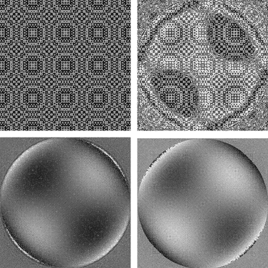
The technique presented here is based on multi-plane phase retrieval, but we include a diffractive binary phase element which acts like a micro lens array (Figure ). The lens array introduces an additional intensity variation in the recorded intensity distribution even for smooth phase distributions. The combined phase of the object wave and the diffractive micro lens array is recovered reliably after only a few iterations. For the reconstruction of the object phase, a calibration measurement of the diffractive micro lens array alone has to be carried out before and its phase can be numerically eliminated from the combined phase. Figure illustrates the complete reconstruction steps. The iteration kernel is scalar light propagation based on angular spectrum propagation as shown in the following:
Figure 1 Measurement setup for phase retrieval with a binary diffractive micro lens array, inserted between the sample and the first plane of intensity recordings.
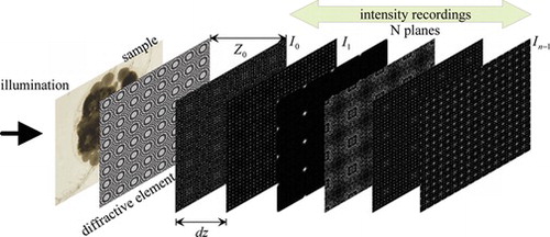
Figure 2 Reconstruction steps: 1. Reconstruction of diffractive micro lens array alone (top left); 2. Reconstruction of the superposition of object and diffractive micro lens array (top right); 3. Elimination of the diffractive micro lens array phase (bottom left); 4. Backward propagation of the reconstructed complex field into the object plane (bottom right).
Initially we have used a refractive micro lens array, which has also the effect of increasing the intensity variation, therefore accelerating the convergence of the algorithm. By using a refractive micro lens array, however, another problem occurs: The intensity along the optical axis of each lens is insensitive to a constant phase bias added to the micro lens phase. Therefore, in the reconstruction, there is an unknown constant phase bias, which varies from micro lens to micro lens. For continuous phase distributions, this unknown phase bias in each micro lens window can in principle be removed by stitching adjacent windows, i.e., by adding a constant phase which restores continuity across the edges. This method will, however, fail in the case of discontinuous phase distributions.
The phase bias problem can be removed completely, if the apertures of the micro lenses overlap, thus introducing a coupling between adjacent micro lens windows. Such an overlap can only be realized using diffractive optical elements. Using a computer designed diffractive micro lens array, we can adjust the focal length and the overlap factor freely and therefore can control the coupling between neighboring lenses in a wide range. Using diffractive overlapping micro lens array (DOMLA), we were also able to increase the intensity variation, thus accelerating the convergence of the algorithm, but without the problem of unknown phase bias.
For these overlapping diffractive lenses, the diameter, the focal distance f and the numerical aperture N A design can be chosen independently of the lens pitch P. The cell NA is defined by the aperture angle arising from one single micro lens period P:
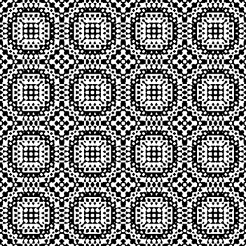
Figure 3 Phase distribution of the binary diffractive micro lens array with overlapping apertures. The overlap factor is 3. White corresponds to a phase level of π and black to a phase level of 0.
For the fabrication of the DOMLAs, we applied a photo resist with a thickness of 500 nm (1 PI - phase shift) and used a laser lithography for structuring. More detailed information about design and fabrication of such diffractive lenses can be found in.[ Citation22-24 ] Traditionally, diffractive elements show a strong wavelength dependence. For the DOMLAs used in our application, the coherence requirements are much less stringent in such a way that a wavelength change has mostly an effect on focal length and only very little effect on diffraction efficiency. Changing the illumination wavelength from red to blue, e.g., shifts the focal spot to a larger distance away from the phase element. Yet, the overall focal pattern remains almost unchanged in comparison to using monochromatic illumination. By using white light, the focal spot is spread longitudinally into a spectrum from red (near) to blue (far). In fact we used a red LED light source for illumination, and it showed almost the same intensities as using a monochromatic illumination, but the incoherent nature of the LED avoids the speckle noise that would be observable with a laser.
For tolerance analysis, the number of planes N plane , the initial distance of the first plane to the diffractive element Z 0 and the distance between two adjacent planes dz were varied in a set of numerical simulations. The amplitude quantization caused by a camera was also taken into account in our numerical simulations by saving the intensities in 8-bit-image files. The reconstructions revealed that with more than 7 planes the algorithm converges quickly after 3 iterations. The distance between two adjacent planes should be larger than f/30. To utilize the dynamic range of the camera, the intensities should not be recorded too close to the focal plane of the diffractive micro lens. Therefore the initial position Z 0 should be considerably less or larger than the focal length f.
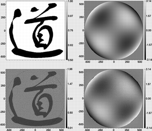
In order to estimate the achievable accuracy of phase measurement, we simulated a complex wave front reconstruction. The object phase was the standard Zernike polynomial Z13 and the object amplitude was the picture of a chinese character. Figure shows a comparison between original and reconstructed amplitude and phase correspondingly. The measured standard phase deviation in this computer experiment was less than λ/22, demonstrating the applicability of phase retrieval also for interferometric applications.
Figure 4 Reconstruction simulation of a complex wave front, with Standard Zernike Z13 as the phase: original amplitude (top left), original phase (top right), reconstructed amplitude (bottom left) and reconstructed phase (bottom right). The standard deviation of the phase is below λ/22.
The DOMLA approach, proposed here may appear very similar to an alternative approach of using a random amplitude or phase element to improve phase retrieval. Note however, that the DOMLA is a regular structure, while the random phase plate is a stochastic structure. The approach of using a random amplitude or phase element was suggested by several authors before.[ Citation25-28 ] E.g., the papers by Almoro and Hanson used a random phase plate to improve convergence. In order to analyze the differences, we compared the reconstructions using a binary random phase plate to our technique. The main observation was that the random phase plate may produce phase anomalies in the iterative reconstruction. With the overlapping lenses we have never seen such anomalies.
3. EXPERIMENTAL RESULTS
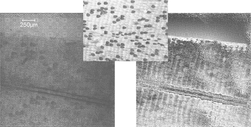
Figure and show the reconstructed amplitude and phase of real experimental samples. In Figure a dog flea was used as object and in Figure a piece of a butterfly wing was reconstructed. The small images in the top centre were recorded with a conventional wide-field microscope. All the reconstructions were carried out in four iterations and the relevant parameters used in the measurement were: N planes = 10, dz = 750 µm (distance between two adjacent planes) and Z 0 = f/5 (distance between DOMLA and the first measurement plane). The reconstructed complex amplitude allows a numerical refocusing. Thus the volume nature of these objects could also be verified. For the dog flea example, we could focus at different parts of the leg and observe how these parts were moving in and out of focus.
Figure 5 Experimental reconstruction of a dog flea: microscope image (top); reconstructed amplitude (left bottom); reconstructed phase (right bottom).
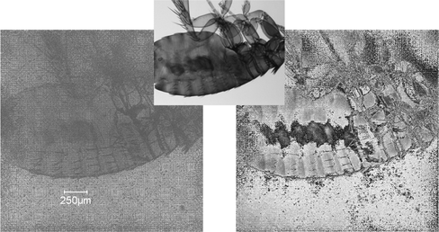
Figure 6 Experimental reconstruction of a piece of butterfly wing: microscope image (top); reconstructed amplitude (left bottom); reconstructed phase (right bottom).
4. SUMMARY
By introducing a DOMLA in the standard multi-plane phase retrieval method, the amplitude and the phase of arbitrary wave fronts can be reconstructed reliably. Unlike conventional multi-plane phase retrieval, which works only for rough phase distributions, this method also performs well for smooth phase distributions. Because amplitude and phase are reconstructed with high accuracy, the method represents an alternative to digital holography as well as to interferometry. The optical set-up is robust and simple and does not require any imaging lenses. Thus no image aberrations or geometric distortions are introduced in the measurement. The field of view is only limited by the sensor size and the resolution by the pixel size of the sensor. Unlike holography, no laser light is needed; therefore the results are speckle free. No additional spatial bandwidth has to be reserved for the reference wave like in off-axis holography.
Notes
Color versions of one or more of the figures in the article can be found online at www.tandfonline.com/uopt.
REFERENCES
- Foucault , L.M. Memoire sur la construction des telescopes en verre argente . Ann. Observ. Paris 1859 , 5 , 197 – 237 .
- Malacara , D. Optical Shop Testing. , 2nd Edition . John Wiley and Sons : New York , 1992 , 472 – 484 .
- Teague , M.R. Deterministic phase retrieval: A Green's function solution . J. Opt. Soc. Am. 1983 , 73 , 1434 – 1441 .
- Streibl , N. Phase imaging by the transport equation of intensity . Opt. Commun. 1984 , 49 , 6 – 10 .
- Ragazzoni , R. Pupil plane wavefront sensing with an oscillating prism . J. Mod. Opt. 1996 , 43 , 289 – 293 .
- Brenner , K.-H. ; Lohmann , A.W. Wigner distribution function display of complex 1D signals . Opt. Commun. 1982 , 42 , 310 – 314 .
- Lohmann , A.W. Image rotation, Wigner rotation, and the fractional Fourier transform . J. Opt. Soc. Am. A 1993 , 10 , 2181 – 2186 .
- Raymer , M.G. ; Beck , M. ; McAlister , D.F. Complex wave-field reconstruction using phase-space tomography . Phys. Rev. Lett. 1994 , 72 , 1137 – 1140 .
- McAlister , D.F. ; Beck , M. ; Clarke , L. ; Mayer , A. ; Raymer , M.G. Optical phase retrieval by phase-space tomography and fractional-order Fourier transformation . Opt. Lett. 1995 , 20 , 1181 – 1183 .
- Brenner , K.-H. ; Lohmann , A.W. ; Ojeda-Castaneda , J. The ambiguity function as a polar display of the OTF . Opt. Commun. 1983 , 44 , 323 – 326 .
- Tu , J. ; Tamura , S. Wave field determination using tomography of the ambiguity function . Phys. Rev. E 1997 , 55 , 1946 – 1949 .
- Dragoman , D. ; Dragoman , M. ; Brenner , K.-H. Amplitude and phase recovery of rotationally symmetric beams . Appl. Opt. 2002 , 41 , 5512 – 5518 .
- Dragoman , D. ; Dragoman , M. ; Brenner , K.-H. Tomographic amplitude and phase recovery of vertical-cavity surface-emitting lasers by use of the ambiguity function. Opt. Lett. 2002, 27, 1519–1521.
- Liu , X. ; Brenner , K.-H. Reconstruction of two-dimensional complex amplitudes from intensity measurements . Opt. Commun. 2003 , 225 , 19 – 30 .
- Dragoman , D. Redundancy of phase-space distribution functions in complex field recovery problems . Appl. Opt. 2003 , 42 , 1932 – 1937 .
- Semichaevsky , A. ; Testorf , M. Phase-space interpretation of deterministic phase retrieval . J. Opt. Soc. Am. A 2004 , 21 , 2173 – 2179 .
- Liu , X. ; Hruscha , C. ; Brenner , K.-H. Efficient reconstruction of two-dimensional complex amplitudes utilizing the redundancy of the ambiguity function . Appl. Opt. 2008 , 47 , E1 – E7 .
- Gerchberg , R.W. ; Saxton , W.O. A practical algorithm for the determination of phase from image and diffraction plane pictures . Optik 1972 , 35 , 237 – 246 .
- Fienup , J.R. Phase retrieval algorithms: a comparison . Appl. Opt. 1982 , 21 , 2758 – 2769 .
- Pedrini , G. ; Osten , W. ; Zhang , Y. Wave-front reconstruction from a sequence of interferograms recorded at different planes . Opt. Lett. 2005 , 30 , 833 – 835 .
- Almoro , P. ; Pedrini , G. ; Osten , W. Complete wavefront reconstruction using sequential intensity measurements of a volume speckle field . Appl. Opt. 2006 , 45 , 8596 – 8605 .
- Brenner , K.-H. ; Buschlinger , R. Parallel image scanning with binary phase grating . J. Europ. Opt. Soc. Rap. Publ. 2011 , 6 , 11024 .
- Hulsken , B. ; Vossen , D. ; Stallinga , S. High NA diffractive array illuminators and application in a multi-spot scanning microscope . J. Europ. Opt. Soc. Rap. Publ. 2012 , 7 , 12026 .
- Liu , X. ; Stenau , T. ; Brenner , K.-H. Diffractive micro lens arrays with overlapping apertures. In Proc. 11th Euro-American Workshop on Information Optics. 2012 .
- Zhang , F. ; Pedrini , G. ; Osten , W. Phase retrieval of arbitrary complex-valued fields through aperture-plane modulation . Phys. Rev. A 2007 , 75 , 043805 .
- Anand , A. ; Pedrini , G. ; Osten , W. ; Almoro , P. Wavefront sensing with random amplitude mask and phase retrieval . Opt. Lett. 2007 , 32 , 1584 – 1586 .
- Almoro , P.F. ; Hanson , S.G. Random phase plate for wavefront sensing via phase retrieval and a volume speckle field . Appl. Opt. 2008 , 47 , 2979 – 2987 .
- Almoro , P.F. ; Pedrini , G. ; Gundu , P.N. ; Osten , W. ; Hanson , S.G. Appl. Opt. 2010 , 35 , 1028 – 1030 .
- Color versions of one or more of the figures in the article can be found online at www.tandfonline.com/uopt.